Slow release protein polymers
a protein and slow release technology, applied in the direction of prosthesis, parathyroid hormones, metabolic disorders, etc., can solve the problems of inability to use the sustained release route of drug delivery, the active form of protein is difficult to form in biodegradable polymers, and the delivery of protein to the systemic and local circulation is still relatively rapid, so as to achieve the effect of low burst
- Summary
- Abstract
- Description
- Claims
- Application Information
AI Technical Summary
Benefits of technology
Problems solved by technology
Method used
Image
Examples
example 1
General Preparation of a Macromer Solution
[0133]The protein was weighed out, and the following components were added to the protein: (i) 90 mM TEOA / PBS, pH 8.0; (ii) 35% n-vinyl pyrrolidinone (n-VP); and (iii) 1000 ppm Eosin. The resulting mixture was stirred well using a spatula. The solution was kept in the dark for about 10 minutes, or until the macromer had absorbed all of the solution, or until the solution was homogenous.
[0134]Macromer solutions having the following ingredients were prepared.
[0135]
AmountAmountAmountAmount90 mM35%1000 ppmAmountAmountTotalProteinTEOAn-VPEosin3.4kL52kL5amount15 mg57 mg15 mg3 mg45 mg 0 mg135 mg15 mg57 mg15 mg3 mg 0 mg45 mg135 mg
example 2
Precipitation of hGH and Formulating into Hydrogel Microspheres
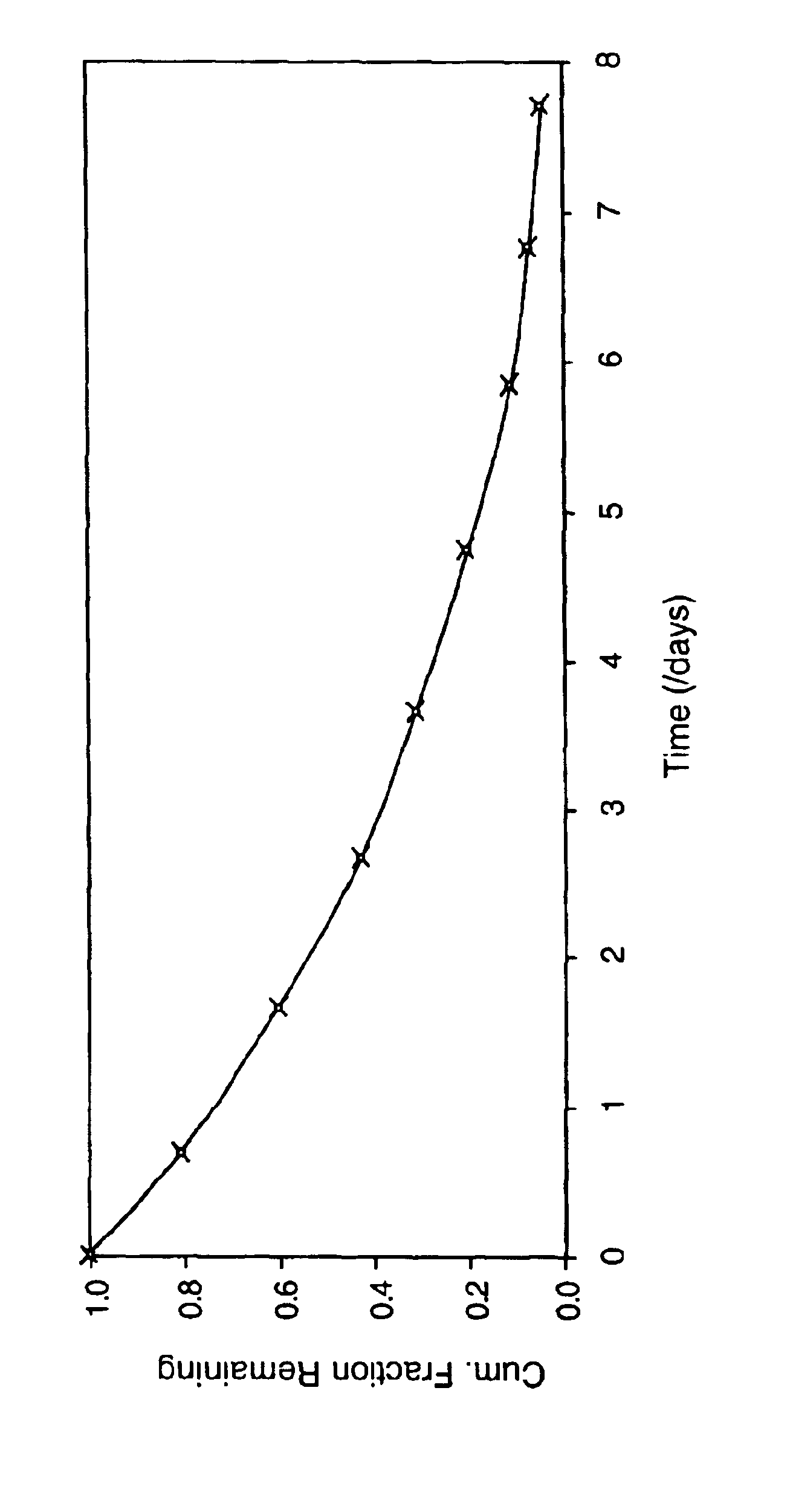
[0136]To a 100 mg / ml hGH solution in 5 mM ammonium hydrogen carbonate buffer 77 μl of a 1300 mM triethanolamine, pH 8, solution was added. Upon the addition of 400 mg of PEG 2K to the above solution, a fine precipitate of hGH was formed. The sample was centrifuged at 4000 rpm for several minutes and 0.9 ml of the supernatant was removed. To the precipitated mixture, 1 g of 4.2kL5-A3 macromer was added, followed by the addition of 0.1 ml of a 10 mM Eosin Y solution. The mixture was then emulsified in an oil phase to form microspheres which were polymerized using an argon laser. The in vitro release characteristics of this formulation are shown in FIG. 1. No burst was observed and release continued for at least 5 days.
example 3
Micronization of Freeze-Dried Human Growth Hormone and Formulation into Hydrogel Microspheres
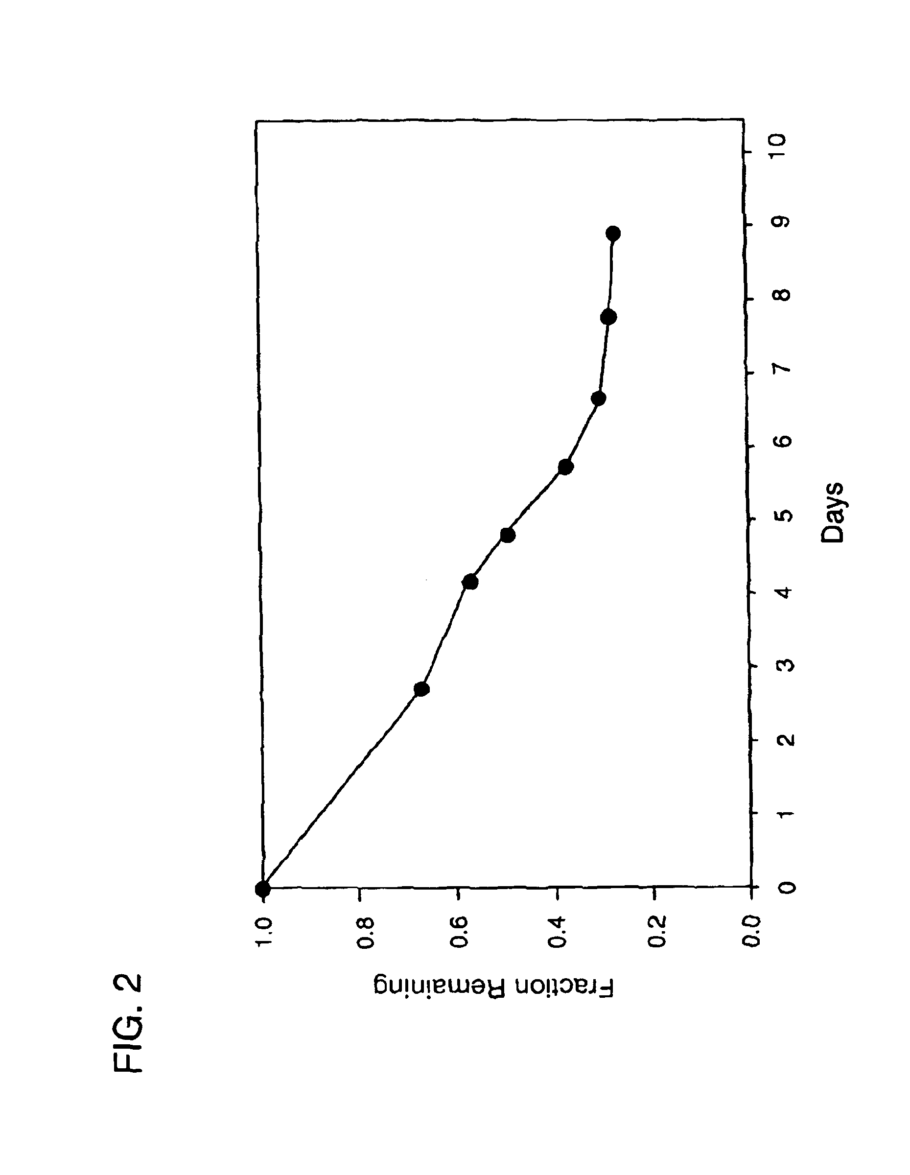
[0137]A 10 mg / ml solution of hGH ammonium acetate was frozen and freeze-dried, resulting in a dry cake of pure hGH. The following macromer solution was prepared: 1 g 2kL5, 1.2 g phosphate buffered saline (pH 7), 0.4 g of a solution of 25% trehalose and 0.4% F-68 in water, 0.24 g of a 10% solution of 2,2-dimethoxy 2 phenyl-acetophenone in tetrahydrofuran. To this solution was added 0.2 g of the freeze-dried hGH. Following mixing to disperse the hGH, the hGH in suspension was further micronized by 20 passages through a 1.5 inch 20 g needle. This suspension was then emulsified in an oil phase to form microspheres which were polymerized by exposure to UV light (365 nm) for 3 min. The in vitro release characteristics of this micropheres are shown in FIG. 2.
PUM
| Property | Measurement | Unit |
|---|---|---|
| Length | aaaaa | aaaaa |
| Fraction | aaaaa | aaaaa |
| Fraction | aaaaa | aaaaa |
Abstract
Description
Claims
Application Information
 - R&D
- Intellectual Property
- Life Sciences
- Materials
- Tech Scout
- Unparalleled Data Quality
- Higher Quality Content
- 60% Fewer Hallucinations
Browse by: Latest US Patents, China's latest patents, Technical Efficacy Thesaurus, Application Domain, Technology Topic, Popular Technical Reports.
© 2025 PatSnap. All rights reserved.Legal|Privacy policy|Modern Slavery Act Transparency Statement|Sitemap|About US| Contact US: help@patsnap.com


