
On-line correction of patient motion in three-dimensional positron emission tomography

a three-dimensional positron emission tomography and patient technology, applied in tomography, instruments, applications, etc., can solve the problems of blurred pet image, time-consuming reprocessing of file data, and mode approach to motion de-blurring

Inactive Publication Date: 2005-09-20
SIEMENS MEDICAL SOLUTIONS USA INC
View PDF7 Cites 12 Cited by
  • Summary
  • Abstract
  • Description
  • Claims
  • Application Information

AI Technical Summary

Benefits of technology

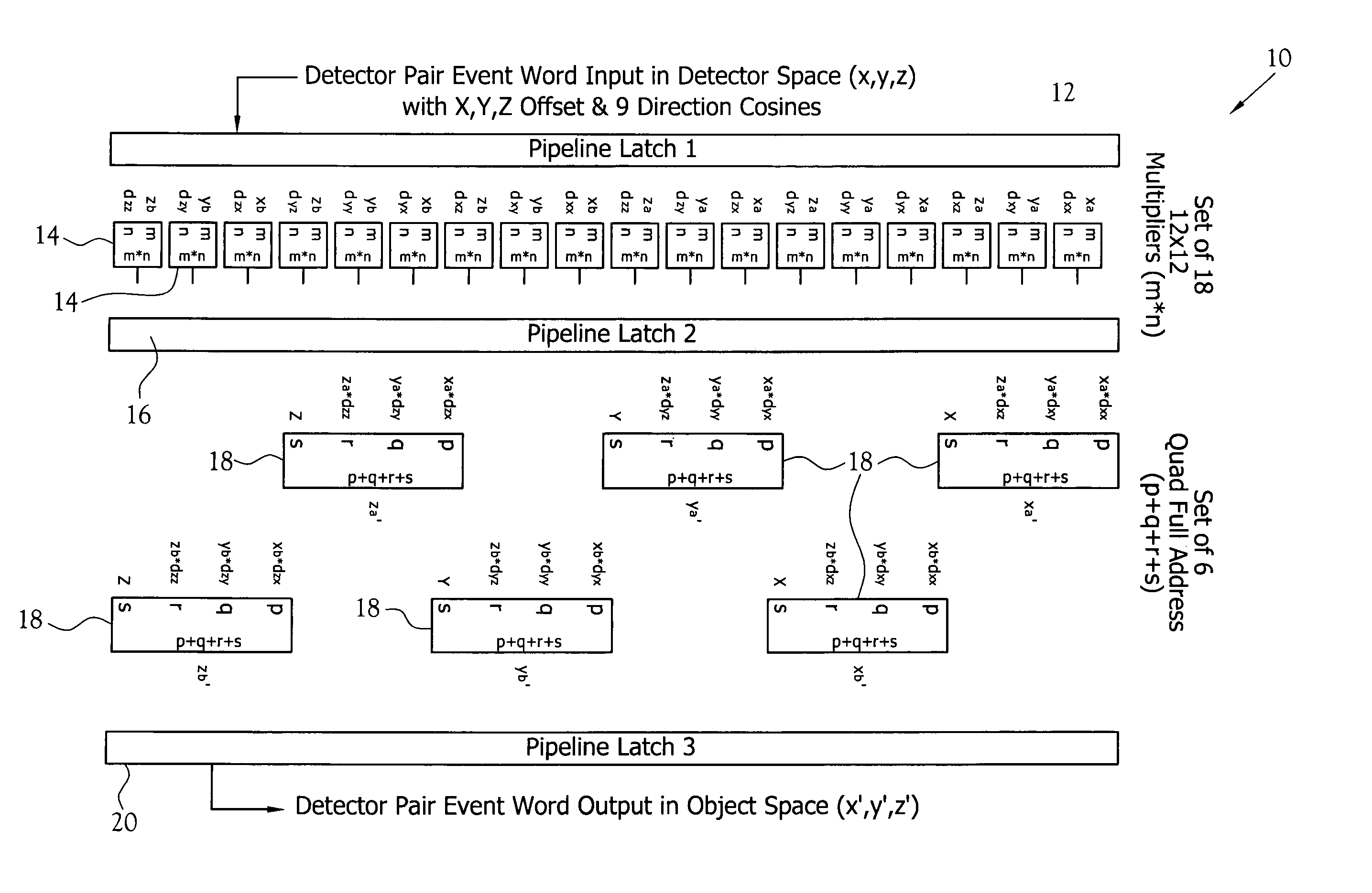
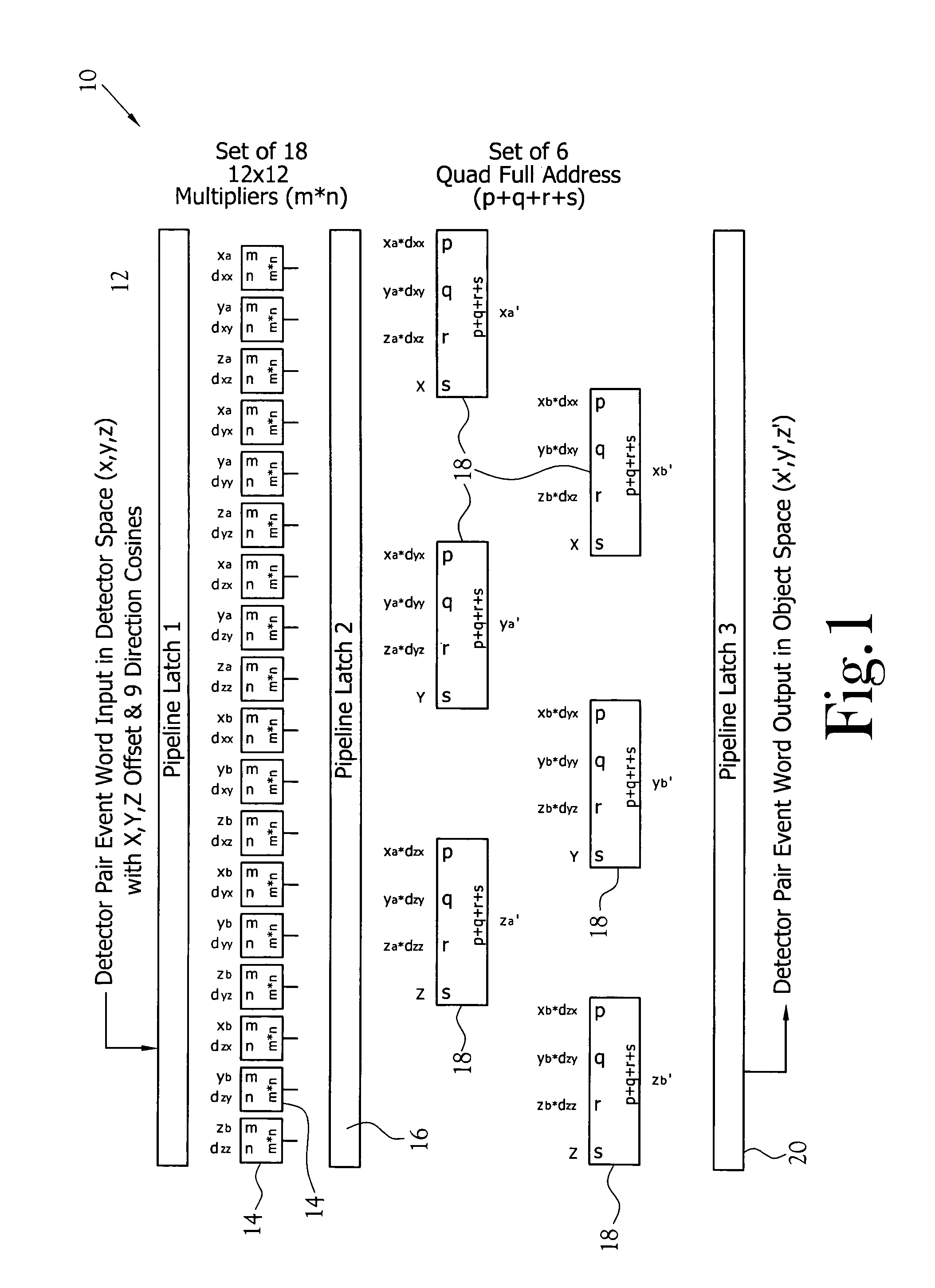
[0022]While data for a specific LOR and a current object orientation are input to the first digital pipeline latch, processing for a different LOR and an earlier object orientation (limited to the 3 translation offsets, X Y Z) are stored in the second digital pipeline latch. Additionally, fully tra...

Problems solved by technology

However, if the patient or object in the field of view (FOV) does not remain stationary, the PET image is blurred.
While this event-by-event list mode approach corrects for the motion of, for example, a patient's head, the reprocessing of the file data is a time consuming task.
The aforementioned list mode approach to motion de-blurring is problematic in view of these economic pressures in that more operator interaction and unacceptably long processing times are required.
However, whole body PET scans also suffer from a lack of correction for motion.
While problems limit effectiveness compared to brain scans, gross motions of the whole human body may also be de-blurred.
Difficult technical challenges remain to more effectively correct for the relative motions between and within individual organs such as, among others, the heart, lungs, liver, and lymph nodes.
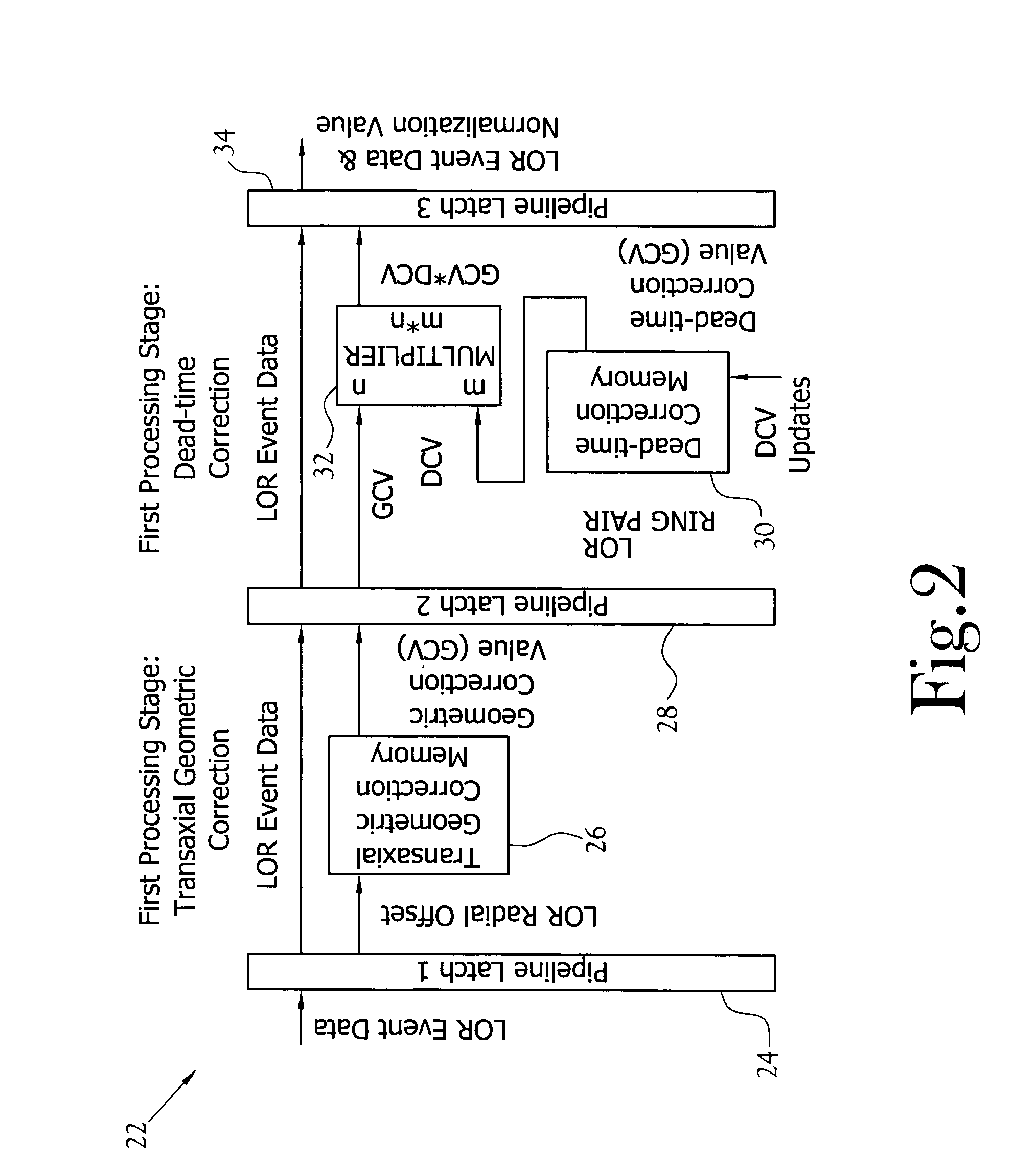
Another known obstacle to on-line motion correction in the context of most existing PET applications is on-line normalization.
The known digital electronic technique of pipelining is applied to arithmetic operations such that the speed of the electrical circuit is limited not by the whole computation but instead by the slowest individual piece of the computation.
However, such technique has not been shown in the prior art to be successful in the environment of on-line correction of patient data in a PET scan to account for patient motion.

Method used


Image

  • On-line correction of patient motion in three-dimensional positron emission tomography
  • On-line correction of patient motion in three-dimensional positron emission tomography
  • On-line correction of patient motion in three-dimensional positron emission tomography

Examples


Embodiment Construction

[0028]A device and method for on-line correction of patient motion in three-dimensional positron emission tomography (3D PET) incorporating various features of the present invention is illustrated generally at 10 in the figures. The device for on-line correction of patient motion in 3D PET, or device 10, is designed to apply a correction for patient motion in an on-line and real-time manner. Moreover, the device 10 provides a means whereby as each PET coincidence event is detected, each line of response (LOR) is re-mapped or rebinned in real time from a stationary 3-D reference space of the PET detector array into a virtual 3-D reference space which moves dynamically with the patient.

[0029]Discussion of the present invention will be directed primarily toward correction of motion of the patient's head. However, it will be understood the present invention is applicable to other portions of a patient's body as well. As discussed previously, with respect to PET, the skull and brain are ...


PUM


Abstract

A device and method for on-line correction of patient motion in three-dimensional positron emission tomography. The devices encompass an on-line hardware pipelining architecture to support 3D translation, normalization, and weighted histogramming as required. For the 3D translation circuit, a first digital pipeline latch is provided for receiving data as it is collected by the PET scanner. A bank of multiplier circuits receives the PET scan data. Each multiplier circuit receives and multiplies a portion of the entire scan data simultaneous with each other multiplier circuit. The product of each multiplier circuit is output to a second digital pipeline latch. The data is then passed to a bank of adders, each of which supports four input variables. While a specific LOR and a current object orientation are input to the first digital pipeline latch, processing for a different LOR and an earlier object orientation are stored in the second digital pipeline latch. Additionally, fully transformed coordinates from a third LOR are loaded into a third digital pipeline latch. As the banks may each complete their respective tasks under a threshold time limit, the pipelining technique permits the entire 3D transformation for the AB pair to take place in real time. Further, on-line normalization and on-line weighted histogramming are also performed in real time.

Description

CROSS-REFERENCE TO RELATED APPLICATIONS[0001]Not Applicable.STATEMENT REGARDING FEDERALLY SPONSORED RESEARCH OR DEVELOPMENT[0002]Not Applicable.BACKGROUND OF THE INVENTION[0003]1. Field of Invention[0004]This invention relates to the field of positron emission tomography (PET). More specifically, the present invention is related to a device and method for improving PET resolution through on-line correction of patient motion.[0005]2. Description of the Related Art[0006]In the art of PET detection, it is known that the resolution of a scan is in part dependent upon the amount of motion of the patient during the scan, and specifically the motion of the portion of the body being scanned. It is known that typical PET scans last from fifteen (15) minutes to three (3) hours. Ideally, if the patient could lie motionless during a PET scan, a high resolution PET system such as the CTI Pet Systems, Inc., EXACT HR+ is capable of 4 to 5 mm 3-D image resolution. However, if the patient or object ...

Claims


Application Information

Patent Timeline
IPC IPC(8): A61B5/11G01T1/29
CPCA61B6/037A61B5/418G01T1/2985A61B5/415A61B5/1114A61B5/1127
Inventor JONES, WILLIAM F.
Owner SIEMENS MEDICAL SOLUTIONS USA INC
Learn More