Transfection method and uses related thereto

Inactive Publication Date: 2005-10-04
WHITEHEAD INST FOR BIOMEDICAL RES
View PDF19 Cites 48 Cited by
  • Summary
  • Abstract
  • Description
  • Claims
  • Application Information

AI Technical Summary

Benefits of technology

[0013]After drying is complete (has occurred to a sufficient extent that the DNA remains affixed to the surface under the conditions used in the subsequent steps of the method), eukaryotic cells into which the DNA is to be reverse transfected are placed on top of the surfaces onto which the DNA-containing mixture has been affixed. Actively growing cells are generally used and are plated, preferably at high density (such as 1×10⁵ / cm2), on top of the surface containing the affixed DNA-containing mixture in an appropriate medium, such as Dulbecco's Modified Eagles Medium (DMEM) containing 10% heat-inactivated fetal serum (IFS) with L-glutamine and penicillin / streptomycin (pen / strep). Other media can be used and their components can be determined based on the type of cells to be transfected. The resulting slides, which contain the dried lipid-DNA mixture and cells into which the DNA is to be reverse transfected, are maintained under conditions appropriate for growth of the cells and entry of DNA, such as an entry of an expression vector containing the DNA, into cells. In the present method, approximately one to two cell cycles are sufficient for reverse transfection to occur, but this will vary with the cell type and conditions used and the appropriate length of time for a specific combination can be determined empirically. After sufficient time has elapsed, slides are assessed for reverse transfection (entry of DNA into cells) and expression of the encoded product or effect of the introduced DNA on reverse-transfected cells, using known methods. This can be done, for example, by detecting immunofluorescence or enzyme immunocytochemistry, autoradiography, in situ hybridization, or other means of detecting expression of the DNA or an effect of the encoded product or of the DNA itself on the cells into which it is introduced. If immunofluorescence is used to detect expression of an encoded protein, an antibody that binds the protein and is fluorescently labeled is used (e.g., added to the slide under conditions suitable for binding of the antibody to the protein) and the location (spot or area of the surface) containing the protein is identified by detecting fluorescence. The presence of fluorescence indicates that reverse transfection has occurred and the encoded protein has been expressed in the defined location(s) which show fluorescence. The presence of a signal, detected by the method used, on the slides indicates that reverse transfection of the DNA into cells and expression of the encoded product or an effect of the DNA in recipient cells has occurred in the defined location(s) at which the signal is detected. As described above, the identity of the DNA present at each of the defined locations is known; thus, when expression occurs, the identity of the expressed protein is also known.
[0014]Thus, the present invention relates, in one embodiment, to a method of expressing defined DNA, such as cDNA or genomic DNA, in defined locations or areas of a surface onto which different DNAs, such as DNA in a vector, such as an expression vector, has been affixed, as described herein. Because each area of the surface has been covered / spotted with DNA of known composition, it is a simple matter to identify the expressed protein. In addition, the present method is useful to identify DNAs whose expression alters (enhances or inhibits) a pathway, such as a signaling pathway in a cell or another property of a cell, such as its morphology or pattern of gene expression. The method is particularly useful, for example, as a high-throughput screening method, such as in a microarray format. It can be used in this format for identifying DNAs whose expression changes the phosphorylation state or subcellular location of a protein of interest or the capacity of the cell to bind a reagent, such as a drug or hormone ligand. In a second embodiment, which is also useful as a high-throughput screening method, DNA reverse transfected into cells has an effect on cells or interacts with a cellular component(s) without being expressed, such as through hybridization to cellular nucleic acids or through antisense activity.

Problems solved by technology

The emerging challenge is to uncover the functional roles of the genes and to quickly identify gene products with desired properties.

Method used


Image

  • Transfection method and uses related thereto
  • Transfection method and uses related thereto
  • Transfection method and uses related thereto

Examples


example 1

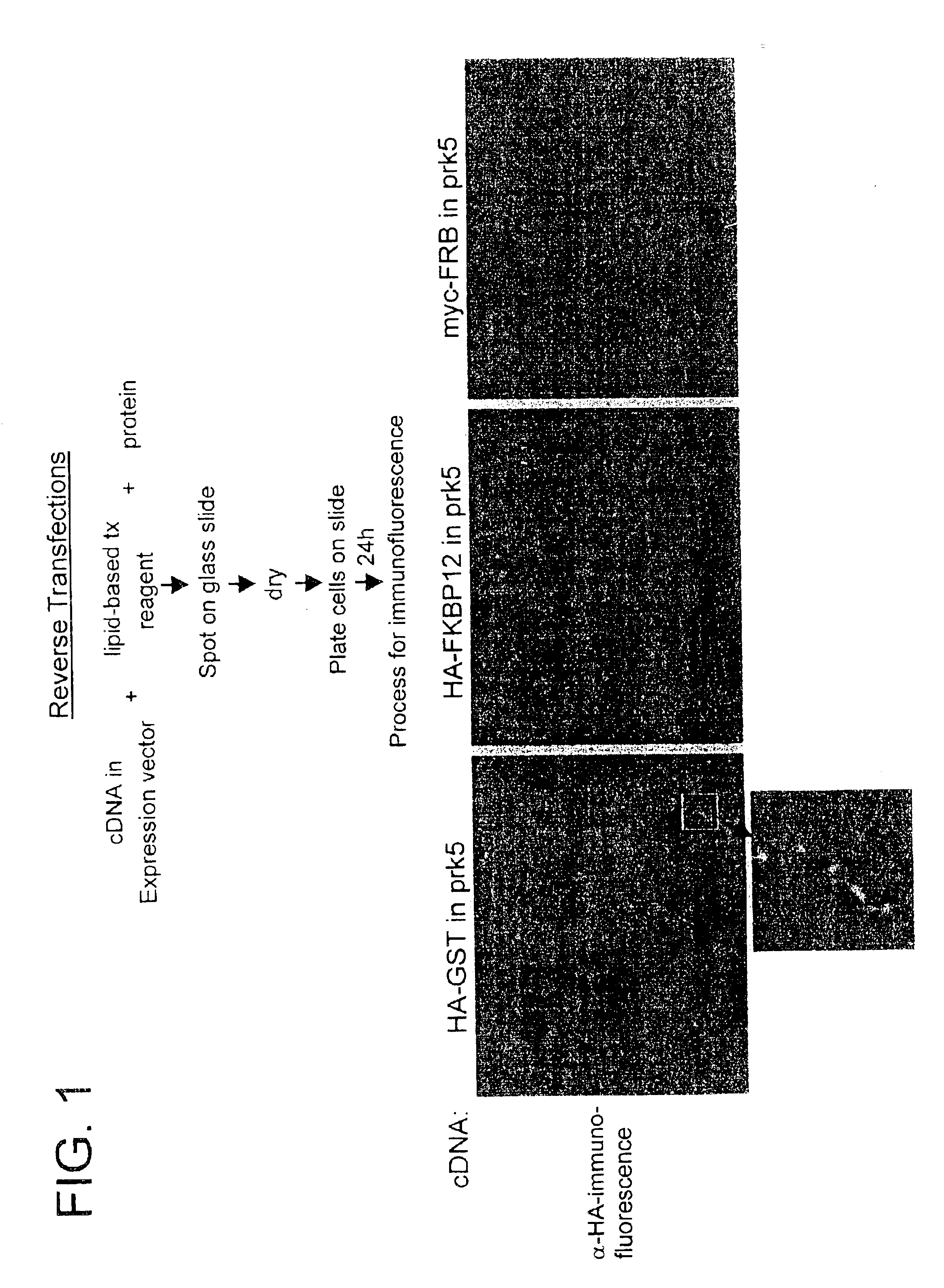
Reverse Transfection: “Gelatin-DNA” Method

Materials

[0186][DNA]: 1 μg / μL (e.g., HA-GST pRK5, pBABE CMV GFP)[0187]Gelatin (ICN, cat. #901771): 0.2% stock in ddH2O, all dilutions made in PBS-0.20% gelatin=0.5 g gelatin+250 mL ddH2O[0188]EFFECTENE™ Transfection Kit (Qiagen, cat. #301425)[0189]Plasmid-DNA: grown in 100 mL L-amp overnight from glycerol stock, purified by standard QIAPREP Miniprep or Qiagen Plasmid Purification Maxi protocols[0190]Cell Type: HEK 293T cultured in DMEM / 10%IFS with L-glut and pen / strep

Diluting and Spotting DNA[0191]Dilute DNA in 0.2% gelatin* to give final [DNA]=0.05 μg / μL**

[0192]* range of gelatin concentration that worked under the conditions used=0.05% to 0.5%

[0193]** range of DNA concentrations that worked under the conditions used0.01 μg / μl to 0.10 μg / μl [0194]Spot DNA / gelatin mix on Σ poly-L-lysine slides using arrayer[0195]Allow slides to dry in vacuum-dessicator overnight***

[0196]*** range of drying time=2 hours to 1 week

Adding Tx. Reagents to Gelat...

example 2

Reverse Transfection: “Lipid-DNA” Method

Materials

[0209][DNA]: 1 μg / μL (e.g., HA-GST pRK5, pBABE CMV GFP)[0210]Gelatin (ICN, cat.#901771): 0.2% stock in ddH2O, all dilutions made in PBS− 0.05% gelatin=250 μL 0.2%+750 μL PBS−[0211]EFFECTENE™ Transfection Kit (Qiagen, cat.#301425):[0212]EC Buffer in 0.4M sucrose=273.6 μL 50% sucrose+726.4 μL EC Buffer[0213]Plasmid-DNA: grown in 100 mL L-amp overnight from glycerol stock, purified by standard QIAPREP™ Miniprep or Qiagen Plasmid Purification Maxi protocols[0214]Cell Type: HEK 293T cultured in DMEM / 10%IFS with L-glut and pen / strep

Reverse Transfection Protocol with Reduced Volume[0215]Aliquot 1.6 μg DNA in separate eppendorf tubes[0216]Add 15 μL of pre-made DNA-condensation buffer (EC Buffer) with 0.4M sucrose* to tubes

[0217]* range of sucrose that worked under the conditions used=0.1M to 0.4M [0218]Add 1.6 μL of Enhancer solution and mix by pipetting several times. Incubate at room temperature for 5 minutes[0219]Add 5 μL of EFFECTENE™ Tra...

example 3

Transfected Cells Microarrays: A Genomics Approach for the Analysis of Gene Products in Mammalian Cells

Lipid-DNA Method

I. Gelatin Preparation and DNA Purification

Materials:

[0229]Gamma-Amino Propyl Silane (GAPS) slides (Corning catalog #2550),[0230]Purified cDNA,[0231]Gelatin, Type B: 225 Bloom (Sigma, catalog #G-9391),

Methods

[0232]0.2% Gelatin was made by incubation in a 60° C. water bath for 15 minutes. The gelatin was cooled slowly to 37° C. at which point it was filtered through 0.45 μm cellular acetate membrane (CA).

[0233]Bacterial clones with DNA plasmids were grown in a 96 Deep-Well Dish for 18 to 24 hours in 1.3 mL of terrific broth (TB) shaking at 250 rpm at 37° C. The plasmids were miniprepped and optical density (OD) was taken. DNA purity, as indicated by final 280 nm / 260 nm absorbance ratio, was greater than 1.7.

Storage

[0234]For storage purposes, gelatin was kept at 4° C. and miniprepped DNA kept at −20° C.

II. Sample Preparation and Array Printing

Materials

[0235]EFFECTENE™...


PUM

PropertyMeasurementUnit
concentrationaaaaaaaaaa

Abstract

The invention features a method of introducing nucleic acid molecules into eukaryotic cells by (a) depositing a nucleic acid molecule-containing mixture onto a surface, (b) affixing the nucleic acid molecule-containing mixture to the surface, and (c) plating eukaryotic cells onto the surface under appropriate conditions for entry of the nucleic acid molecules into the cells.

Description

CROSS-REFERENCE TO RELATED APPLICATIONS[0001]This is a continuation of U.S. patent application Ser. No. 09 / 817,003, filed Mar. 22, 2001 (now pending), which is a continuation-in-part of U.S. patent application Ser. No. 09 / 664,297, filed Sep. 18, 2000 (now U.S. Pat. No. 6,544,790), which in turn claims the benefit of U.S. Provisional Application No. 60 / 193,580, filed Mar. 30, 2000, and U.S. Provisional Application No. 60 / 154,737, filed Sep. 17, 1999 (each of which is now abandoned). The entire teachings of each of the above-referenced applications are incorporated herein by reference.BACKGROUND OF THE INVENTION[0002]Genome and expressed sequence tag (EST) projects are rapidly cataloging and cloning the genes of higher organisms, including humans. The emerging challenge is to uncover the functional roles of the genes and to quickly identify gene products with desired properties. The growing collection of gene sequences and cloned cDNAs demands the development of systematic and high-th...

Claims


Application Information

Patent Timeline
Patent Type & Authority Patents(United States)
IPC IPC(8): C12Q1/68C12N15/87C12N15/88G01N33/53C12M1/00C12M1/34C12N5/10C12N15/09G01N33/566G01N37/00
CPCC12N15/87C12N15/88C12Q1/6806C12Q2565/518
Inventor SABATINI, DAVID M.
Owner WHITEHEAD INST FOR BIOMEDICAL RES
Why Eureka
Try Eureka