Surgical biopsy device

a biopsy device and tissue technology, applied in the field of surgical devices, can solve the problems of increasing morbidity, increasing scarring, and increasing scarring, and requiring long recovery times, and achieve the effect of enhancing ultrasound imaging

Inactive Publication Date: 2005-11-01
REX MEDICAL LP
View PDF108 Cites 34 Cited by
  • Summary
  • Abstract
  • Description
  • Claims
  • Application Information

AI Technical Summary

Benefits of technology

[0022]The apparatus may also include an ultrasonic transducer at a distal end of the housing to enhance ultrasound imaging during the biopsy procedure.

Problems solved by technology

In this procedure, large tissue samples are surgically removed, requiring long recovery times, risking disfigurement of the breast, increased scarring and increased morbidity.
One disadvantage of this technique is that numerous cell samples are required to be taken from the tissue to obtain a sufficient mass for testing, thereby requiring numerous needle sticks and increasing the time required for the procedure.
Another disadvantage is that careful locational tracking of the tissue cells removed is required for accurate analysis.
Also, with these devices there is a greater potential for false negatives due to the small sized specimens being removed without removal of sufficient surrounding areas of healthy tissue for comparison.
These devices also have the disadvantage that the spring force cutting action may displace malignant cells into the adjacent normal tissue.
Also, the amount of false negatives can be high because of inadequate removal of surrounding healthy tissue.
Another disadvantage common to both fine needle and core needle biopsy devices is that the entire lesion cannot be removed.
Besides the additional cost and surgeon time, this can have an adverse psychological affect on the patient who must await the second surgical procedure.
Although overcoming some disadvantages such as reducing the number of needle sticks, the Mammotome still fails to overcome many of the other drawbacks since careful tracking is required, success is skill dependent, and a second surgery is necessary if the lesion is malignant, with the attendant expenses and trauma.
However, a major disadvantage of this instrument is that if pathology indicates the lesion is benign, then a large tissue mass would have been unnecessarily removed, resulting in more pain, a larger scar, and possible disfigurement of the breast.
Thus, ironically, the instrument is more beneficial if the tumor is malignant, and disadvantageous if the tumor is benign.
In either case, the instrument has the further disadvantages of causing additional bleeding because of the large incision and requiring closure of a larger incision, thereby increasing scarring, lengthening patient recovery time, and adding to the cost, time and complexity of the procedure.

Method used


Image

  • Surgical biopsy device
  • Surgical biopsy device
  • Surgical biopsy device

Examples


Embodiment Construction

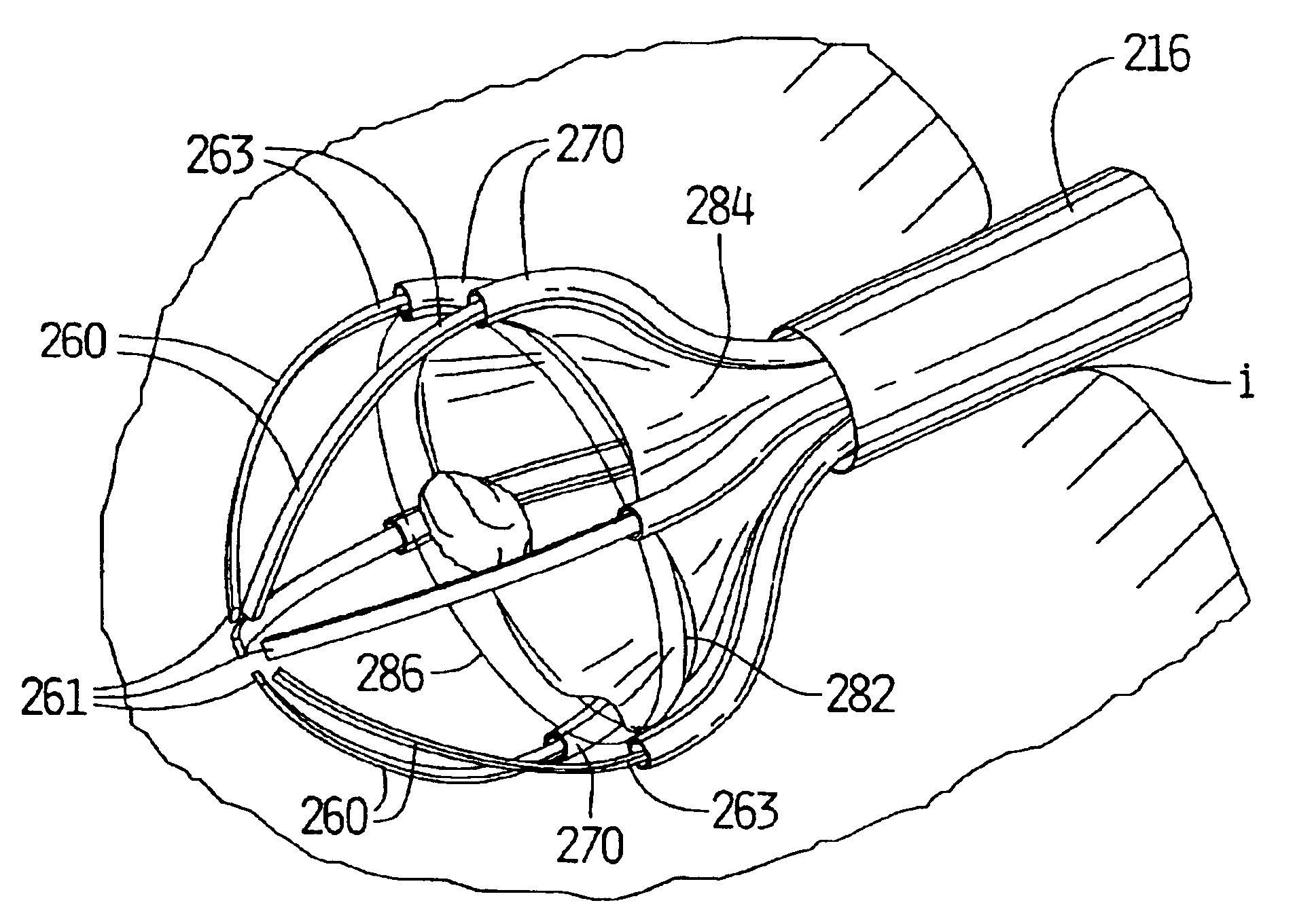
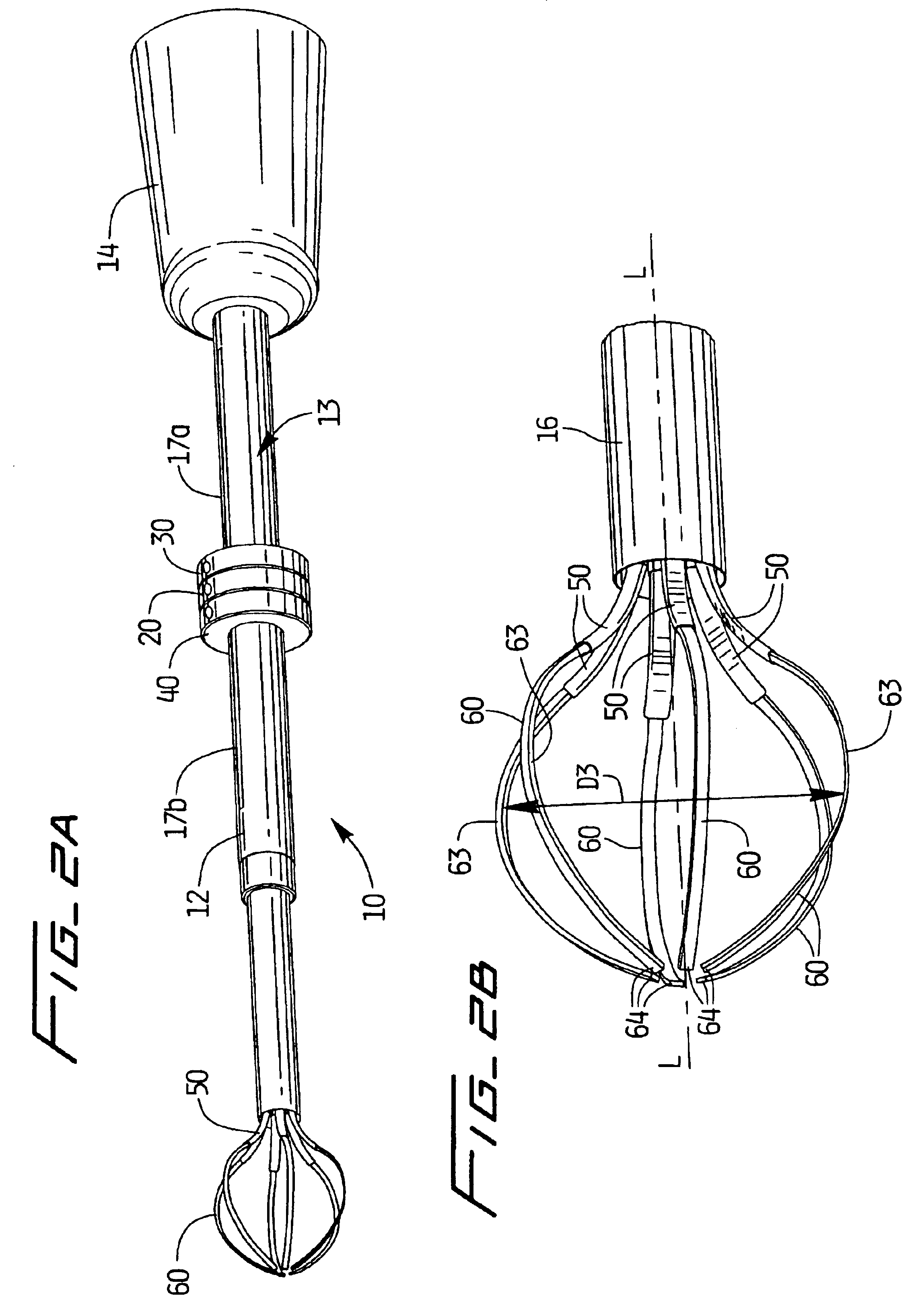
[0075]Referring now in detail to the drawings where like reference numerals identify similar or like components throughout the several views, the surgical apparatus for removing tissue is designated generally by reference numeral 10 in FIG. 1. The apparatus 10 of the present invention is particularly designed for removing breast tissue, however use of the apparatus for removal, i.e. biopsy, of other body tissue is contemplated.

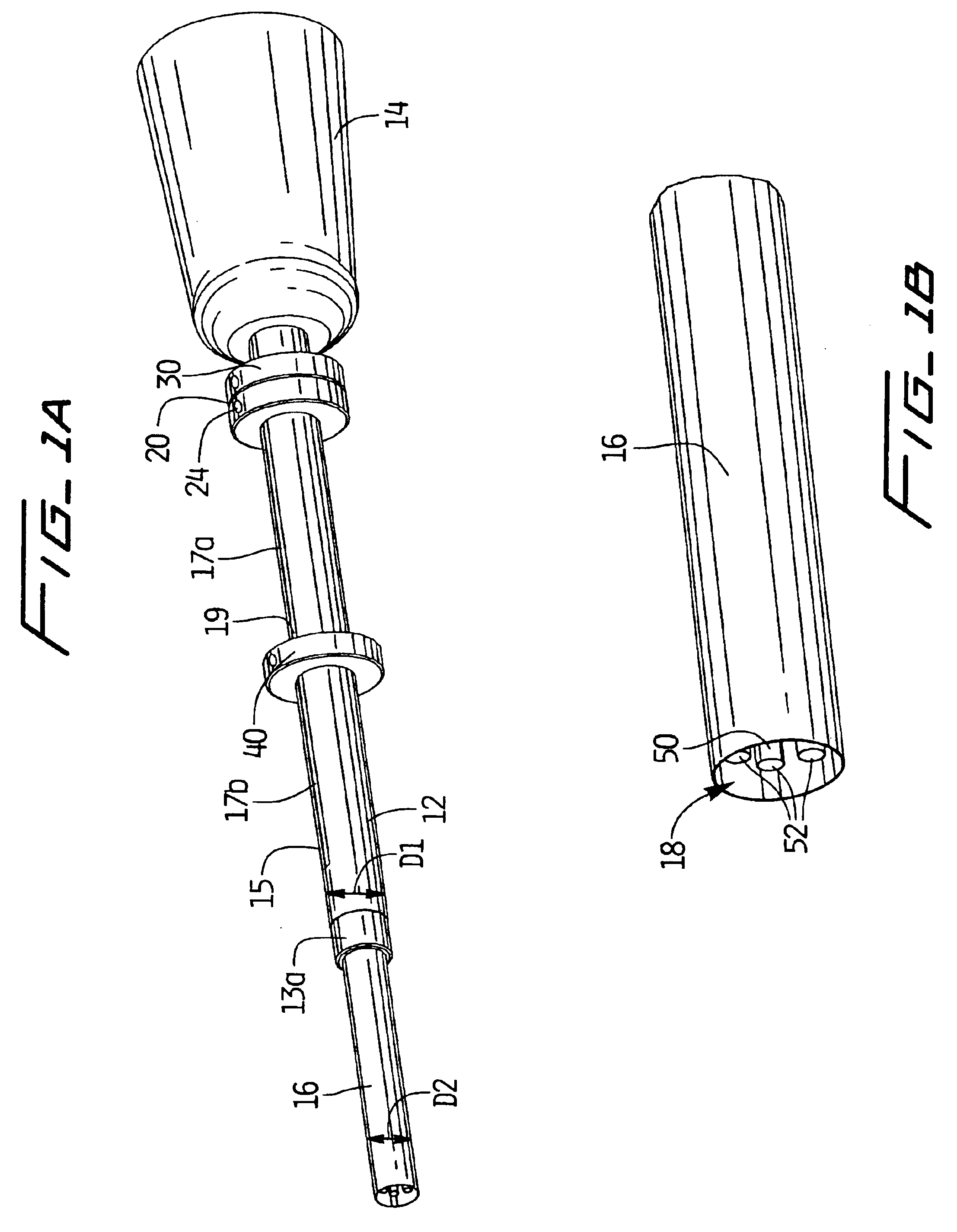
[0076]Referring to FIGS. 1A and 2A, apparatus 10 has a housing or cannula 12, a series of deployment rings 20, 30, 40, and a handle portion 14. The ring 20 deploys outer (female) or first rails 50 and ring 30 deploys inner (male) or second rails 60. As shown, outer rails (or outer tissue penetrating members) 50 and second rails (or inner tissue penetrating members) 60 are deployed from an initial position retracted within lumen 18 of cannula 12 as shown in FIGS. 1A and 1B to a deployed position where outer rails 60 encircle the tissue to be biopsied. The tissu...


PUM


Abstract

A surgical biopsy apparatus for cutting tissue comprising a housing having a longitudinal axis, first and second members movable from a retracted position to an extended position with respect to the housing, a third member slidably positioned and extendable with respect to the first member, a fourth member slidably positioned and extendable with respect to the second member, and an electrocautery cutting wire slidable with respect to the third and fourth members to surround a region of tissue positioned between the third and fourth members to cut the tissue.

Description

[0001]This application is a continuation of Ser. No. 09 / 844,729 filed Apr. 27, 2001 now U.S. Pat. No. 6,626,903 which is a continuation-in-part of U.S. patent application Ser. No. 09 / 122,185, filed Jul. 23, 1998 now U.S. Pat. No. 6,280,450 which claims priority from U.S. provisional application Ser. No. 60 / 053,664, filed Jul. 24, 1997. The contents of both these applications are incorporated herein by reference.BACKGROUND[0002]1. Technical Field[0003]This application relates to a surgical device for removing tissue and more particularly relates to a surgical tissue biopsy device insertable through a small incision in the body.[0004]2. Background of Related Art[0005]Over 150,000 women in the United States alone are diagnosed each year with breast cancer. A biopsy of breast tissue is indicated when a breast abnormality is found, allowing removal of the tissue and testing to determine whether the abnormality is malignant and further surgery is necessary. Early diagnosis and removal of ...

Claims


Application Information

Patent Timeline
Patent Type & Authority Patents(United States)
IPC IPC(8): A61B17/32A61B10/00A61B17/22A61B18/20A61B19/00A61B18/14A61B18/22A61B17/00A61B17/02A61B10/02A61B18/12
CPCA61B10/02A61B10/0266A61B17/221A61B17/320016A61B17/32056A61B10/0041A61B17/0218A61B17/320725A61B18/14A61B18/1482A61B19/38A61B19/54A61B2017/00287A61B2017/00796A61B2017/008A61B2017/2212A61B2017/2215A61B2017/32004A61B2017/320072A61B2018/1407A61B2018/2238A61B90/39A61B90/40
Inventor MCGUCKIN, JR., JAMES F.HINCHLIFFE, PETER W. J.DEFONZO, STEPHAN A.
Owner REX MEDICAL LP
Why Eureka
Try Eureka