Split-screen display system and standardized methods for ultrasound image acquisition and processing for improved measurements of vascular structures

a display system and ultrasound technology, applied in the field of ultrasound image acquisition and processing, can solve the problems of inability to perform asymptomatic persons, limited imaging techniques, morbidity and mortality in industrialized nations, etc., and achieve the effect of accurate and cost-effective screening

Inactive Publication Date: 2005-12-27
UNIV OF SOUTHERN CALIFORNIA +1
View PDF3 Cites 35 Cited by
  • Summary
  • Abstract
  • Description
  • Claims
  • Application Information

AI Technical Summary

Benefits of technology

[0011]This needs to be accomplished at the earliest time possible, such that practitioners can more accurately and cost-effectively screen individuals at r

Problems solved by technology

Cardiovascular disease remains the leading cause of morbidity and mortality in industrialized nations.
However, this imaging technique is limited to luminal dimensions and only inferences about the extent of arterial wall involvement can be made.
Further, since angiographic procedures are invasive, they cannot be performed in asymptomatic persons.
Other than B-mode ultrasonography, there is no reliable non-invasive technique available to directly measure atherosclerosis or its progression.
The accuracy, reproducibility and precision of IMT measurements, determined by manual identification methods, are limited by human variability in the operation of coordinate designation devices and by the resolution of the displayed ultrasoun

Method used


Image

  • Split-screen display system and standardized methods for ultrasound image acquisition and processing for improved measurements of vascular structures
  • Split-screen display system and standardized methods for ultrasound image acquisition and processing for improved measurements of vascular structures
  • Split-screen display system and standardized methods for ultrasound image acquisition and processing for improved measurements of vascular structures

Examples


Embodiment Construction

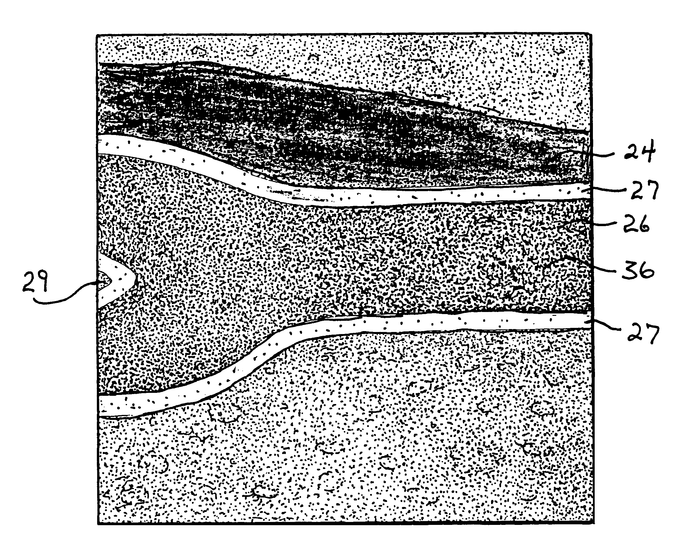
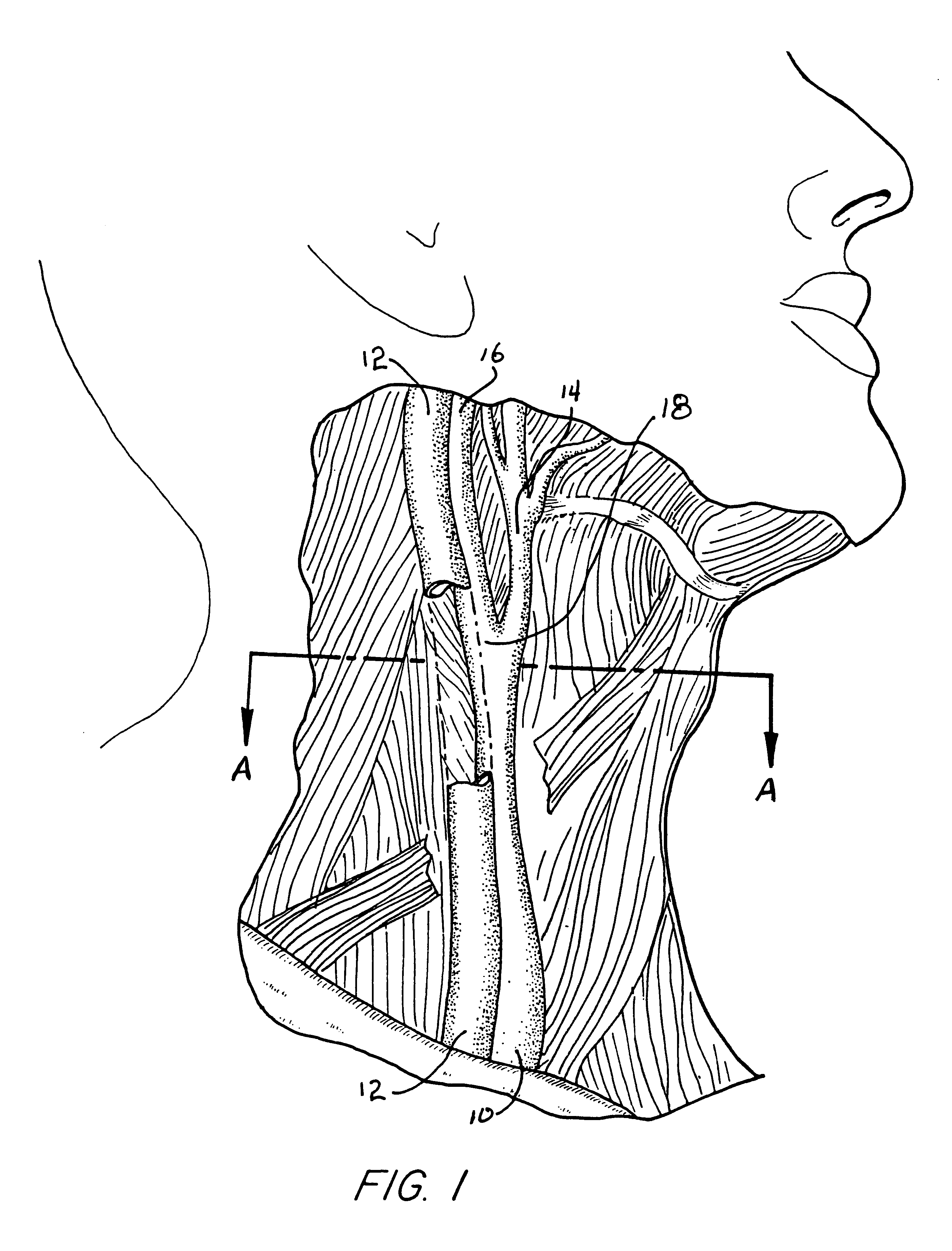
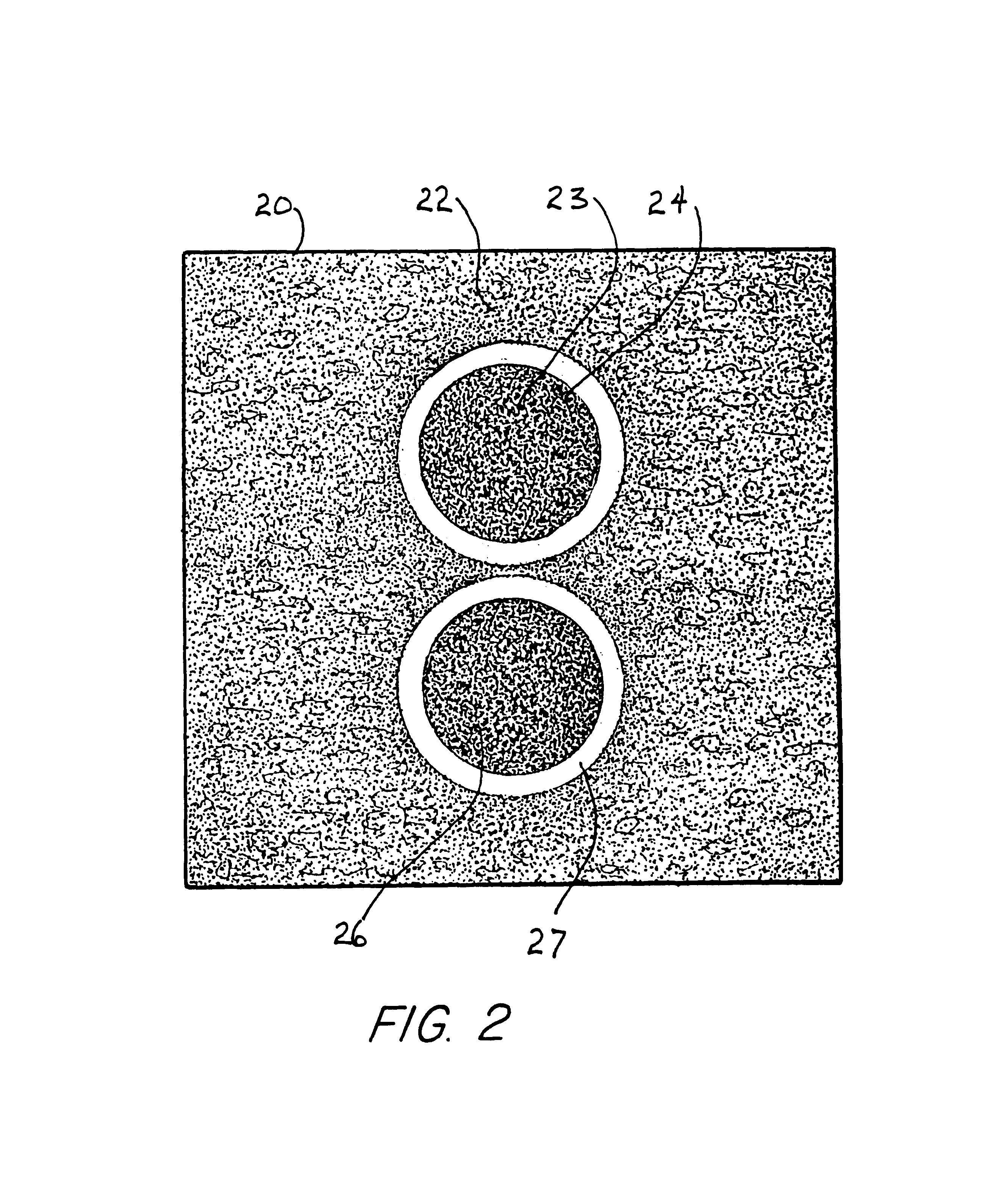
[0038]B-mode ultrasound has gained popularity as a non-invasive method for direct visualization of superficial vessels. With B-mode ultrasound, intima-media thickness (IMT) and arterial stiffness can be directly measured since image acquisition of the arterial wall thickness and vessel diameter can be obtained simultaneously in a dynamic fashion throughout the cardiac cycle. The present invention relates generally to a standardized method of carotid artery B-mode ultrasound image acquisition with a computerized split-screen system, and carotid arterial diameter and intima-media thickness (IMT) measurements from B-mode ultrasound images which utilizes computerized edge tracking. Multi-frame image processing automatically measures arterial diameter and IMT in multiple sequential frames spanning several cardiac cycles. Further, in accordance with the invention, a computerized methodology assists operators to accurately replicate images obtained from a particular individual over several...


PUM


Abstract

A standardized acquisition methodology assists operators to accurately replicate high resolution B-mode ultrasound images obtained over several spaced-apart examinations. The methodology utilizes a split-screen display in which the arterial ultrasound image from an earlier examination is displayed on one side of the screen while a real-time “live” ultrasound image from a current examination is displayed next to the earlier image on the opposite side of the screen. A computerized echo edge recognition and tracking methodology automatically identifies ultrasound echo boundaries of the intima-media complex and automatically extracts IMT and arterial dimension measurements, without introducing human measurement error. Measurement accuracy is enhanced by use of sub-pixel resolution for echo edge boundary definition. Utilizing this methodology, measurement of vascular dimensions, such as carotid arterial IMT and diameter, the coefficient of variation is substantially reduced to values approximating from about 1.0% to about 1.25%. Dynamic material properties of arterial structures are measured in a standardized region in accordance with a standardized methodology in order to promote measurement repeatability.

Description

CROSS REFERENCE TO RELATED APPLICATIONS[0001]The present application is related to and takes priority from U.S. provisional patent application Ser. No. 60 / 279,229, filed Mar. 27, 2001, entitled MOTORIZED ROTATOR DEVICE FOR ULTRASOUND PROBE, and is further related to U.S. Patent applications entitled SPLIT-SCREEN DISPLAY SYSTEM AND STANDARDIZED METHODS FOR ULTRASOUND IMAGE ACQUISITION AND MULTI-FRAME DATA PROCESSING, SYSTEM AND METHOD FOR IMPROVING ULTRASOUND IMAGE ACQUISITION AND REPLICATION FOR REPEATABLE MEASUREMENTS OF VASCULAR STRUCTURES, and MOTORIZED ULTRASOUND TRANSDUCER FOR REPEATABLE MEASUREMENTS OF VASCULAR STRUCTURES, all filed on instant date herewith, all commonly owned by the assignee of the present invention, the entire contents of all of which are expressly incorporated herein by reference.FIELD OF THE INVENTION[0002]The present invention is directed to the field of ultrasonographic vascular visualization and vascular structure measurements and, more particularly, to...

Claims


Application Information

Patent Timeline
IPC IPC(8): A61B5/107A61B8/02A61B8/08
CPCA61B5/02007A61B5/1075A61B8/0858A61B8/463A61B8/485
Inventor SELZER, ROBERT H.HODIS, HOWARD N.
Owner UNIV OF SOUTHERN CALIFORNIA
Why Eureka
Try Eureka