Hyperspectral retinal imager

a retinal imager and hyperspectral technology, applied in the field of hyperspectral retinal imagers, can solve the problems of increasing the risk of vision loss, inability to simultaneously record the entire spectral signature of an imaged area before the eye moves, and the above described instrumentation has a number of significant limitations, and achieves high spatial resolution and high spectral resolution

Inactive Publication Date: 2006-01-31
KESTREL CORP
View PDF8 Cites 48 Cited by
  • Summary
  • Abstract
  • Description
  • Claims
  • Application Information

AI Technical Summary

Benefits of technology

[0009]One aspect of the present invention is that it provides an optics system with both high spectral resolution and high spatial resolution.
[0010]Another aspect of the present invention provides a hyperspectral retinal image having improved quality from a clinical device, to thereby enable an improved level of opthalmic healthcare.
[0013]Another aspect of the present invention provides a controllable fundus illumination light source that allows selection of both the duration of the light and the spectral bandwidth of the light.
[0023]It is yet another aspect of the present invention to increase the sensitivity of retinal diagnosis.
[0031]In one embodiment of the present invention, the scene camera includes a movable mirror that diverts the image from the fundus imager away from the hyperspectral imager to a high resolution imager whose output is used by the operator to align the imager to the eye. The light source includes two illumination sources that are optically coupled to the fundus imager. The first light source is a low power continuously-controlled intensity source filtered to prevent any harmful infrared and ultraviolet light from being directed to the eye by the fundus imager. The light directed into the eye from this first light source is reflected by the retina and this reflected light is imaged by the scene camera for alignment purposes. The second light source is a high brightness continuous source optically filtered to remove harmful infrared and ultraviolet light and then optically filtered to provide selectable optical bandwidth light that is directed to the eye by the fundus imager. The light directed into the eye from this second light source is reflected by the retina and this reflected light is used by the hyperspectral imager optics to collect a hyperspectral image of the selected region of the retina.
[0032]The light sources contains appropriate shutters to control the duration of the illumination, prevent simultaneous illumination by both sources, and provide fail safe control of the light to prevent accidental hazardous illumination of the subject. A first optically coupled reference for monitoring the spectral radiance of the bright light source is used to access a sample of the bright light prior to its being directed to the eye by the fundus imager. This sample is inserted into the Fourier transform hyperspectral image at the aperture. A second optically coupled reference for calibrating the spectral performance of the hyperspectral imager connects a calibration light source, such as a gas discharge lamp or laser, to the aperture of the hyperspectral imager. The optical set used to adapt the instrument to various sized eyes includes replaceable field and aperture stops, field lens, hole coupling optics, and illumination source geometry modifying optics.

Problems solved by technology

There is strong evidence linking certain retinal biochemical and physical changes to increased risk of developing vision losses.
A major problem for the imagers disclosed is the inability to simultaneously record the entire spectral signature for an imaged area before the eye has moved.
The above described instrumentation has a number of significant limitations.
Numerous exposures are required and it takes a long time to record a complete spectra.

Method used


Image

  • Hyperspectral retinal imager
  • Hyperspectral retinal imager
  • Hyperspectral retinal imager

Examples


Embodiment Construction

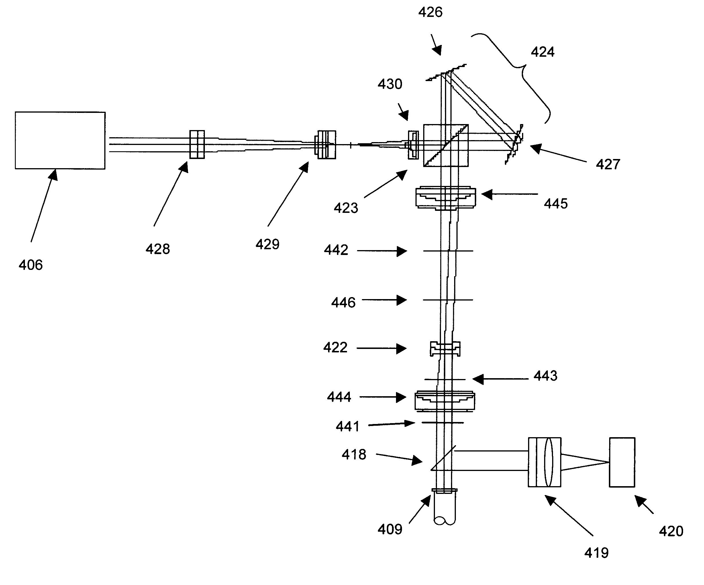
[0051]As noted above, the present invention is a system and method for obtaining hyperspectral images of a field of view provided by an optical system such as a fundus retinal imaging system. The present invention provides hyperspectral images of the retina to permit early diagnosis of various pathologies of the eye.

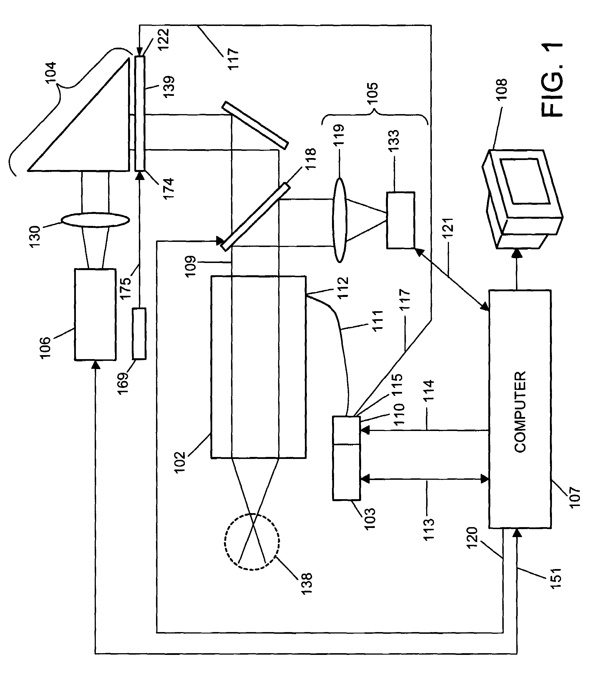
[0052]Referring to FIG. 1, a hybrid block diagram and optical schematic of one embodiment of the present invention is illustrated. A fundus retinal imager 102 observes an eye 138 and provides a collimated output 109. The fundus retinal imager 102 may be a JST ZOMZ, Model KFG 2, Zeiss FF4, FF5, FF450, the Topcon TRC-50 series, the Canon CF-60 series or CR5-45 and others can also be used, as one skilled in the art will appreciate. A light source 103 and an optical filter assembly 110 are connected to the fundus retinal imager 102 via a fiber optic cable 111 and a fiber optic port 112 provided on fundus retinal imager 102. The light source 103 includes two continuously lit ...


PUM


Abstract

An ophthalmic instrument (for obtaining high resolution, wide field of area hyperspectral retinal images for various sized eyes) includes a fundus retinal imager, (which includes optics for illuminating and imaging the retina of the eye); apparatus for generating a real time image of the area being imaged and the location of the hyperspectral region of interest; a high efficiency spatially modulated common path Fourier transform hyperspectral imager, a high resolution detector optically coupled to the hyperspectral and fundus imager optics; and a computer (which is connected to the real time scene imager, the illumination source, and the high resolution camera) including an algorithm for recovery and calibration of the hyperspectral images.

Description

CROSS REFERENCE TO RELATED APPLICATIONS[0001]This application claims benefit under 35 U.S.C. § 119(e) of provisional application No. 60 / 407,330, filed Aug. 29, 2002, which is incorporated by reference herein, in its entirety, for all purposes.STATEMENT OF GOVERNMENT INTEREST[0002]This invention was made with government support under Contract No. 2000-153-KESTREL-SOLIZ awarded by the National Medical Technology Testbed, Inc. The government has certain rights in the invention.FIELD OF THE INVENTION[0003]The present invention relates to obtaining hyperspectral images of a field of view provided by an optical system. More particularly, the present invention relates to a fundus retinal imaging system that provides early diagnosis of various pathologies of the eye.BACKGROUND OF THE INVENTION[0004]The ability to resolve the fine spectral and spatial details on the retina can play a key role in the early diagnosis of vision loss. There is strong evidence linking certain retinal biochemical ...

Claims


Application Information

Patent Timeline
Patent Type & Authority Patents(United States)
IPC IPC(8): G01B9/02A61B3/12G01J3/28G01J3/453
CPCA61B3/12G01J3/453G01J3/2823G01J3/28
Inventor SOLIZ, PETEROTTEN, III, LEONARD JOHNTRUITT, PAUL WILEY
Owner KESTREL CORP
Why Eureka
Try Eureka