Peptides recognized by melanoma-specific A1-, A2- and A3-restricted cytoxic lymphocytes, and uses therefor
a cytoxic lymphocyte and peptide technology, applied in the direction of peptides/protein ingredients, peptides, immunoglobulins, etc., can solve the problems of difficult definition of individual peptides that comprise specific ctl epitopes, and the inability to recognize peptides by the other ctl lines tested
- Summary
- Abstract
- Description
- Claims
- Application Information
AI Technical Summary
Benefits of technology
Problems solved by technology
Method used
Image
Examples
examples
Materials and Methods—Cell Lines and HLA Typing
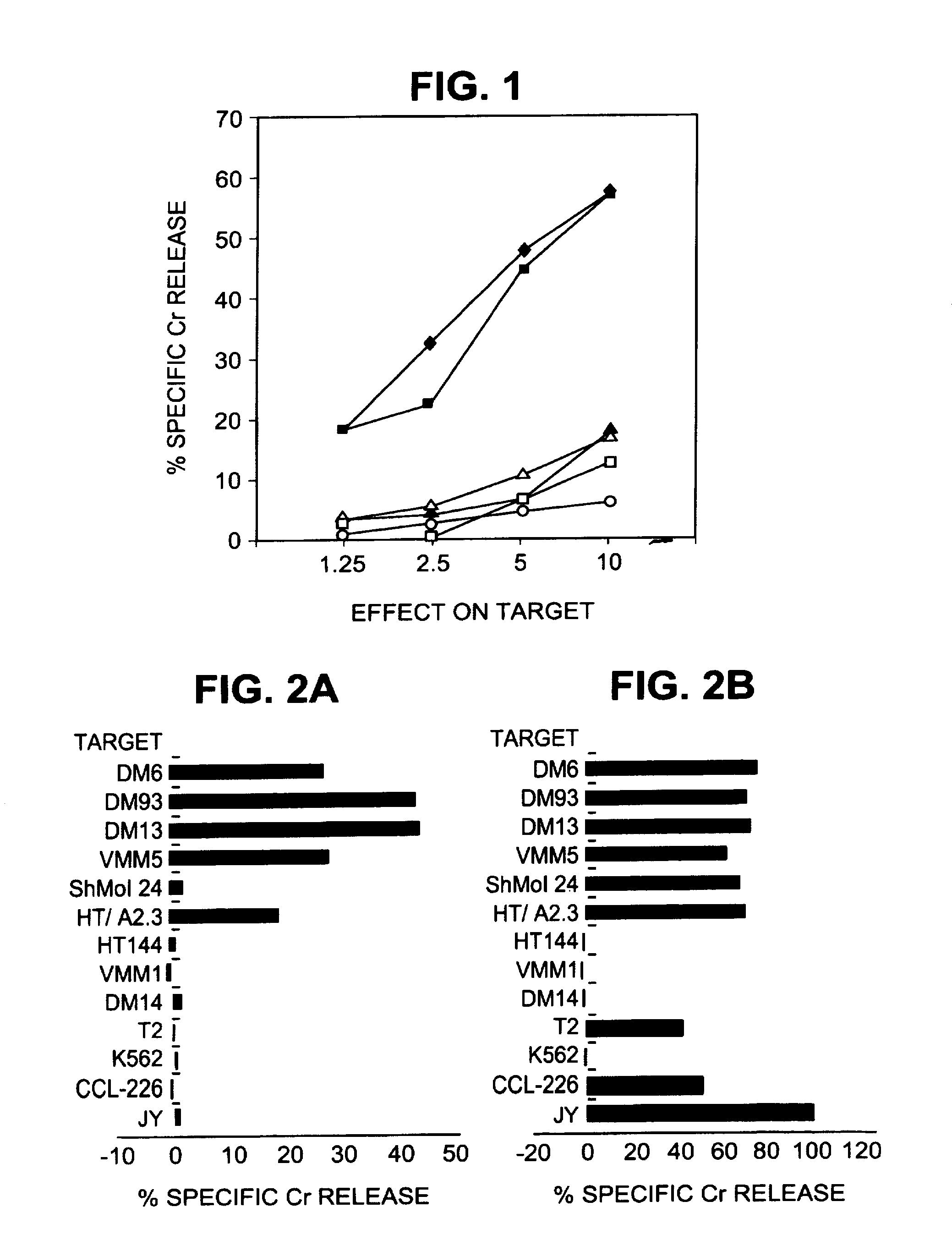
[0213]All tumor cell lines were of human origin. Melanoma cell lines HT144 and Sk-MeI-24, osteosarcoma 143b, colon cancer CCL-228 (SW480), and breast cancer MDA-MB-468 were obtained from the American Type Culture Collection (Bethesda, Md.). Fibroblasts GM126 were obtained from the National Institute of General Medical Sciences Human Genetic Mutant Cell Repository, Bethesa, Md. Melanoma lines DM6, DM13, DM14, and DM93 were the gift of Drs. Hilliard F. Siegler and Timothy L. Darrow. VMM1 and VMM5 are melanoma cell lines established from metastatic melanoma resected from patients at the University of Virginia (Charlottesville, Va.). VBT2 (squamous cell lung carcinoma), VA01 (adenocarcinoma of the ovary), and VAB5 (adenocarcinoma of the breast) are cell lines also established at this institution. JY, MICH, MWF, 23.1, RPMI 1788, and Herluff are EBV-transformed B lympho-blastoid lines. K562 is a NK-sensitive human erythroleukemia line. The ce...
example i
Extraction of HLA-A2.1-Associated Peptides from Melanoma Cells
[0219]The human melanoma cells DM6 and DM93 were cultured in 10-chamber cell factories (Nunc, Thousand Oaks, Calif.) in MEM supplemented with 1% FCS and Pen-Strept. In initial experiments, the cells were harvested with 0.03% EDTA in PBS, whereas in later experiments 0.025% trypsin was also included. Trypsinization resulted in more complete harvests and in higher cell viability without any evident change in reconstitution of epitopes or in the peptide profile (data not shown). Peptides bound to the A2 molecules were acid eluted and isolated by centrifuge filtration using a modification of the protocol described by Hunt et al. Cells were washed three times in cold PBS and solubilized in 20 ml, per 109 cells, of 1% NP-40, 0.25% sodium deoxycholate, 174 microg / ml PMSF, 5 microg / ml aprotinin, 10 microg / ml leupeptin, 16 microg / ml pepstatin A, 33 microg / ml iodoacetamide, 0.2% sodium azide, and 0.03 microg / ml EDTA. The mixture wa...
example ii
HPLC Fractionation of Peptide Extracts
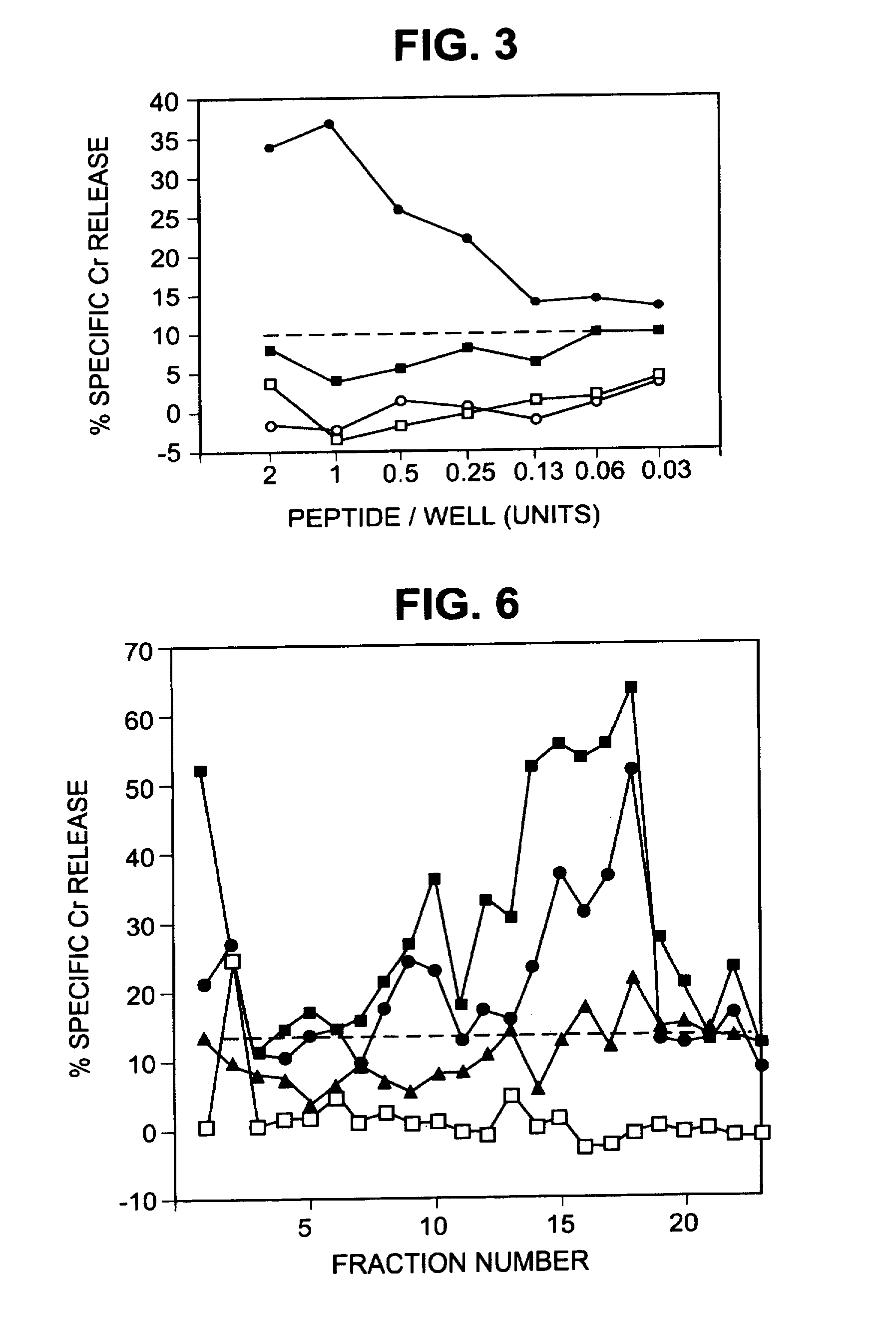
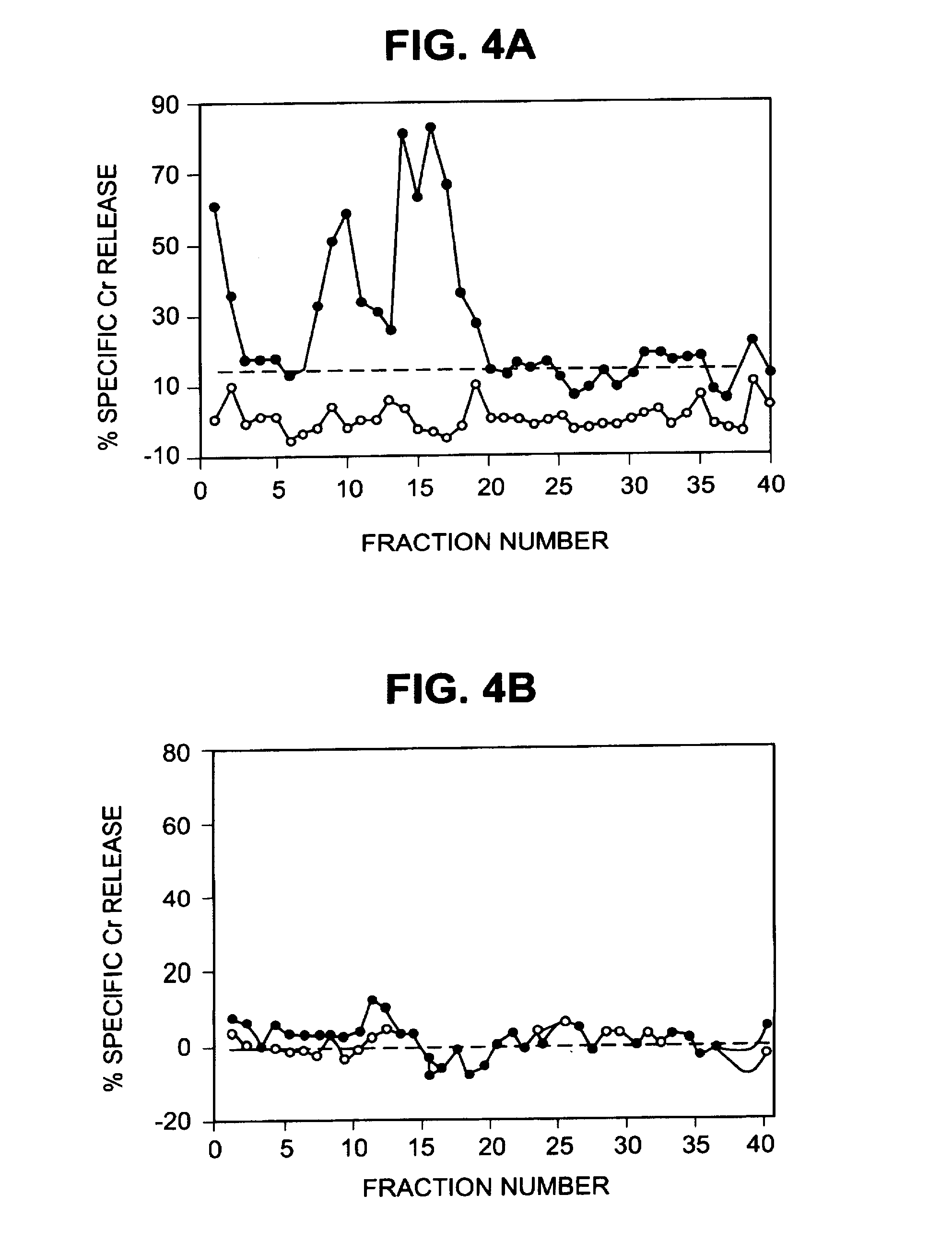
[0220]The peptide extracts were fractionated by reversed phase high performance liquid chromatography (HPLC) on an Applied Biosystems model 130A (Foster City, Calif.) separation system. Peptide extracts were concentrated to 100 microl on a Speed Vac, injected onto a Brownlee narrow-bore C-18 Aquapore column (2.1 mm×3 cm, A, 7 microm) and eluted with a 40-min gradient of 0 to 60% (v / v) acetonitrile / 0.085% trifluoracetic acid (TFA) in 0.1% TFA. Fractions were collected at 1-min intervals. Cytotoxicity assays were performed to identify fractions that reconstituted CTL epitopes. For some experiments, reconstituting fractions were divided into two equal parts. The first was injected onto a Brownlee narrow-bore C-18 Aqua-pore column (2.1 mm×3 cm, 300 A, 7 microm) and eluted with a 40-min gradient of 0 to 60% (v / v) acetonitrile in 0.1% HFA that had been adjusted to pH 8.1 with 14.8 M ammonium hydroxide. The second half was injected onto the same type o...
PUM
| Property | Measurement | Unit |
|---|---|---|
| incubation time | aaaaa | aaaaa |
| temperature | aaaaa | aaaaa |
| temperature | aaaaa | aaaaa |
Abstract
Description
Claims
Application Information
 - R&D
- Intellectual Property
- Life Sciences
- Materials
- Tech Scout
- Unparalleled Data Quality
- Higher Quality Content
- 60% Fewer Hallucinations
Browse by: Latest US Patents, China's latest patents, Technical Efficacy Thesaurus, Application Domain, Technology Topic, Popular Technical Reports.
© 2025 PatSnap. All rights reserved.Legal|Privacy policy|Modern Slavery Act Transparency Statement|Sitemap|About US| Contact US: help@patsnap.com


