Region extracting method for medical image

a region extraction and medical image technology, applied in the field of medical image region extraction, can solve the problems of insufficient automatic extraction in the hard to extract regions, inability to detect in the liver area using conventional methods, and inability to extract the whole liver region. the effect of efficiency improvemen

Inactive Publication Date: 2006-05-16
AZE
View PDF6 Cites 19 Cited by
  • Summary
  • Abstract
  • Description
  • Claims
  • Application Information

AI Technical Summary

Benefits of technology

[0012]In view of the foregoing circumstances, it is an object of the present invention to provide a region extracting method for a medical image by which a region of a predetermined organic part can be extracted uniquely in a stable fashion from images captured for medical use.

Problems solved by technology

Therefore, in the CT of liver in which hundreds of images are taken at one time, enormous labors are necessary in terms of time and mental efforts to extract the whole liver region.
However, automatic extractions in regions hard to extract have not yielded sufficient results yet.
However, these conventional methods may not be reliable in the detection in areas about the liver such as parts in contact with surroun

Method used


Image

  • Region extracting method for medical image
  • Region extracting method for medical image
  • Region extracting method for medical image

Examples


Embodiment Construction

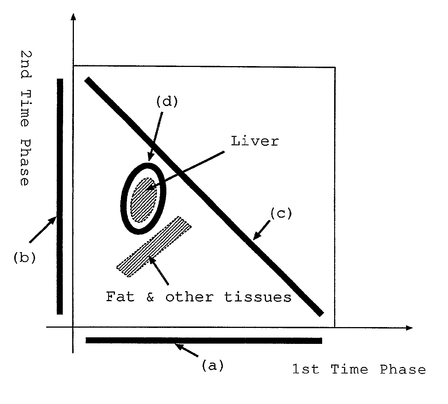
[0043]In the following, the region extracting method for a medical image in accordance with an embodiment of the present invention will be explained with reference to the drawings.

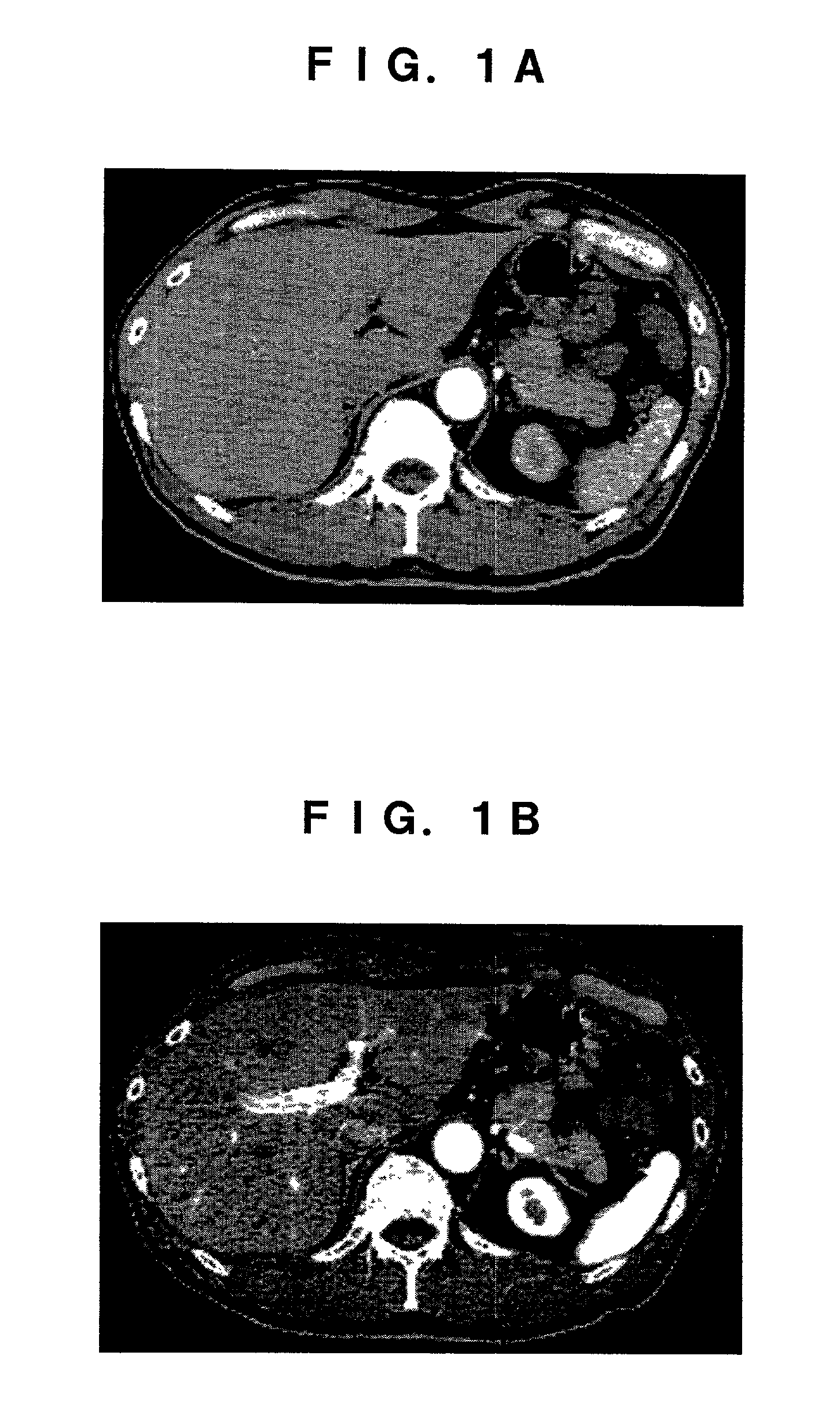
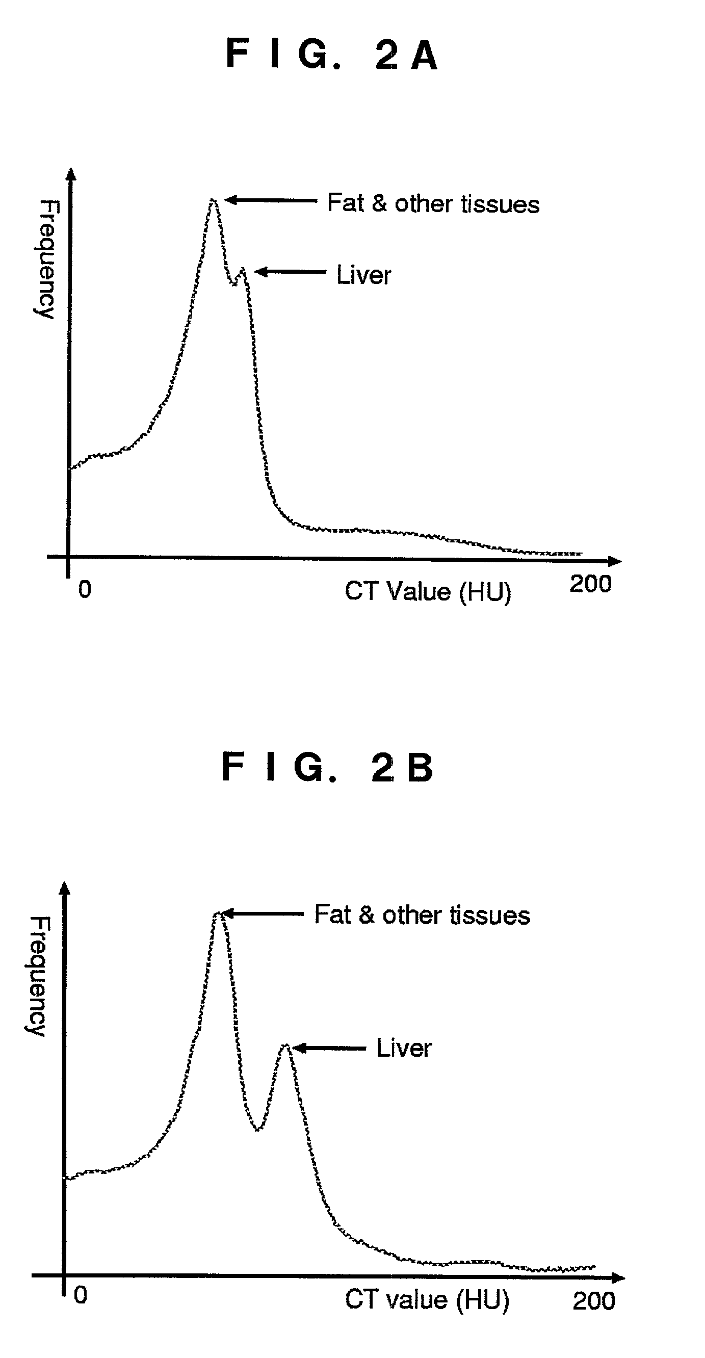
[0044]The method in accordance with this embodiment is one automatically extracting a liver region from multislice CT images in which an abdomen is captured. As images for extracting the region, two image series taken in respective time phases different from each other, specifically, an image series of a first arterial phase captured immediately after a contrast medium is injected into a liver (hereinafter referred to as first time phase) and an image series of a second arterial phase captured after several tens of seconds from the contrast medium injection (hereinafter referred to as second time phase), are employed. The individual images of these two image series are captured in a single respiration-holding time, whereas two images corresponding to each other (capturing the same sectional position) betwe...


PUM


Abstract

From CT images having captured an abdomen, a two-dimensional characteristic space is assumed, in which X and Y axes indicate CT values of image data taken in first and second time phases different from each other. In this space, a two-dimensional histogram concerning respective pixels located at the same position in the two time phases is taken as a sample distribution of pixels. A two-variable distribution function is applied to this sample distribution, so as to estimate a matrix distribution of pixels corresponding to the whole liver region. According to this matrix distribution, a range of CT value of pixels corresponding to the liver region is estimated.

Description

RELATED APPLICATIONS[0001]This application claims the priority of Japanese Patent Application No. 2001-152523 filed on May 22, 2001, which is incorporated herein by reference.BACKGROUND OF THE INVENTION[0002]1. Field of the Invention[0003]The present invention relates to a region extracting method for medical images in which regions of organic parts such as organs including liver and pancreas, blood vessels, tumors, and the like are extracted by image processing from medical images taken by use of radiation diagnosis systems such as CT (computed tomography), MRI (magnetic resonance imaging), nuclear medicine, CR (computed radiography), DSA (digital subtraction angiography), and DR (real-time radiography).[0004]2. Description of the Prior Art[0005]While CT and MRI, for example, have conventionally been known as radiation medical diagnosis systems, the technical progress in these fields has recently made it possible to take high-resolution images in a short time. In the field of CT, f...

Claims


Application Information

Patent Timeline
IPC IPC(8): G06K9/00A61B6/03A61B5/055G06T1/00G06T5/00G06T7/00
CPCG06T7/0081G06T2207/30101G06T2207/30056G06T2207/10081G06T7/11
Inventor MASUMOTO, JUNSATO, YOSHINOBUHORI, MASATOSHIMURAKAMI, TAKAMICHIJOHKOH, TAKESHINAKAMURA, HIRONOBUTAMURA, SHINICHIMIYAMOTO, MASAKI
Owner AZE
Why Eureka
Try Eureka