Imaging system having preset processing parameters adapted to user preferences

a preset processing and user-friendly technology, applied in the field of ultrasonic imaging, can solve the problems of many image processing parameters that are not available, not always effective, and cannot be adjusted by the user

Inactive Publication Date: 2006-06-13
GE MEDICAL SYST GLOBAL TECH CO LLC
View PDF7 Cites 31 Cited by
  • Summary
  • Abstract
  • Description
  • Claims
  • Application Information

AI Technical Summary

Benefits of technology

[0016]As the system operator uses the above extended adaptive gray mapping feature in clinical scanning, th

Problems solved by technology

However, this is not always effective since the user can scan any anatomy in any application, acoustic data varies from patient to patient, and the raw pixel intensity values depend on other system settings such as dynamic range and gain.
In conventional scanners, there are also many image processing parameters that are not available in the preset menu; i.

Method used


Image

  • Imaging system having preset processing parameters adapted to user preferences
  • Imaging system having preset processing parameters adapted to user preferences
  • Imaging system having preset processing parameters adapted to user preferences

Examples


Embodiment Construction

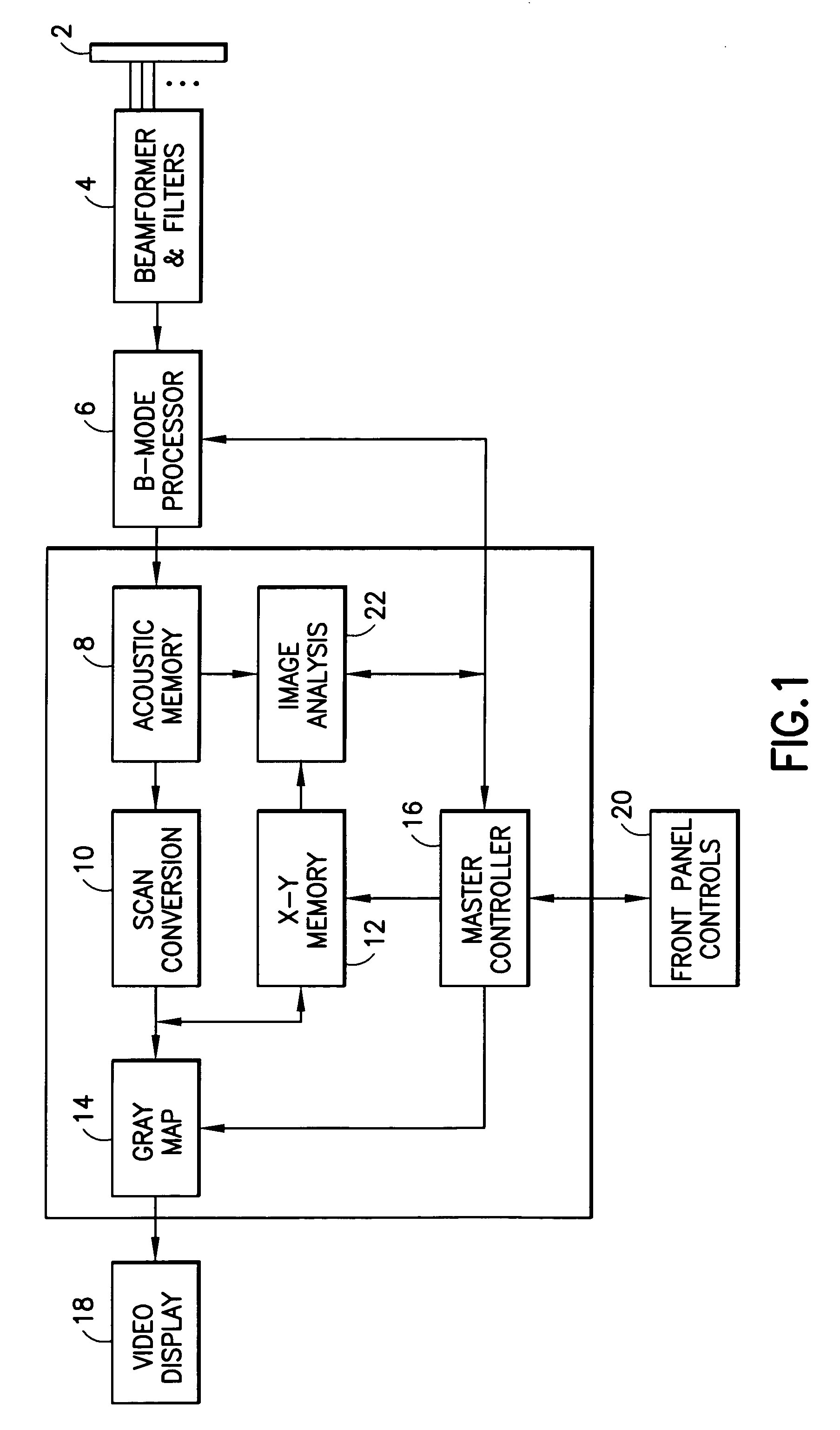
[0020]Referring to FIG. 1, an ultrasound imaging system in accordance with one preferred embodiment of the invention comprises a transducer array 2, and a beamformer 4 including filters. The transducer array 2 comprises a multiplicity of piezoelectric elements which are activated by a transmitter in beamformer 4 to transmit an ultrasound beam focused at a transmit focal position. The return RF signals are detected by the transducer elements and then dynamically focused at successive ranges along a scan line by a receiver in beamformer 4. The receive beamformer produces RF or equivalent I / Q data representing the echoes along each imaging beam or vector. This raw acoustic data is output to a B-mode processor 6. The B-mode processor in accordance with the preferred embodiment detects the power (I2+Q2) of the complex signal, and performs other standard filtering and transmit zone splicing operations on a vector by vector basis. The magnitude (i.e., intensity) of the signal output by the...


PUM


Abstract

An imaging system has a computer which monitors changes to any given front-panel control setting for a particular image processing parameter (e.g., dynamic range, transmit foci position, Doppler wall filter setting, etc.) as compared to its default or preset value. For each user ID (e.g., based on the system user's name or any user code) and each application type, if, over a number of exam cycles, the user is found to consistently adjust a particular control setting to values that deviate significantly from the preset value, the system computer will automatically adjust the preset value to, e.g., the average setting of that image processing parameter over the past N trials, where N is a predefined integer number.

Description

FIELD OF THE INVENTION[0001]This invention generally relates to ultrasound imaging for the purpose of medical diagnosis. In particular, the invention relates to methods for imaging tissue and blood flow by detecting ultrasonic echoes reflected from a scanned region of interest in a human body.BACKGROUND OF THE INVENTION[0002]Conventional ultrasound scanners are capable of operating in different imaging modes. In the B mode, two-dimensional images can be generated in which the brightness of each display pixel is derived from the value or amplitude of a respective acoustic data sample representing the echo signal returned from a respective focal position within a scan region.[0003]In the B-mode imaging, an ultrasound transducer array is activated to transmit beams focused at respective focal positions in a scan plane. After each transmit firing, the echo signals detected by the transducer array elements are fed to respective receive channels of a receiver beamformer, which converts th...

Claims


Application Information

Patent Timeline
IPC IPC(8): G06F3/01G16H30/20
CPCA61B8/461G01S7/5205G01S7/52084G06T5/007G06T5/40G01S15/899G06T2207/30004G06T2207/10016G06T2207/10132G06F19/321G16H30/20
Inventor MO, LARRY Y. L.BROUWER, DEAN W.DUESTERHOEFT, TERRY J.
Owner GE MEDICAL SYST GLOBAL TECH CO LLC
Why Eureka
Try Eureka