Endoscopic retractor instrument and associated method

Active Publication Date: 2006-10-17
GRANIT MEDICAL INNOVATION
View PDF9 Cites 33 Cited by
  • Summary
  • Abstract
  • Description
  • Claims
  • Application Information

AI Technical Summary

Benefits of technology

[0042]Where a sheath is used as an attachment device for an endoscopic retractor, in accordance with the invention, the sheath provides the additional benefit of keeping the endoscope relatively free from contact with the patient's body fluids or fecal material. The sheath is made of a thin flexible material that is split from its proximal end to the distal end or to a point close to the distal end. One side of the split line has a permanent adhesive component that sufficiently overlaps the other side of the sheath to enable fixation of the sheath to an endoscope insertion member. The adhesive can be covered by a removable protective strip that is simply peeled off to expose the adhesive layer. The sheath is then wrapped over the endoscope and adhered to the non-adhesive side of the sheath, making a secure and fluid tight bond the entire length of the sheath. In order to easily apply a long and narrow sheath in accordance with the invention to an endoscope, the non-adhesive part of the sh

Problems solved by technology

One problem frequently encountered by endoscopists is that folds of tissues in the digestive tract and particularly the colon can obscure polyps so that they can be overlooked.
However, if too much air is introduced, the colon could perforate.
Also, the patient is made quite uncomfortable with introduction of substantial amounts of air.
Another problem with visualization during an endoscopic procedure is that polyps sometimes bleed when they are severed

Method used


Image

  • Endoscopic retractor instrument and associated method
  • Endoscopic retractor instrument and associated method
  • Endoscopic retractor instrument and associated method

Examples


Embodiment Construction

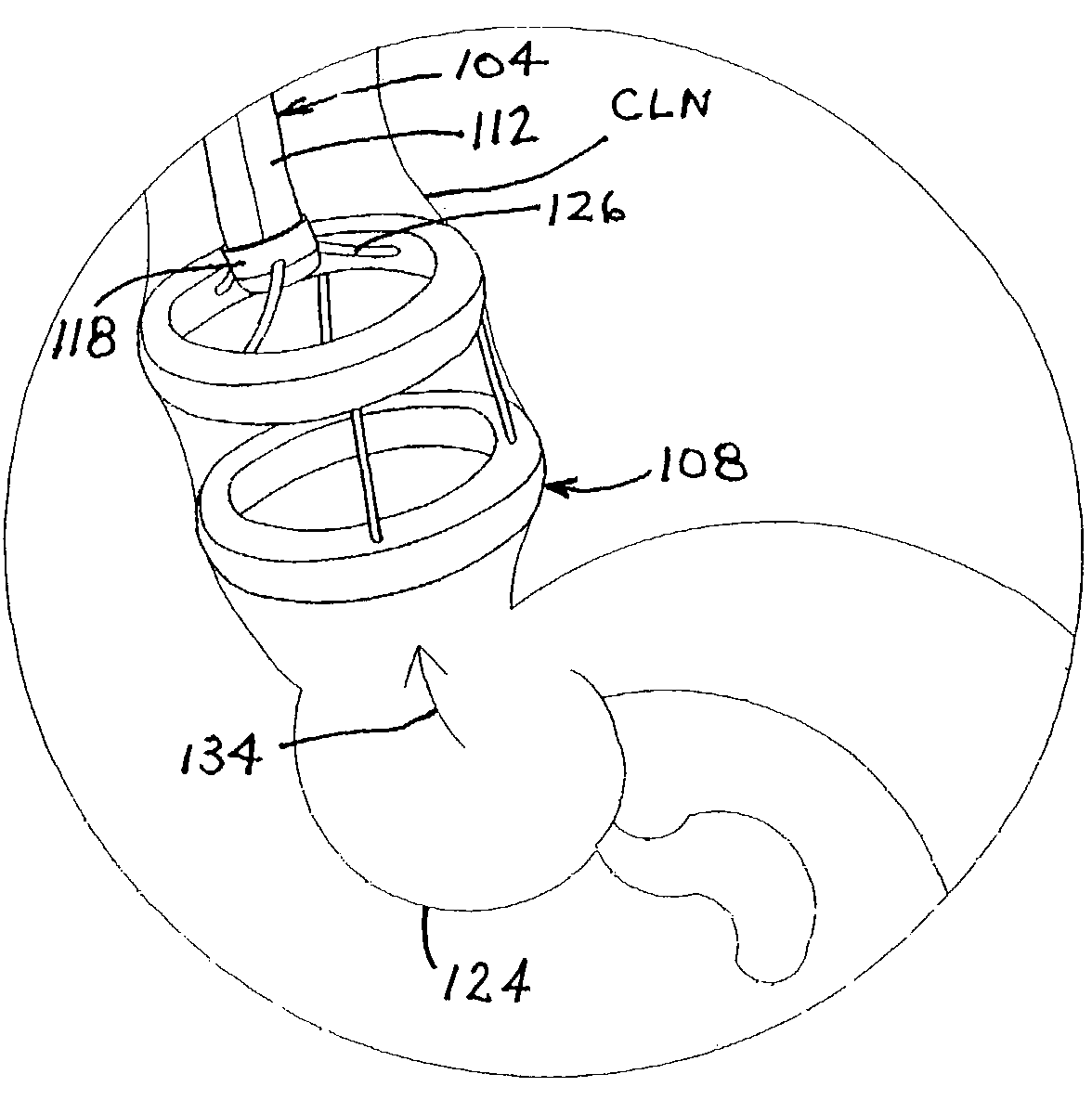
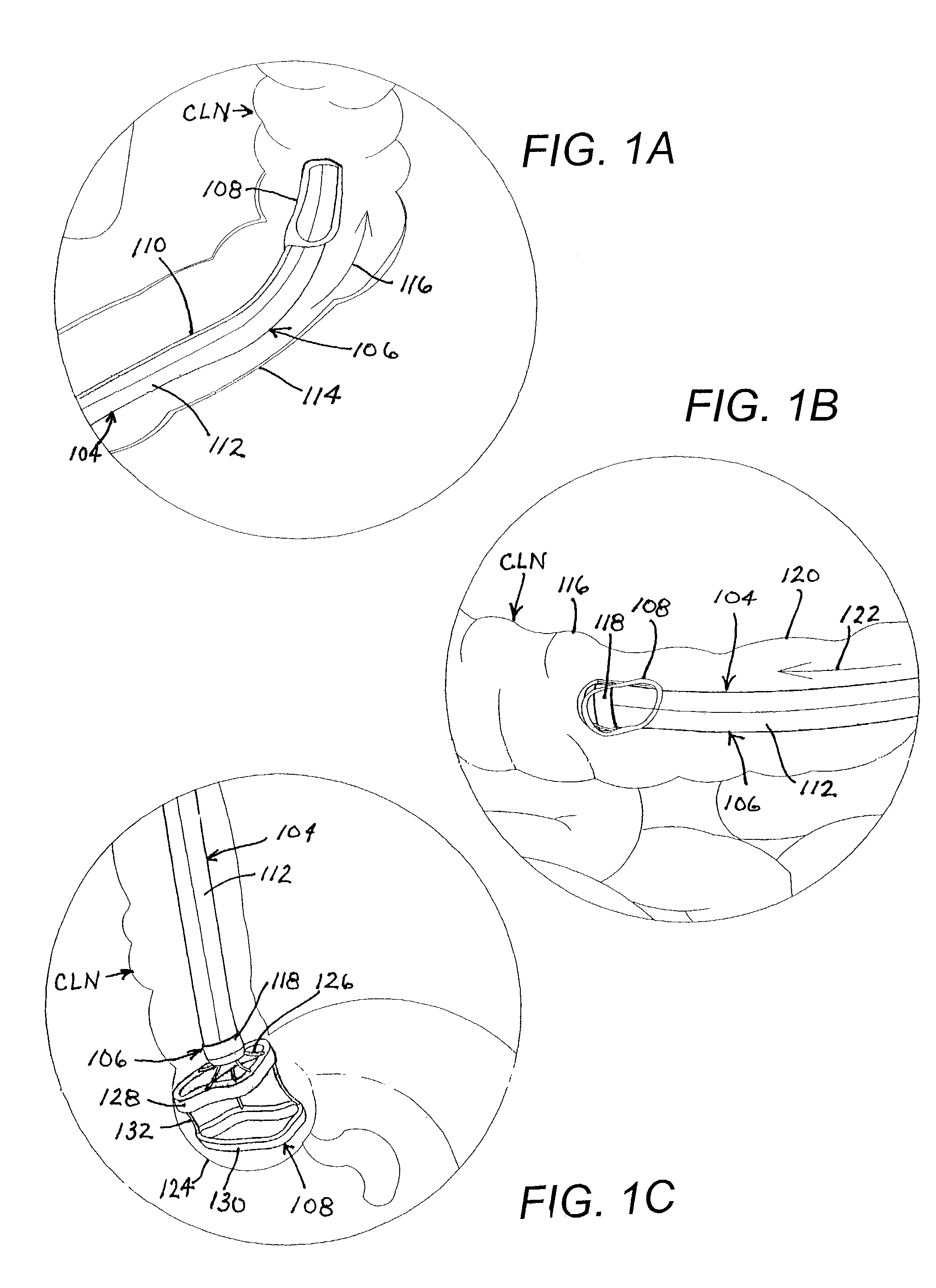
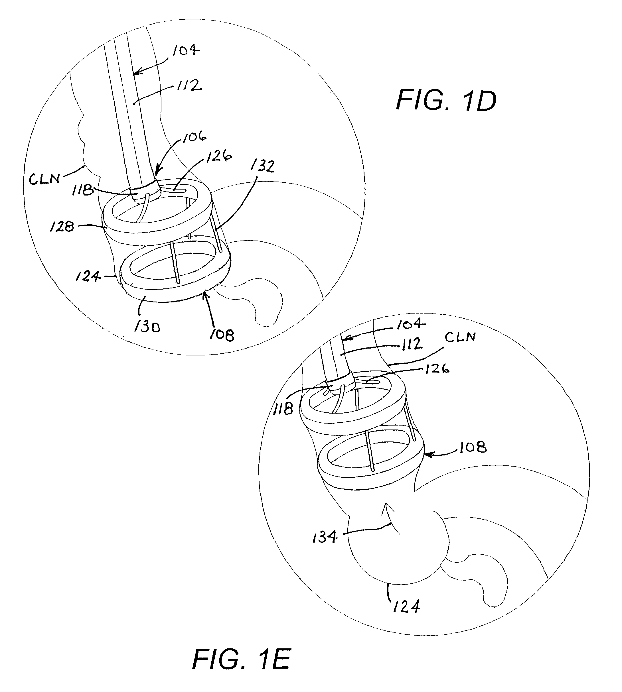
[0057]FIG. 1A shows an endoscope insertion member or shaft 104 encased in a sheath assembly 106 that carries, at a distal end, a balloon-type retractor 108 in a collapsed configuration. Retractor balloon 108 is essentially identical to that disclosed above with reference to FIGS. 1 and 2 and is connected to an inflation tube 110 extending longitudinally back along endoscope insertion member 104. Tube 110 may be a part of or connected to a sheath 112 of assembly 106. Alternatively, tube 110 may extend separately alongside sheath 112 and endoscope insertion member 104. In another alternative, tube 110 extends through a biopsy channel of endoscope insertion member 104.

[0058]It is to be noted that retractor balloon 108 is configured in its collapsed insertion configuration so as not to interfere with insertion of the endoscope member 104 or visualization through the endoscope. Thus, the collapsed retractor balloon 108 is situated outside the visual field of the endoscope lens (not shown...


PUM


Abstract

An endoscopic retractor instrument assembly includes a balloon having a pair of expandable or inflatable end portions and at least one expandable or inflatable spacer portion connecting the end portions to one another. A fastener is provided for securing the balloon to a distal end of an endoscopic insertion member, with the balloon being disposed in a collapsed configuration along an outer surface of the endoscopic insertion member. An inflation element is operatively coupled with the balloon for enabling an inflating of the balloon from the collapsed configuration to an expanded use configuration in which the spacer portion pushes the end portions apart from one another and in which the balloon extends at least partially in a distal direction away from the distal end of the endoscopic insertion member for spreading internal tissues of a patient to facilitate access to the tissues via the endoscopic insertion member.

Description

BACKGROUND OF THE INVENTION[0001]This invention relates to a medical device and to an associated medical technique. More particularly, this invention relates to an endoscopic instrument and to an endoscopic method.[0002]Flexible endoscopes are inserted into the digestive tract for diagnostic and therapeutic purposes. Endoscopes generally include a light guide for transmitting optical-wavelength electromagnetic radiation into the patient. Images are captured, typically via lenses and an optical fiber bundle or a charge-coupled device, whereby a user can visually inspect the inner walls or surfaces of the digestive tract. One common objective of endoscopic investigations in the digestive tract is to detect the presence of polyps. Where a polyp is visually detected, particularly in the colon, it should be severed, captured, and removed from the patient. Alternatively, particularly where the polyp may be a malignant cancer, the polyp may be severed and captured for extraction from the p...

Claims


Application Information

Patent Timeline
IPC IPC(8): A61B1/32A61M29/00A61B1/00A61B1/12A61B1/31A61B17/00A61B17/02
CPCA61B1/00082A61B1/00142A61B1/31A61B5/6886A61B17/0218A61B2017/00269A61B2017/00557
Inventor NAKAO, NAOMI L.
Owner GRANIT MEDICAL INNOVATION
Why Eureka
Try Eureka