
System and method for monitoring cardiac function via cardiac sounds using an implantable cardiac stimulation device

Inactive Publication Date: 2006-11-21
PACESETTER INC
View PDF18 Cites 125 Cited by
  • Summary
  • Abstract
  • Description
  • Claims
  • Application Information

AI Technical Summary

Benefits of technology

[0020]The predictor values vary in accordance with changes in actual cardiac function and hence are effective as bases for adjusting pacing parameters to optimize overall cardiac function. Changes in the predictors for cardiac function are also useful as a means for tracking progression of heart failure. Contractility is proportional to max dP/dt and so P_max_dP/dt also serves as a predictor or proxy for contractility.
[0021]In accordance with a second aspect of the invention, a technique is provided for performing heart sound monitoring using an implantable cardiac pacing device wherein heart sound signals are detected, parameters representative of overall cardiac function are evaluated and then control parameters are automatically adjusted based on the cardiac function parameters. In an exemplary embodiment, control parameters are adjusted to optimize cardiac function as represented by one or more predictors of stroke volume, cardiac output, and max dP/dt, which are derived from the heart sound signals using the techniques summarized above. Adjustments are triggered following any significant change in max dP/dt, stroke volume or cardiac output. Changes can also be triggered be significant changes in atrioventricular conduction interval, interventricular conduction interval, heart rate

Problems solved by technology

One factor that contributes to heart failure is asynchronous activation of the ventricles such that the mechanical contraction is not coordinated effectively thus compromising cardiac function.
As a result, the pumping ability of the heart is diminished and the patient experiences shortness of breath, fatigue, swelling, and other debilitating symptoms.
The weakened heart is also susceptible to potentially lethal ventricular tachyarrhythmias.
A decrease in cardiac function can result in a progression of heart failure.
However, this evaluation and programming requires an office visit and is therefore a timely and expensive process.
It also restricts the evaluation to a resting state, commonly with the patient in a supine position.
As such, the system is not necessarily optimized for activity, for the upright position, for other times of day since there may also be a circadian rhythm to cardiac function.
Moreover, when relying on any external hemodynamic monitoring system, the control parameters of the pacemaker or ICD cannot be automatically adjusted to respond to on-going changes in patient cardiac function.
However, high frequency heart noises due to rapid turbulent flow are not detected.
Although the filter-based technique of Kadhiresan may be effective for recording certain types of heart sound data for diagnostic purposes, it does not provide for the on-board evaluation of overall cardiac function or for the automatic adjustment of pacing control parameters to permit ventricular resynchronization therapy.
Moreover, by filtering out noises associated with turbulent flow, the technique does not permit detection of heart murmurs and the like, which can be important in determination of appropriate therapy.
Although the technique of Baumann et al. provides for automatic adjustment of certain pacing parameters (such as AV intervals) based on heart sound signals, it does not appear to provide for ventricular resynchronization therapy.
Also, although Baumann et al. discuss the adjustment of certain pacing parameters so as to optimize cardiac performance, the technique does not appear to provide any capability for actually evaluating overall cardiac output in terms of, e.g., stroke volume, cardiac output, or max dP / dt.
Hence, overall cardiac function is not actually tracked and optimization of pacing parameters based on overall cardiac function is not achieved.

Method used


Image

  • System and method for monitoring cardiac function via cardiac sounds using an implantable cardiac stimulation device
  • System and method for monitoring cardiac function via cardiac sounds using an implantable cardiac stimulation device
  • System and method for monitoring cardiac function via cardiac sounds using an implantable cardiac stimulation device

Examples


Embodiment Construction

[0032]The following description includes the best mode presently contemplated for practicing the invention. This description is not to be taken in a limiting sense but is made merely to describe general principles of the invention. The scope of the invention should be ascertained with reference to the issued claims. In the description of the invention that follows, like numerals or reference designators will be used to refer to like parts or elements throughout.

Overview of Stimulation Device

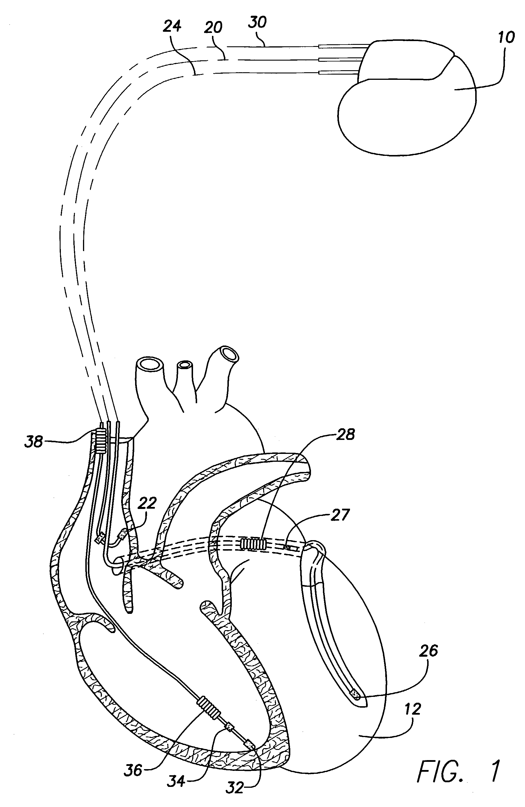
[0033]FIG. 1 illustrates a stimulation device 10 in electrical communication with a patient's heart 12 by way of three leads 20, 24 and 30 suitable for delivering multi-chamber stimulation and shock therapy. To sense atrial cardiac signals and to provide right atrial chamber stimulation therapy, the stimulation device 10 is coupled to an implantable right atrial lead 20 having at least an atrial tip electrode 22, which typically is implanted in the patient's right atrial appendage.

[0034]To sense ...


PUM


Abstract

Techniques are provided for performing internal measurement of heart sounds to estimate patient cardiac function in terms of stroke volume, cardiac output, or a maximum rate of change of aortic pressure with time (max dP / dt). Control parameters of the medical device are then automatically adjusted so as to optimize overall cardiac function or to provide for ventricular resynchronization therapy. In one example, heart sound signals are derived from acceleration signals received from an accelerometer. The heart sound signals are analyzed to identify S1 and S2 heart sounds as well as ejection period and isovolumic interval (ISOV). Proxies for max dP / dt, stroke volume and cardiac output are then derived from the S1 and S2 heart sounds, the ejection period and the ISOV. Alternative techniques, not requiring detection of ISOV, are employed for use if the patient has heart value regurgitation.

Description

FIELD OF THE INVENTION[0001]The invention relates generally to implantable cardiac stimulation devices, such as pacemakers or implantable cardioverter-defibrillators (ICDs), and in particular to techniques for monitoring overall cardiac function using heart sounds and other cardiac sounds and for automatically adjusting pacing parameters to improve cardiac function.BACKGROUND OF THE INVENTION[0002]Cardiac function is a measure of the overall effectiveness of the cardiac system of a patient and is typically represented in terms of, one or more of, stroke volume, cardiac output, end-diastolic volume, end-systolic volume, ejection fraction or cardiac output index. Stroke volume is the amount of blood ejected from the left ventricle during systole. Cardiac output is the volume of blood pumped by the left ventricle per minute (or stroke volume times the heart rate). End-diastolic volume is the volume of blood in the chamber at the end of the diastolic phase, when the chamber is at its fu...

Claims


Application Information

Patent Timeline
IPC IPC(8): A61B5/02A61N1/365
CPCA61B5/02A61B5/04004A61B5/0402A61B7/04A61N1/36585A61B5/7239A61N1/3627A61B5/30A61B5/318
Inventor MIN, XIAOYILEVINE, PAUL A.PARK, EULJOON
Owner PACESETTER INC
Features
  • Generate Ideas
  • Intellectual Property
  • Life Sciences
  • Materials
  • Tech Scout
  • Unparalleled Data Quality
  • Higher Quality Content
  • 60% Fewer Hallucinations
Learn More