Device for containing and analyzing surgically excised tissue and related methods

Active Publication Date: 2007-02-06
DUKE UNIV
View PDF17 Cites 28 Cited by
  • Summary
  • Abstract
  • Description
  • Claims
  • Application Information

AI Technical Summary

Benefits of technology

[0015]The invention is directed to a collection device for excised tissue immobilization and transport for radiography and pathological analysis. The device is further used to facilitate removi

Problems solved by technology

More than half of all cancers are susceptible to screening and early detection procedures, with resultant increases in survivability.
However, cancer is not a single disease that responds well to a single treatment regimen.
If so, the preferred course of treatment is often chemotherapy, which can have severe side effects.
However, studies suggest that only 10% of patients treated with chemotherapy actually benefit from the treatment.
Therefore, up to 90% of those undergoing chemotherapy may be suffering the side effects needlessly.
Unfortunately, it is necessary, since gross and microscopic pathological analysis of the tumor cannot differentiate which patients will or will not benefit from the treatment.
Sources of tumor tissue for research are generally limited to tumor banks.
Although these banks have been an immensely valuable source of materials and information for molecular analysis of tumors, for a number of reasons, the amount of useful data that can be collected is limited.
First, a majority of the tissue found in tumor banks is from large tumors.
Since genetic expression changes during different stages of tumor development, testing of larger tumors only does not provide a complete picture of tumor “lifecycles”.
Furthermore, clinical screening for tumors in patients is becoming more precise, and therefore a greater majority of tu

Method used


Image

  • Device for containing and analyzing surgically excised tissue and related methods
  • Device for containing and analyzing surgically excised tissue and related methods
  • Device for containing and analyzing surgically excised tissue and related methods

Examples


Embodiment Construction

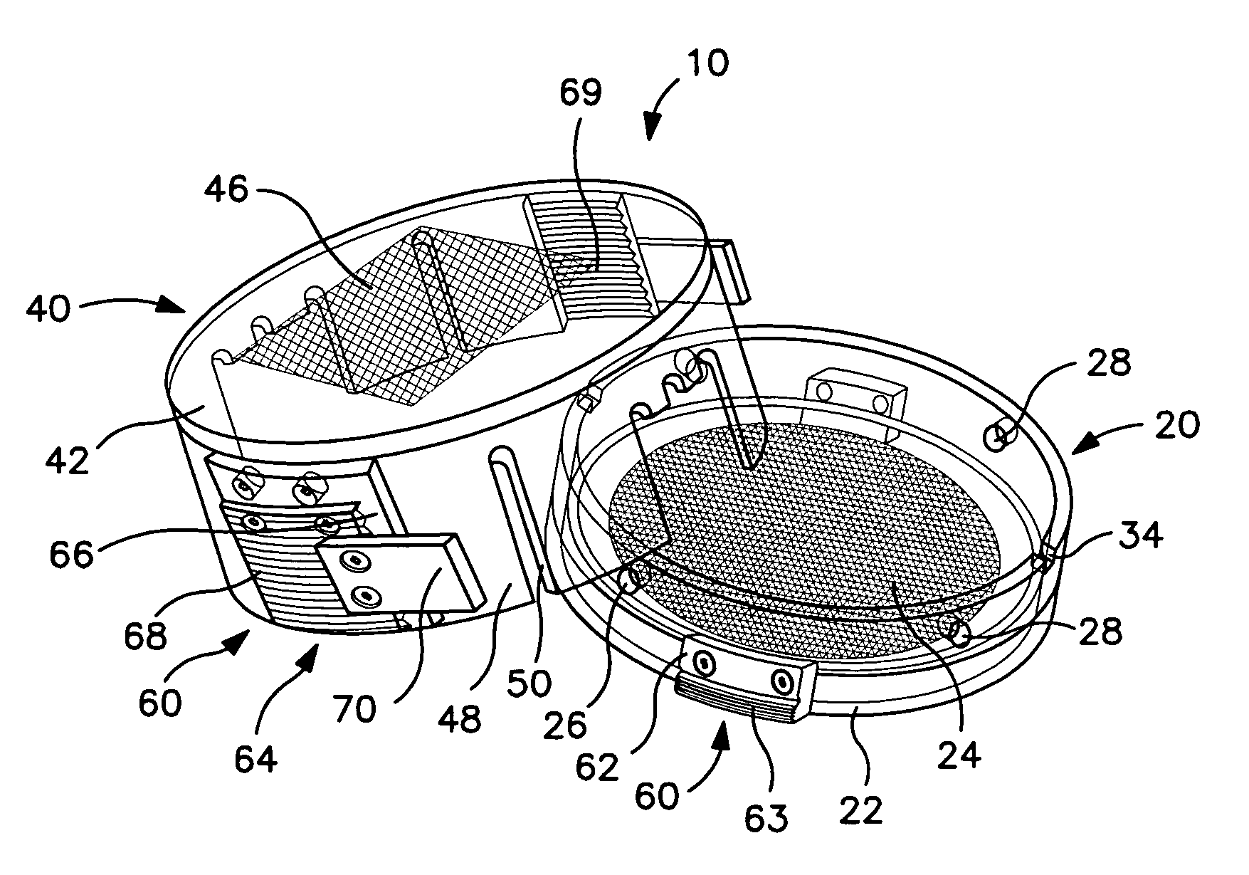
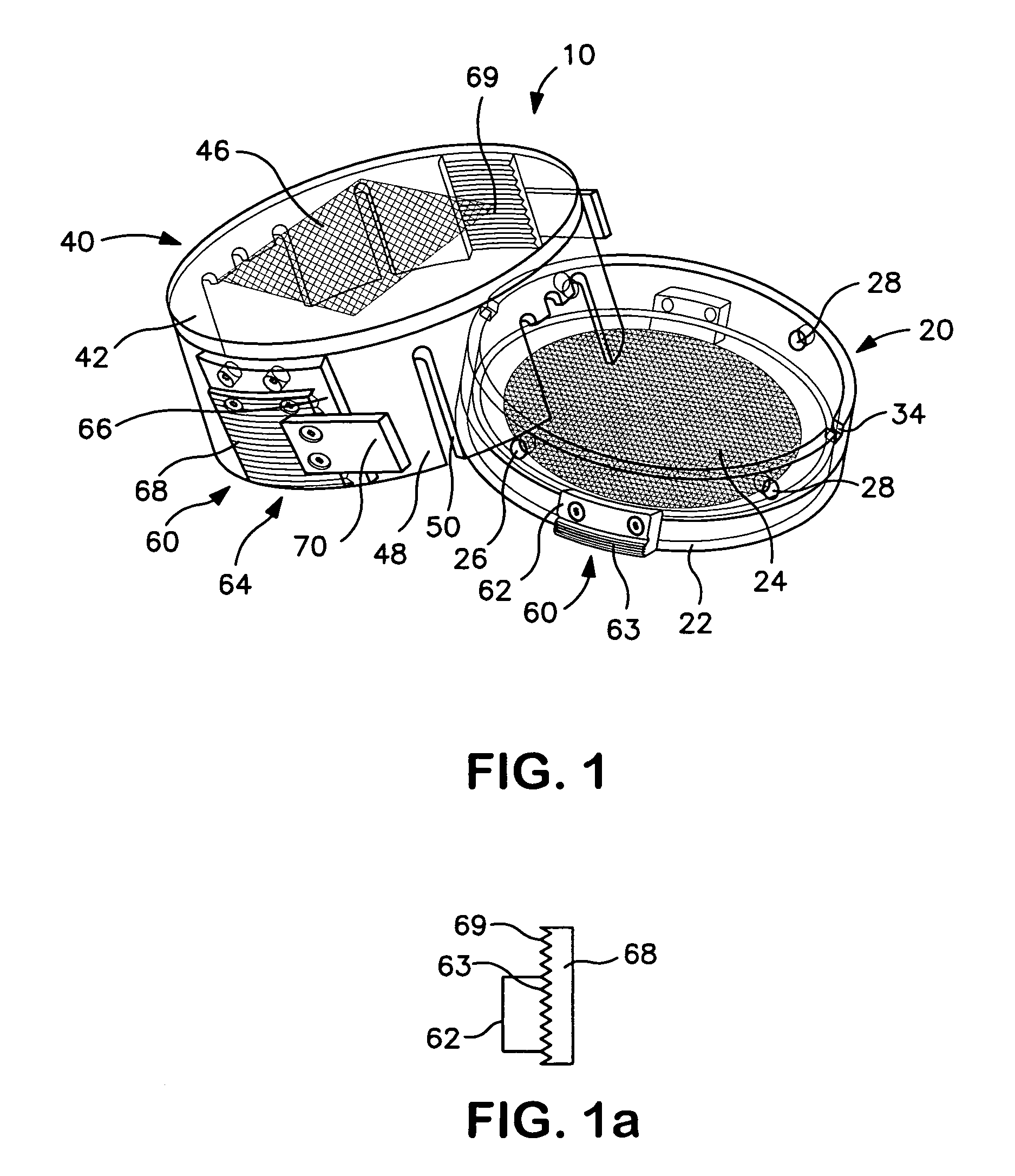
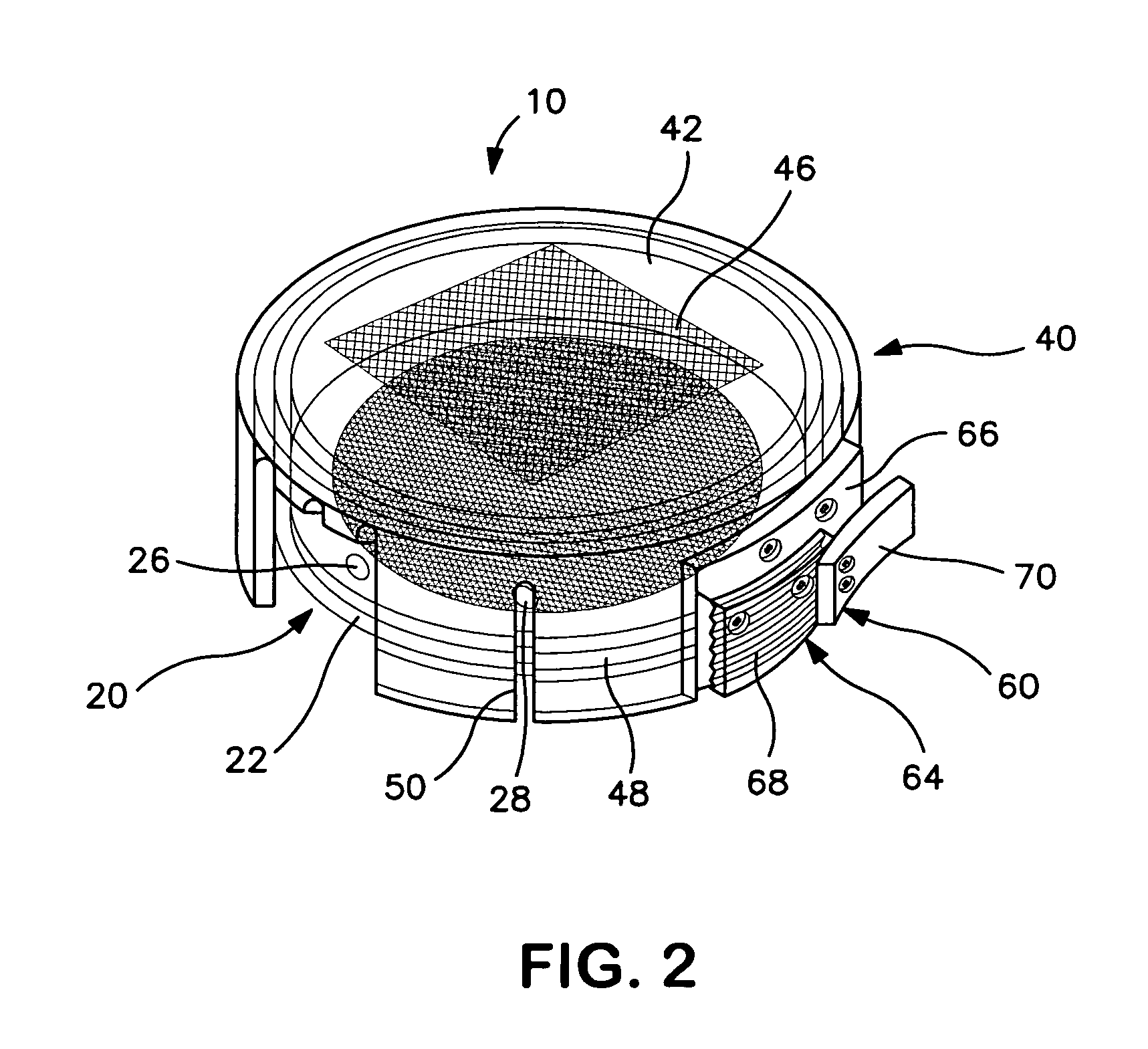
[0032]As shown in FIGS. 1–3, a collection device 10 for containing and immobilizing a surgically excised tissue specimen TS is disclosed. The collection device 10 functions to permit core sampling of tissue specimen TS for molecular or similar analysis without requiring shifting or relocation of immobilized tissue specimen TS. Collection device 10 containing the remainder of tissue specimen TS can then be used as a transport and storage device for specimen TS as it undergoes radiographic and / or pathological analysis. The excised tissue specimen TS may be a surgically or otherwise excised specimen suspected of being abnormal and therefore requiring further diagnostic or therapeutic evaluation or examination, such as, but not limited to, tissue containing a tumor, lesion, cyst or mass of cell. Collection device 10 of the present invention is particularly well suited for containing such specimens undergoing multiple forms of analysis, in particular, specimens where it is desirable to p...


PUM


Abstract

A collection device and method of use for excised tissue immobilization, removal of core samples from immobilized tissue and transport of the tissue for specimen radiography and pathological analysis. The collection device is comprised of a base member having an annular bottom wall and a side wall upwardly extending from the peripheral edge of the side wall, a lid member having a top wall and a skirt circumscribing the side wall and extending downwardly therefrom, and an incremental latching mechanism for securing the lid member to the base member. The side wall of the lid member includes a plurality of apertures positioned around at least a portion of the circumference of the side wall. In use, a tissue specimen placed in the base member is immobilized by compressing the tissue between the top wall of the lid member and the bottom wall of the base member and securing the lid member and base member together with the latching mechanism. A core sample can be removed from the immobilized specimen by inserting a portion of a sampling device through an aperture. The remainder of the specimen can be transported as needed for pathological or radiographic analysis.

Description

TECHNICAL FIELD[0001]The present invention relates to devices for containing tissue specimens, and more particularly to devices that provide for the containment, immobilization, core sampling, transport, analysis, and storage of surgically excised tissues, particularly tissues containing suspected tumor masses, and methods related to using the devices.BACKGROUND ART[0002]Cancer is the second leading cause of death in the U.S. after heart disease. The American Cancer Society predicts that more than 1.3 million new cancers will be diagnosed in 2003. This prediction does not even include non-invasive cancers and basal and squamous cell skin cancers. It is predicted that over 500,000 Americans will die from cancer this year alone.[0003]Tremendous effort and resources are directed toward diagnosing and treating cancer. More than half of all cancers are susceptible to screening and early detection procedures, with resultant increases in survivability. It is estimated that if all cancers a...

Claims


Application Information

Patent Timeline
IPC IPC(8): A61B5/00A61B10/00C12M1/22
CPCC12M1/22A61B10/0096
Inventor OLSON, JR., JOHN A.
Owner DUKE UNIV
Why Eureka
Try Eureka