Methods of identifying and isolating stem cells and cancer stem cells

a stem cell and cancer stem cell technology, applied in the field of stem cell identification and isolating stem cells and cancer stem cells, to achieve the effect of altering the proliferation of cells

Inactive Publication Date: 2007-05-15
THE BOARD OF TRUSTEES OF THE LELAND STANFORD JUNIOR UNIV
View PDF5 Cites 59 Cited by
  • Summary
  • Abstract
  • Description
  • Claims
  • Application Information

AI Technical Summary

Benefits of technology

[0011]In another embodiment of the invention, methods are provided for the alteration of cellular proliferation in hematopoietic cells, by introducing stabilized β-catenin into a hematopoietic cell that is altered in its ability to undergo

Problems solved by technology

In the case of spontaneously arising human leukemias it is likely that stem cells accumulate the mutations that are necessary for neoplastic prol

Method used


Image

  • Methods of identifying and isolating stem cells and cancer stem cells
  • Methods of identifying and isolating stem cells and cancer stem cells
  • Methods of identifying and isolating stem cells and cancer stem cells

Examples


example 1

Sorting and Transplantation of Myeloid Progenitors and Hematopoietic Stem Cell (HSC) Enriched Populations from Leukemic Mice

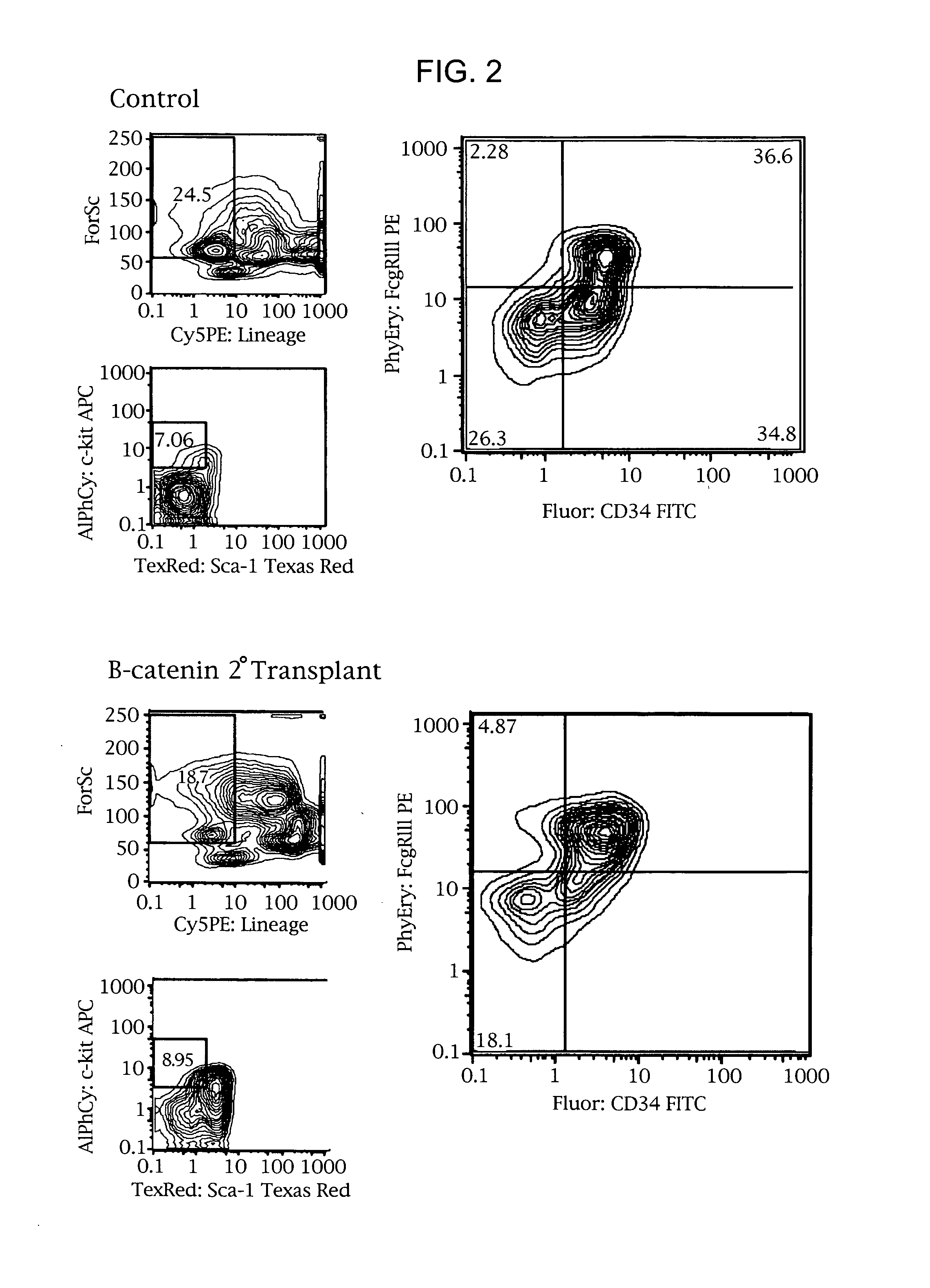
[0081]Leukemic mouse bone marrow progenitor populations were analyzed and sorted using 5 color flow cytometric analysis (FACS Vantage) and compared with those of control animals. Briefly, bone marrow was flushed from the femurs, a single cell suspension was made by passage through a 25 gauge needle and after washing the cells were incubated with a biotinylated lineage antibody cocktail consisting of CD3, 4, 8, B220, IL-7 Receptor, Thy 1.1, Mac-1, Gr-1 and Ter 119 for 30 minutes followed by washing and addition of Dynabeads for 30 minutes. Lin+ cells were then removed using a Dynal magnetic particle concentrator. Progenitors were stained with anti-CD34 FITC, c-Kit APC, Sca-1 Texas Red and FcγRIII PE for 30 minutes followed by staining with Avidin Cy5 PE for 30 minutes and finally the addition of propidium iodide. Equivalent numbers (5000 / mouse) of GMP (c-kit+ sc...

example 2

Analysis of β-catenin Expression by Normal Versus Leukemic Cells

[0088]Ficoll-enriched mononuclear populations from normal or CML peripheral blood or bone marrow and the CML blast crisis cell line, K562, were stained with anti-β-catenin Alexa-594 (red) conjugated antibody and counterstained with Hoechst (blue) for nuclear visualization and then analyzed for cytoplasmic versus nuclear localization of β-catenin using a dual photo Zeiss LSM confocal fluorescence microscope. Both cytoplasmic and nuclear β-catenin expression were higher in K562 and CML mononuclear cells compared with normal cells.

[0089]Human hematopoietic stem and myeloid progenitor cell populations were stained, analyzed and sorted with the aid of a FACS Vantage using a modification of previously described methodology (Manz et al P.N.A.S. (2002) 99:11872–11877). Briefly, mononuclear and CD34+ normal or CML chronic phase (CP), accelerated phase (AP) or blast crisis (BC) peripheral blood and bone marrow cells were stained ...

example 3

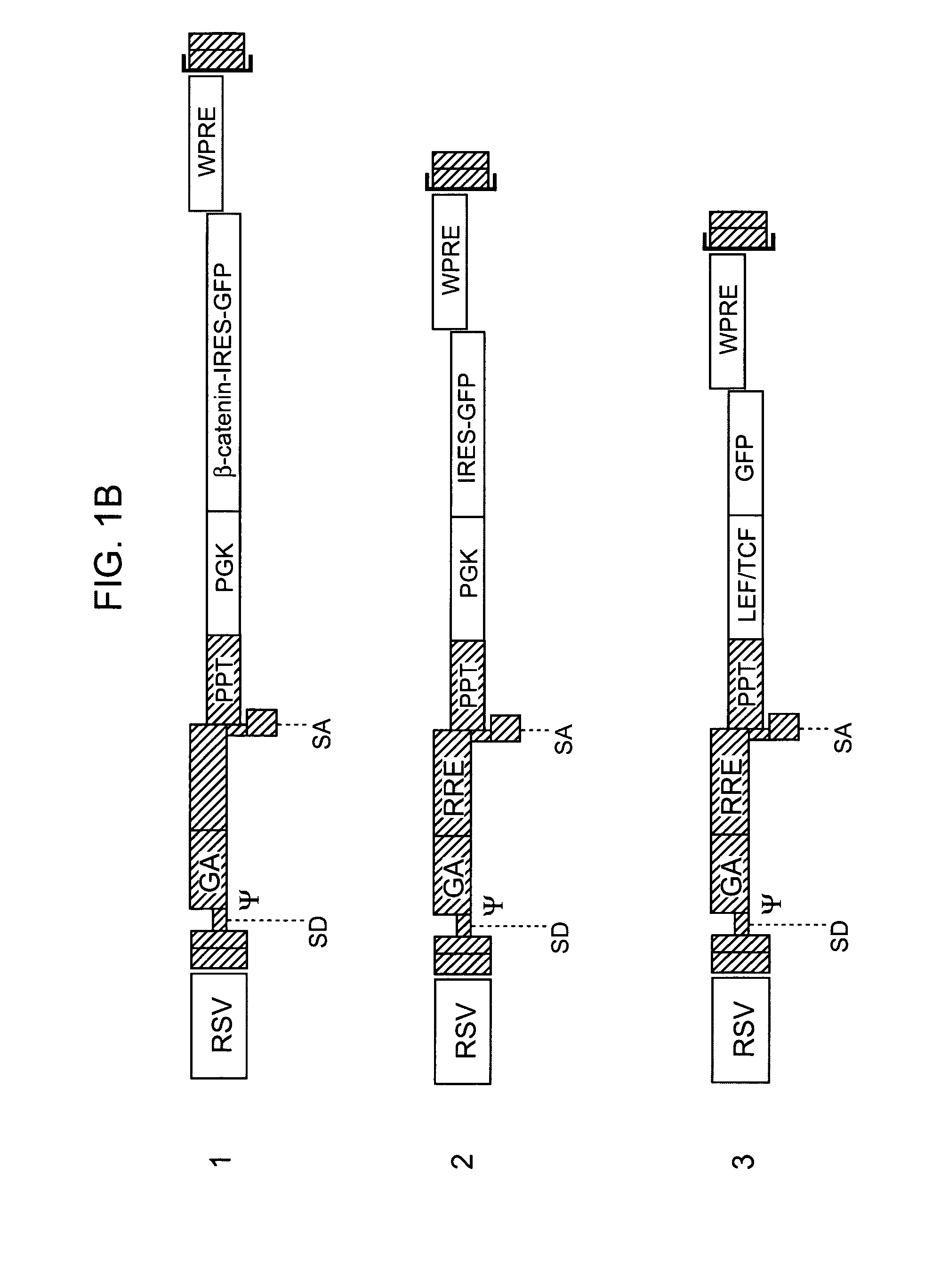
[0091]HSCs in vivo normally signal via LEF / TCF elements. It was determined whether HSCs in vivo utilize signals associated with the Wnt / Fzd / beta-catenin pathway. Sorted KTLS HSCs were infected with vectors carrying the LEF-1-TCF reporter driving expression of destabilized GFP (TOP-dGFP) or control reporter construct carrying mutations in the LEF / TCF binding sites (FOP-dGFP), and transplanted into lethally irradiated mice. The HSCs were then transplanted into groups of irradiated recipient mice and recipient bone marrow examined after 14 weeks to determine whether donor HSCs demonstrated reporter activity. In the representative example shown, donor derived HSCs infected with TOP-dGFP were found to express GFP in 29% of the cells while HSCs from the recipient mouse were negative for GFP. Moreover, only 2.5% of HSCs transduced with the control FOP-dGFP reporter expressed GFP, demonstrating that functional LEF-TCF binding sites were specifically required for KTLS HSC expression of GFP.

[...


PUM

PropertyMeasurementUnit
Timeaaaaaaaaaa
Fluorescenceaaaaaaaaaa

Abstract

Methods and compositions are provided for the identification of stem cells and cancer stem cells. β-catenin is also identified as a target for the development of therapeutic moieties against hematopoietic tumors, i.e. leukemia and lymphoma cells, which may include screening assays directed at β-catenin, or members of the β-catenin signaling pathway. Cellular proliferation in hematopoietic cells can be altered by introducing stabilized β-catenin into a hematopoietic cell that is altered in its ability to undergo apoptosis but which is not fully transformed. The immortalized cells are useful in screening assays, and in the analysis of pathways by which hematopoietic cells undergo transformation.

Description

[0001]This invention was made with Government support under contract NIH CA 86017 awarded by the National Institutes of Health. The Government has certain rights in this invention.BACKGROUND OF THE INVENTION[0002]Basic cancer research has focused on identifying the genetic changes that lead to cancer. This has led to major advances in understanding of the molecular and biochemical pathways that are involved in tumorigenesis and malignant transformation. But understanding of the cellular biology has lagged. While the effect of particular mutations on the proliferation and survival of model cells may be known, it is not known what the effects of such mutations will be on the actual cells involved in particular cancers.[0003]In fact, many observations suggest that analogies between normal stem cells and tumorigenic cells may be appropriate. Both normal stem cells and tumorigenic cells have extensive proliferative potential and the ability to give rise to new (normal or abnormal) tissue...

Claims


Application Information

Patent Timeline
IPC IPC(8): C12N5/00C12N15/09C12N15/74C12N5/02C12Q1/68G01N33/50C12N5/06C12N5/08C12N5/10C12Q1/02G01N33/15G01N33/53G01N33/567
CPCC12N5/0647C12Q1/6881C12Q1/6883C12Q1/6886C12Q1/6897C12Q2600/156C12Q2600/136
Inventor JAMIESON, CATRIONA HELEN M.AILLES, LAURIE ELIZABETHREYA, TANNISHTHAWEISSMAN, IRVING L.
Owner THE BOARD OF TRUSTEES OF THE LELAND STANFORD JUNIOR UNIV
Why Eureka
Try Eureka