
Locally-propelled, intraluminal device with cable loop track and method of use

a technology of intraluminal devices and cable loops, which is applied in the field of medical devices, can solve the problems of extending the mesentery, reducing and affecting the patient's recovery, and achieves the effect of increasing the size of the cable loop

Active Publication Date: 2007-06-05
ETHICON ENDO SURGERY INC
View PDF39 Cites 15 Cited by
  • Summary
  • Abstract
  • Description
  • Claims
  • Application Information

AI Technical Summary

Benefits of technology

[0006]Applicant has recognized the desirability of a low cost, potentially disposable locally-propelled intraluminal device which may provide physicians with a desirable alternative to using a conventional, reusable, flexible endoscope. Eliminating the need for the operator to make constant adjustment of the articulation controls of an endoscope may reduce the skill required to intubate the device, allowing operators other than physicians to use the device. This is advantageous because gastroenterologists currently do not have the capacity to handle all of the patients that need colonoscopies, so equipment that enables other staff, such as nurses, to help with the procedure could increase the capacity and allow gastroenterologists to treat more patients.
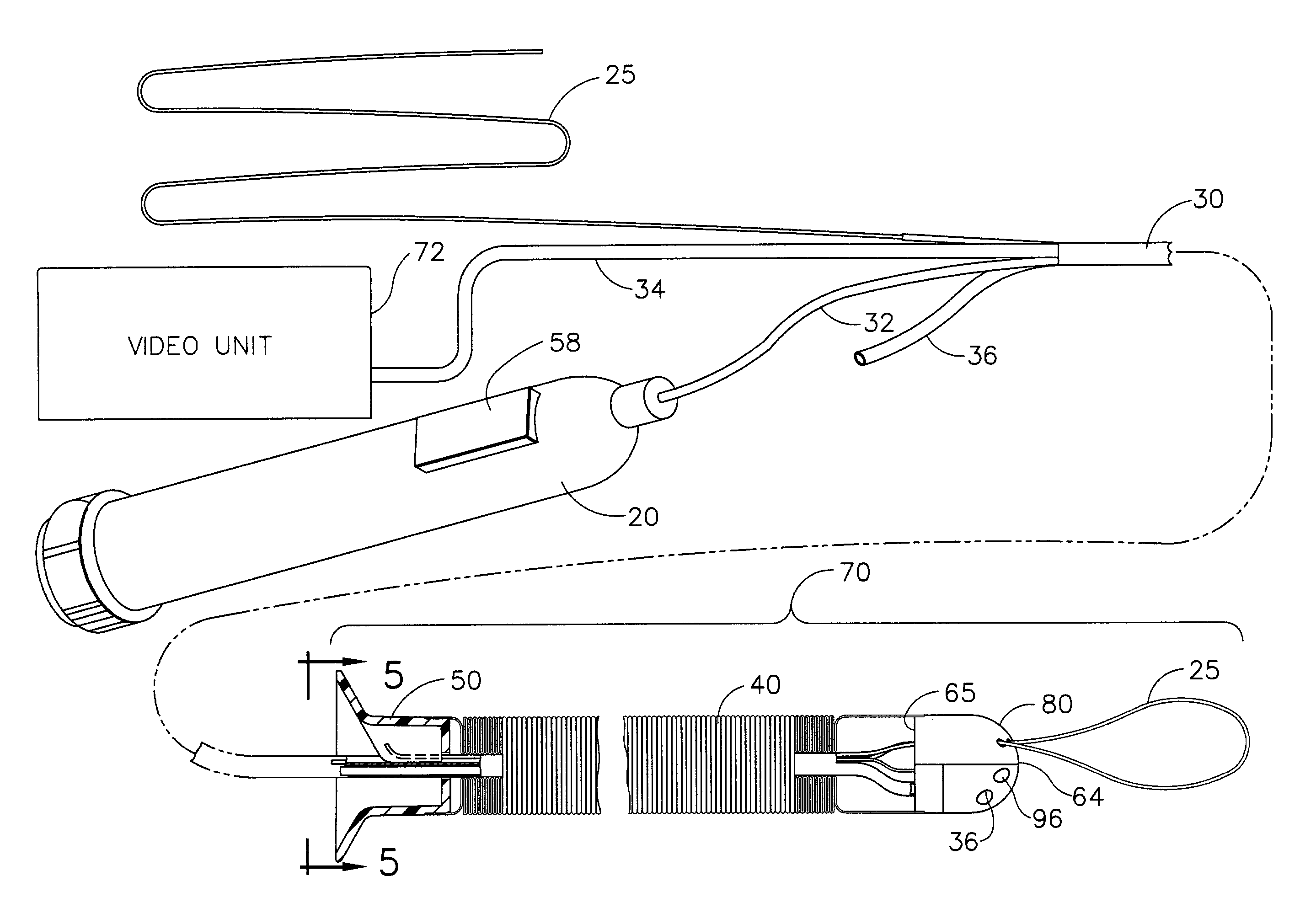
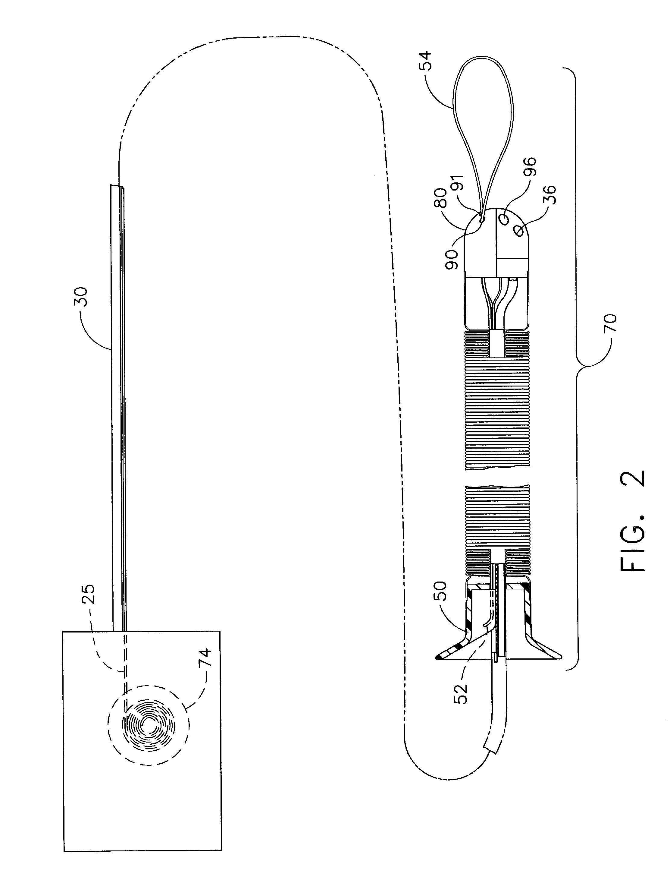
[0012]The invention can be used to assist in the placement of instruments including without limitation balloons, dilators, tissue graspers, tissue cutting devices, tissue stapling devices, tissue staining or treatment devices, vessel ligation devices, and tissue ablation devices. The track, in the form of a cable, can be fed by the operator into the lumen ahead of the locally-propelled capsule to provide a track for the capsule to follow through the tortuous path of the colon, providing a means for advancing or retracting the device. The cable track allows propulsion along a track that is independent of the physical characteristics of the lumen wall, which may be diseased, fatigued, or oddly shaped. As the operator slides more cable through the capsule, the size of the cable loop increases (for instance, because another portion or end of the cable is held stationary, such as by the capsule drive mechanism or by an anchor point). As the loop gets bigger, the loop “unfurls” around bends or other obstructions in the colon, thus creating a path around bends or obstructions in the GI tract without operator manipulation of articulating controls.

Problems solved by technology

Insertion of a flexible endoscope, especially into the colon, is usually a very time-consuming and uncomfortable procedure for the patient, even when sedated with drugs.
Often the flexible endoscope “loops” within the colon, such as at the sigmoid colon or at the splenic flexure of the colon, so that it becomes difficult to further advance the endoscope along the colon.
When a loop is formed, the force exerted to push the scope stretches the mesentery and causes pain for the patient.
Depending on the anatomy of the patient and the skill of the physician in manipulating the flexible endoscope, some portions of the colon may be unexamined, thus increasing the risk of undiagnosed disease.

Method used


Image

  • Locally-propelled, intraluminal device with cable loop track and method of use
  • Locally-propelled, intraluminal device with cable loop track and method of use
  • Locally-propelled, intraluminal device with cable loop track and method of use

Examples


Embodiment Construction

[0024]In one embodiment, the present invention provides a locally-propelled intraluminal medical device. By way of example, the present invention is illustrated and described for application in the colon of the lower GI tract of a human patient. However, the present invention is applicable for use in the body lumens of other hollow organs in humans and in other mammals.

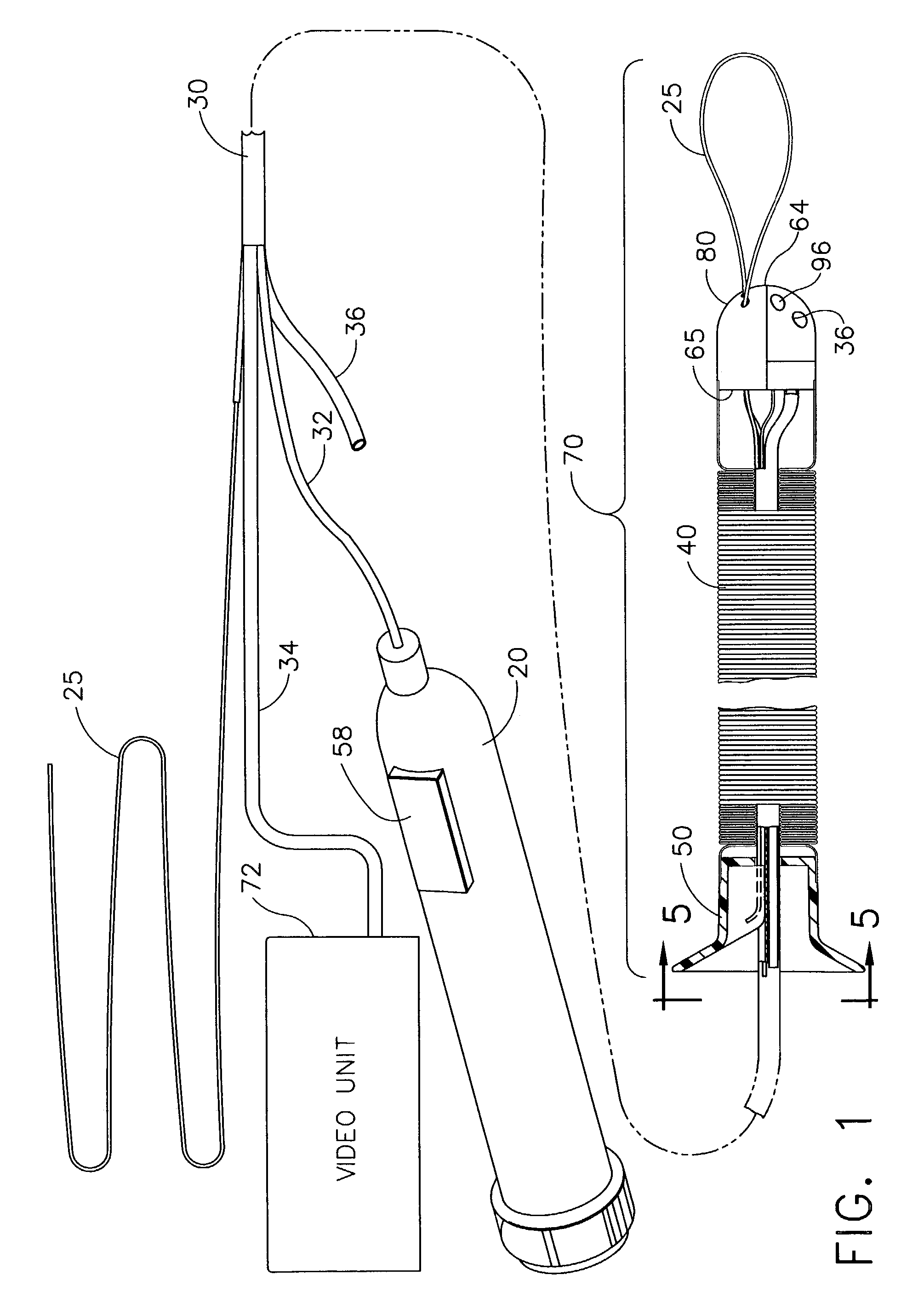
[0025]FIG. 1 shows a medical device 70 of the present invention. The medical device 70 can include a movable apparatus, such as a capsule 80 shaped and sized for movement through a body lumen, a compressible sleeve 40, a fixing plate 50, an umbilicus 30, a cable 25, a video unit 72, and a handpiece 20.

[0026]Capsule 80 generally has a leading end 64 that is smooth for atraumatic passage through a tortuous path of a gastrointestinal (GI) tract, such as a colon. In one embodiment of capsule 80, leading end 64 is hemispherical and a trailing end 65 is flat to accept the contents contained in umbilicus 30. Other shapes of ...


PUM


Abstract

A medical device for performing medical procedures inside a lumen (such as the GI tract) of a patient is provided comprising a capsule, a cable, and a propulsion means. The cable can have one end anchored to the patient, and can extend from this anchored portion to the capsule. The cable can include a loop forward (distal) of the capsule in the GI tract. The propulsion means is operably connected to the cable to vary the length of cable between the anchored end and the capsule, so that the capsule is repositioned inside the GI tract of the patient.

Description

FIELD OF THE INVENTION[0001]The present invention relates to a medical device, and more particularly to a medical device that can be propelled along a cable located within a lumen of a patient's body.BACKGROUND[0002]A physician typically accesses and visualizes tissue within a patient's gastrointestinal (GI) tract with a long, flexible endoscope. For the upper GI, a physician may insert a gastroscope into the sedated patient's mouth to examine and treat tissue in the esophagus, stomach, and proximal duodenum. For the lower GI, a physician may insert a colonoscope through the sedated patient's anus to examine the rectum and colon. Some endoscopes have a working channel, typically about 2.5–3.5 mm in diameter, extending from a port in the handpiece to the distal tip of the flexible shaft. A physician may insert medical instruments into the working channel to help diagnose or treat tissues within the patient. Physicians commonly take tissue biopsies from the mucosal lining of the GI tr...

Claims


Application Information

Patent Timeline
Patent Type & Authority Patents(United States)
IPC IPC(8): A61B1/01A61M25/082A61B17/28A61B1/00A61B1/018A61B1/05A61B1/31A61B17/32A61M25/01
CPCA61B1/00156A61B1/018A61B1/041A61B1/05A61B1/31A61M25/0105A61M25/0116A61M2210/1042A61M2210/106A61M2210/1064A61M2210/1067
Inventor LONG, GARY L.
Owner ETHICON ENDO SURGERY INC
Learn More