Method and apparatus for quantitative three-dimensional reconstruction in scanning electron microscopy

a scanning electron microscopy and three-dimensional reconstruction technology, applied in the direction of fluorescence/phosphorescence, material analysis using wave/particle radiation, instruments, etc., can solve the problems of limiting the wide utilization of fib-based tomographic methods, the density of raw data collection, and the implementation of robust, yet versatile data analysis and volume visualization methods

Active Publication Date: 2007-12-25
CARL ZEISS SMT GMBH
View PDF5 Cites 43 Cited by
  • Summary
  • Abstract
  • Description
  • Claims
  • Application Information

AI Technical Summary

Problems solved by technology

However, 3D tomographic data acquisition and volume visualization through the application of serial FIB (Focused Ion Beam) sectioning has only just begun to emerge as a demonstrable method, with work completed by a small number of researchers.
Factors that limit wider utilization of FIB-based tomographic methods include the ease, speed and density of raw data collection.
Another obstacle is implementing robust, yet versatile data analysis and volume visualization methods suitable for electron imaging.
Yet FIB-based nanotomography has, until now, remained a less practical application due to the time, effort and specialized data reduction expertise involved.

Method used


Image

  • Method and apparatus for quantitative three-dimensional reconstruction in scanning electron microscopy
  • Method and apparatus for quantitative three-dimensional reconstruction in scanning electron microscopy
  • Method and apparatus for quantitative three-dimensional reconstruction in scanning electron microscopy

Examples


Embodiment Construction

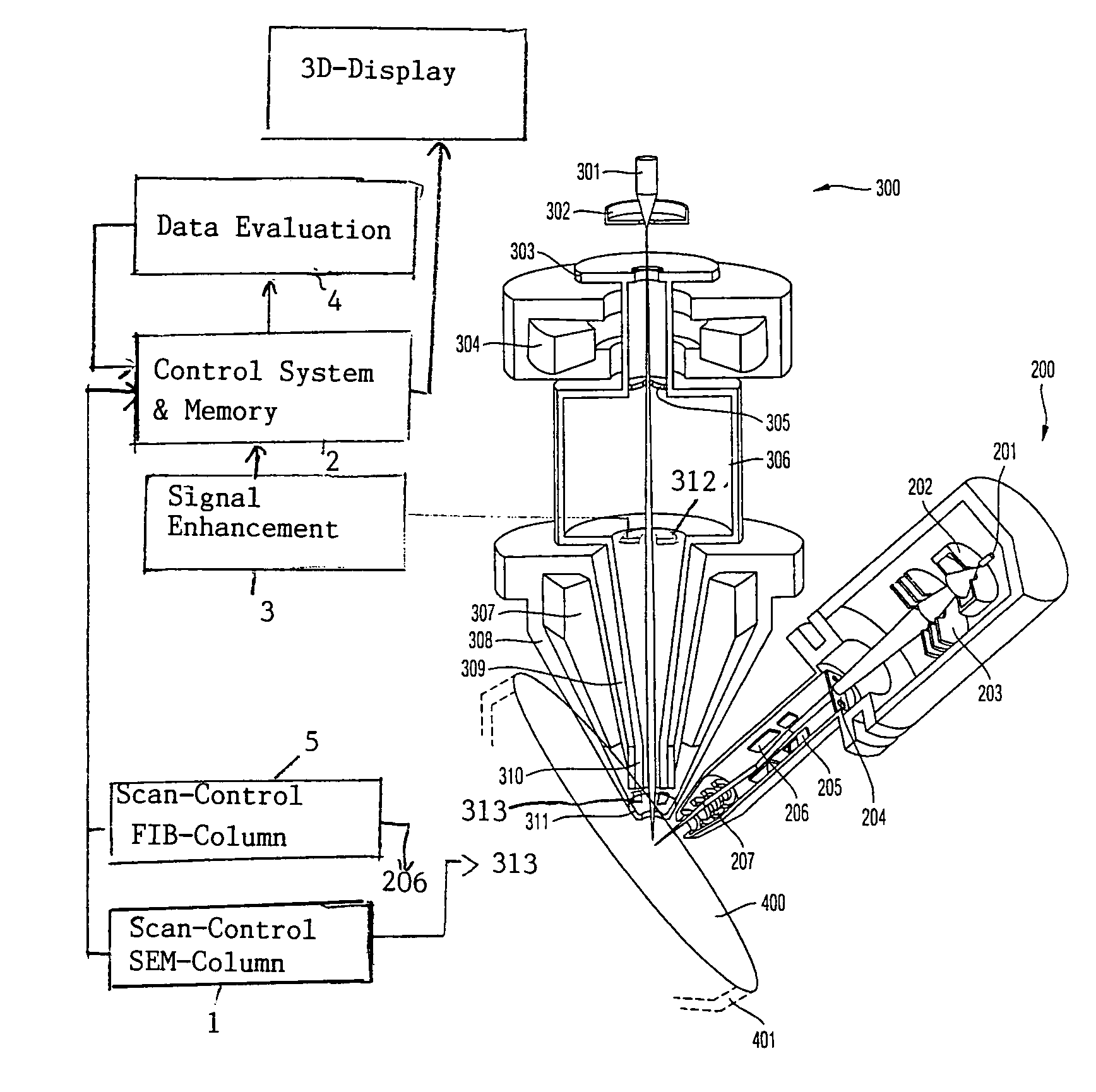
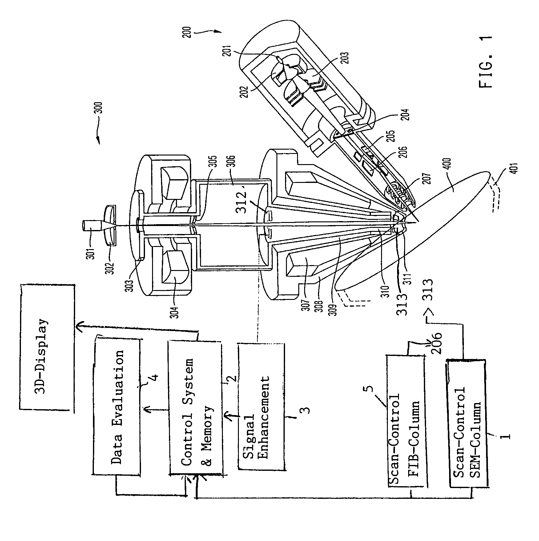
[0021]The charged particle beam system in FIG. 1 comprises a scanning electron beam column 300 and a focused ion beam column 200. As shown in FIG. 1, the optical axis of the electron beam column 300 and of the focused ion beam column 200 intersects substantially in a plane defined by the planar surface of a sample 400. The optical axis of the focused ion beam column extends approximately perpendicularly to this plane of the sample 400 and the ion beam therefore impinges orthogonally on this surface. The angle at which the electron beam impinges on this surface of the sample 400 in this configuration is about 35°.

[0022]In the scanning electron beam column 300, a primary electron beam is generated by an electron source 301, preferably a Schottky field emitter, and an anode 303. The emitted electrons also pass through an extractor electrode 302 disposed between said electron source 301 and the anode 303. The accelerated electron beam then passes through a bore at the bottom of the anod...


PUM

PropertyMeasurementUnit
angleaaaaaaaaaa
depthaaaaaaaaaa
scanning electron microscopeaaaaaaaaaa

Abstract

A method and an apparatus are for three-dimensional tomographic image generation in a scanning electron microscope system. At least two longitudinal marks are provided on the top surface of the sample which include an angle therebetween. In consecutive image recordings, the positions of these marks are determined and are used to quantify the slice thickness removed between consecutive image recordings.

Description

FIELD OF THE INVENTION[0001]The invention relates to the field of generating tomographic image data sets of a sample in scanning electron microscopy and of generating three dimensional views of the sample.BACKGROUND OF THE INVENTION[0002]The value of the ability to acquire, interrogate and display n-dimensional data sets has been well established through various scientific disciplines. The biomedical sciences, in particular, have developed sophisticated methods to visualize volumetric image data. However, 3D tomographic data acquisition and volume visualization through the application of serial FIB (Focused Ion Beam) sectioning has only just begun to emerge as a demonstrable method, with work completed by a small number of researchers. The FIB-SEM and FIB-Auger tomographic methods have demonstrated the ability to provide volumetric data resolution down to 10 nm or less, and thus hold tremendous future potential for both material science and biomedical investigations. Yet even the be...

Claims


Application Information

Patent Timeline
Patent Type & Authority Patents(United States)
IPC IPC(8): G01N23/225H01J37/28
CPCG01N1/32H01J37/28H01J37/3056H01J37/302H01J2237/226
Inventor PRINCIPE, EDWARD
Owner CARL ZEISS SMT GMBH
Why Eureka
Try Eureka