
Peptide biosensors for anthrax protease

a protease and peptide technology, applied in the field of fluorescence-based cell and molecular biochemical assays for toxin detection and drug discovery, can solve the problems of macrophage death, shock and host death, and dramatic hypotension, and achieve the effect of improving both the speed and efficiency of the assay

Inactive Publication Date: 2008-08-12
CELLOMICS
View PDF13 Cites 14 Cited by
  • Summary
  • Abstract
  • Description
  • Claims
  • Application Information

AI Technical Summary

Benefits of technology

[0008]The present invention provides FRET-based protease biosensors, and kits containing them, for detecting the presence of the anthrax protease, as well as methods for using the protease biosensors to detect the presence of Bacillus anthracis in a test sample. The present protease biosensors and assays provide a significant improvement over previous biosensors and assays for detecting the presence of Bacillus anthracis in a sample, by significantly improving both the speed and efficiency of the assay, and by detecting live, virulent strains of Bacillus anthracis. Therefore, the biosensors of the present invention will have fewer false positives, which is desirable for a sensor to be used in a potentially hazardous situation.

Problems solved by technology

Bacillus anthracis is the causative agent of anthrax, which is characterized by the hyperstimulation of host macrophage inflammatory pathways, leading to dramatic hypotension, shock, and death of the host.
It is believed that cleavage of these signaling kinases by LF leads to inhibition of growth-factor response pathways, further leading to macrophage death.
(WO 99 / 50439) lists methods for screening modulators, homologues and mimetics of LF protease activity but they either require laborious analysis methods or are extremely general.
The use of peptides as substrates for LF protease activity was described by Hammond and Hanna ((1998) Infection &Immunity 66, 2374-8); however the peptides were not fluorescently labeled and were analyzed by high-performance liquid chromatography, which is a tedious and costly procedure.

Method used


Image

  • Peptide biosensors for anthrax protease
  • Peptide biosensors for anthrax protease
  • Peptide biosensors for anthrax protease

Examples


examples

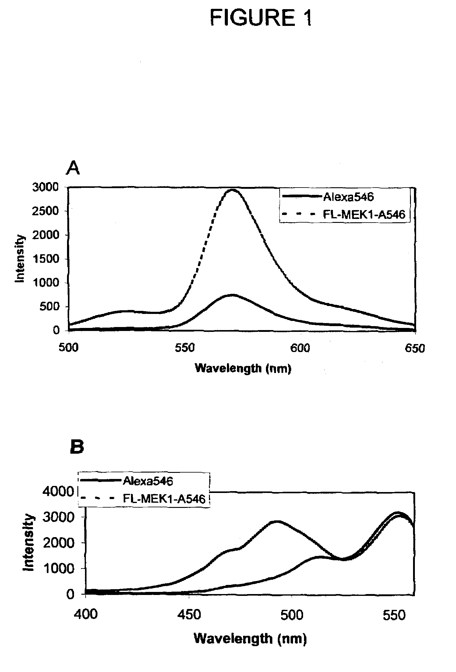
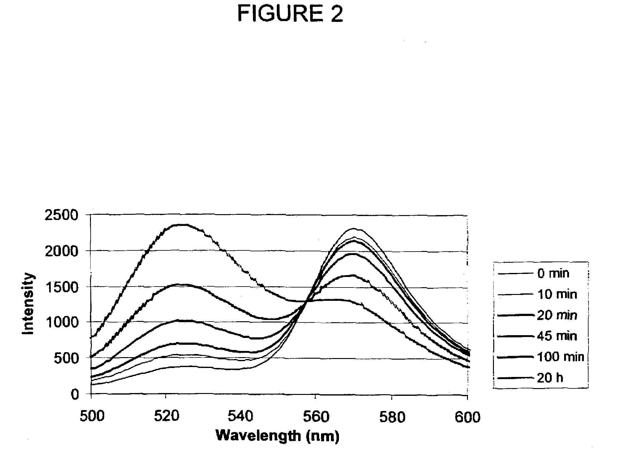
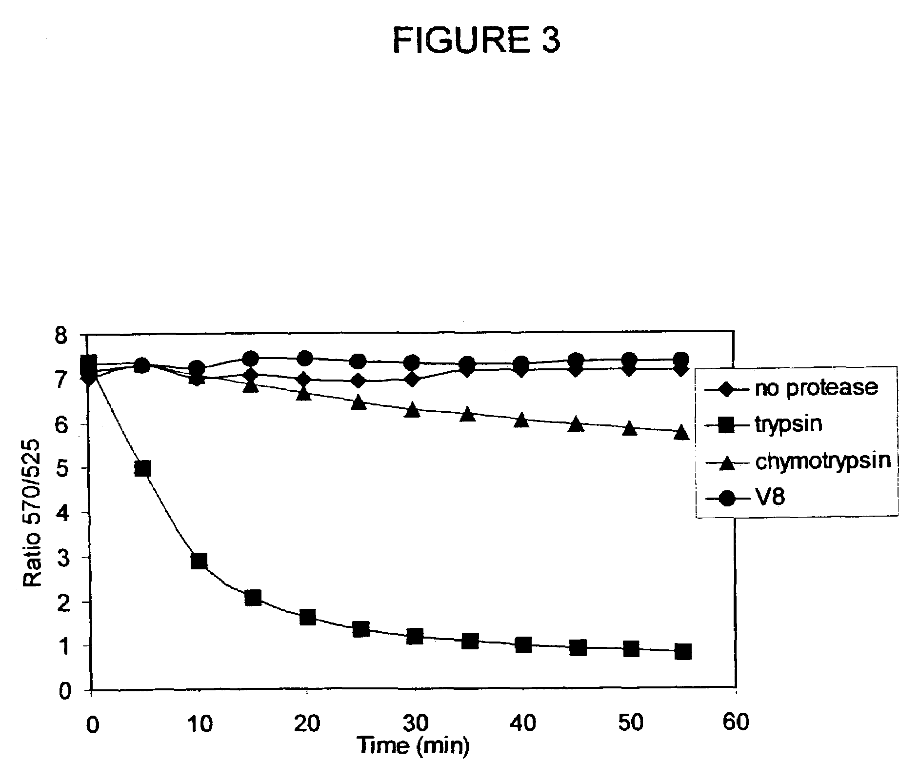
[0106]Design, Synthesis, and Purification of FL-MEK1-A546. Knowing that anthrax protease cleaves MEK1 between proline and isoleucine residue within the first 12 amino acids of the protein, a 13 amino acid sequence was chosen to provide some flanking residues but keeping the length of the peptide short enough to maintain high efficiency FRET. The general strategy for synthesizing the dual-labeled peptide was similar to that described by Contillo et al. (in Techniques in Protein Chemistry, J. W. Crabb, Editor. 1994, Academic Press: San Diego, p. 493-500)] except that the more soluble ALEXA FLUOR® 546 fluorophore was chosen instead of the eosin, which allows for aqueous-phase reaction and purification.

[0107]A cysteine residue was added at the C-terminus to provide a site for thiol-specific labeling with the second fluorophore. The sequence of the peptide is MPKKKPTPIQLNPC (SEQ ID NO:3). The peptide was synthesized by a custom synthesis facility (BioPeptide, San Diego, Calif.) with carb...


PUM

PropertyMeasurementUnit
Digital informationaaaaaaaaaa
Resonance energyaaaaaaaaaa

Abstract

The present invention provides a protease biosensor that can be used to detect the presence of the lethal factor protease from Bacillus anthracis, as well as methods for using the protease biosensor.

Description

CROSS REFERENCE[0001]This application claims priority to U.S. Patent Application Ser. No. 60 / 182,011 filed Feb. 11, 2000, is a US national phase application of PCT / US01 / 04253, filed Aug. 16, 2001 and is related to U.S. patent application Ser. No. 09 / 430,656 filed Oct. 29, 1999, now U.S. Pat. No. 6,756,207.U.S. GOVERNMENT RIGHTS[0002]This invention was made in part with support from the U.S. Government under Contract No. N00014-98-C-0326, awarded by the U.S. Office of Naval Research, an organization of the U.S. Department of Defense. The U.S. Government may have certain nonexclusive rights in this invention.FIELD OF THE INVENTION[0003]This invention is in the field of fluorescence-based cell and molecular biochemical assays for toxin detection and drug discovery.BACKGROUND OF THE INVENTION[0004]Bacillus anthracis is the causative agent of anthrax, which is characterized by the hyperstimulation of host macrophage inflammatory pathways, leading to dramatic hypotension, shock, and death...

Claims


Application Information

Patent Timeline
IPC IPC(8): G01N33/53A61K38/00C12Q1/04C12Q1/37G01N33/542G01N33/569G01N33/58
CPCC12Q1/04C12Q1/37G01N33/56911G01N33/582G01N33/542G01N2333/32
Inventor BURROUGHS-TENCZA, SARAH
Owner CELLOMICS
Learn More