Proteolytic biomarkers for traumatic injury to the nervous system

a traumatic injury and biomarker technology, applied in the field of traumatic injury to the nervous system, can solve the problems of inability to quickly be employed in an emergency room environment, high cost of ct and mri, and the majority of tbi survivors are severely affected, etc., and achieve the effect of reliable detection

Active Publication Date: 2008-11-25
UNIV OF FLORIDA RES FOUNDATION INC +1
View PDF10 Cites 24 Cited by
  • Summary
  • Abstract
  • Description
  • Claims
  • Application Information

AI Technical Summary

Benefits of technology

[0011]The invention provides for the reliable detection and identification of biomarkers that are uniquely produced in brain injury and / or stress, neuronal disorders, organ injury and / or stress, cancer and cancer treatment, and muscle rehabilitation / exercise overtraining and that are important for the diagnosis and prognosis and the monitoring of damage of the same organ / tissue system of interest. The profile of proteolytic products or fragments of organ-enriched or specific proteins / peptides in patients with damage to organ or tissue and cells within them are distinguished from normal controls using inexpensive techniques. These techniques provide simple yet sensitive approaches to diagnosing damage or stress to the nervous system, to other organs or to multiple organs, various cancers, cancer treatment, muscle rehabilitation / exercise over-training and other human pathological or stressed conditions where major organ(s) is compromised or altered using biological fluids.

Problems solved by technology

However, despite dramatically improved emergency triage systems based on these outcome measures, most TBI suffer long term impairment and a large number of TBI survivors are severely affected despite predictions of “good recovery” on the GOS.
In addition, CT and MRI are expensive and cannot be rapidly employed in an emergency room environment.
Accordingly, the neural pathways of a mammal are particularly at risk if neurons are subjected to mechanical or chemical trauma or to neuropathic degeneration sufficient to put the neurons that define the pathway at risk of dying.
Stroke is a leading cause of death in the developed world.
It may be caused by reduced blood flow or ischemia that results in deficient blood supply and death of tissues in one area of the brain (infarction).
The ability to detect such injury is lacking in the prior art.
Mammalian neural pathways also are at risk due to damage caused by neoplastic lesions.
Transformed cells of neural origin generally lose their ability to behave as normal differentiated cells and can destroy neural pathways by loss of function.
Metastatic tumors, which are a significant cause of neoplastic lesions in the brain and spinal cord, also similarly may damage neural pathways and induce neuronal cell death.
Damage to peripheral nerves, such as occurred with diabetic neuropathies or administration of chemo-therapeutic agents such as those used to treat cancer, can also involve proteolytic damage similar to that seen in the CNS.

Method used


Image

  • Proteolytic biomarkers for traumatic injury to the nervous system
  • Proteolytic biomarkers for traumatic injury to the nervous system
  • Proteolytic biomarkers for traumatic injury to the nervous system

Examples


example 1

Detection of Neural Proteins Subjected to Proteolytic Attack 48 h after Traumatic Brain Injury (TBI) in Rats

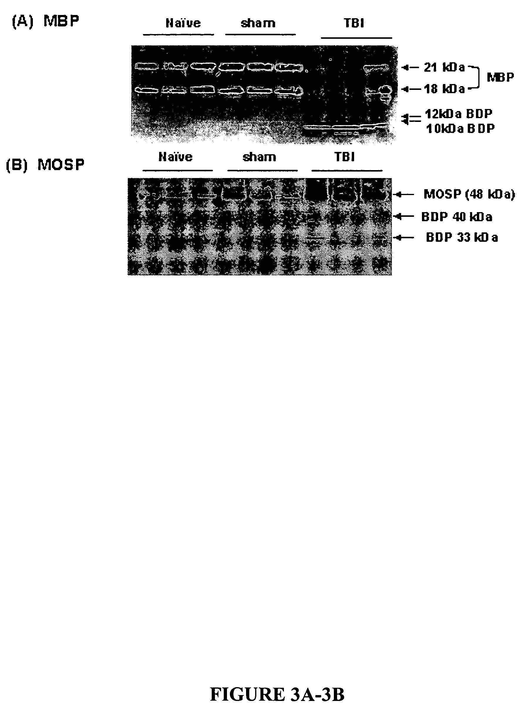
[0290]TBI was induced in rodents as described above. 48 h following TBI or sham operation or naïve rats, samples of CSF were collected and analyzed for presence of five novel neural protein markers (MBP and MOSP) (FIG. 3) were identified to be vulnerable to endogenous proteolytic attack, producing major breakdown products (BDPs) in the ipsilateral hippocampus. Ipsilateral cortical samples were also analyzed and they showed very similar patterns of proteolysis. Based on the unique cleavage site in MBP (DENPVVHFF114K115NIVTPP) SEQ ID NO: 184), we have produced polyclonal and monoclonal that specifically detects the new N-terminal (NH2-KNIVTPP) (SEQ ID NO: 149) of MBP-BDP of 12 kDa and 10 kDa (FIG. 4). These unique BDP's when accumulated in biofluids such as CSF and blood are excellent diagnostic markers for organ-specific (brain, spinal cord or peripheral nerve) injury or stress...

example 2

Detection of Myelin Proteins Subjected to Proteolytic Attack 48 h after Traumatic Brain Injury (TBI) in Rats

[0291]TBI was induced in rodents as described above. 48 h following TBI or sham operation of on naïve rats, samples of CSF were collected and analyzed for presence of two novel myelin sheath protein markers; (MBP and MOSP) (FIG. 3) were identified to be vulnerable to endogenous proteolytic attack, producing major breakdown products (BDPs) in the ipsilateral hippocampus. Ipsilateral cortical samples were also analyzed and they showed very similar patterns of proteolysis. Based on the unique cleavage site in MBP (DENPVVHFF114^K115NIVTPP), (SEQ ID NO: 148) we have produced polyclonal and monoclonal antibodies that specifically detect the new N-terminal (NH2-KNIVTPP), SEQ ID NO: 149) of MBP-BDP of 12 kDa and 10 kDa (FIG. 4). These unique BDP's when accumulated in biofluids such as CSF and blood are excellent diagnostic markers for organ-specific (brain, spinal cord or peripheral n...

example 3

Detection of Synaptic Proteins (Synaptotagmin and Synaptojanin-1) are being Degraded in Rat Cortex and / or Hippocampus 48 hr after TBI in Rats

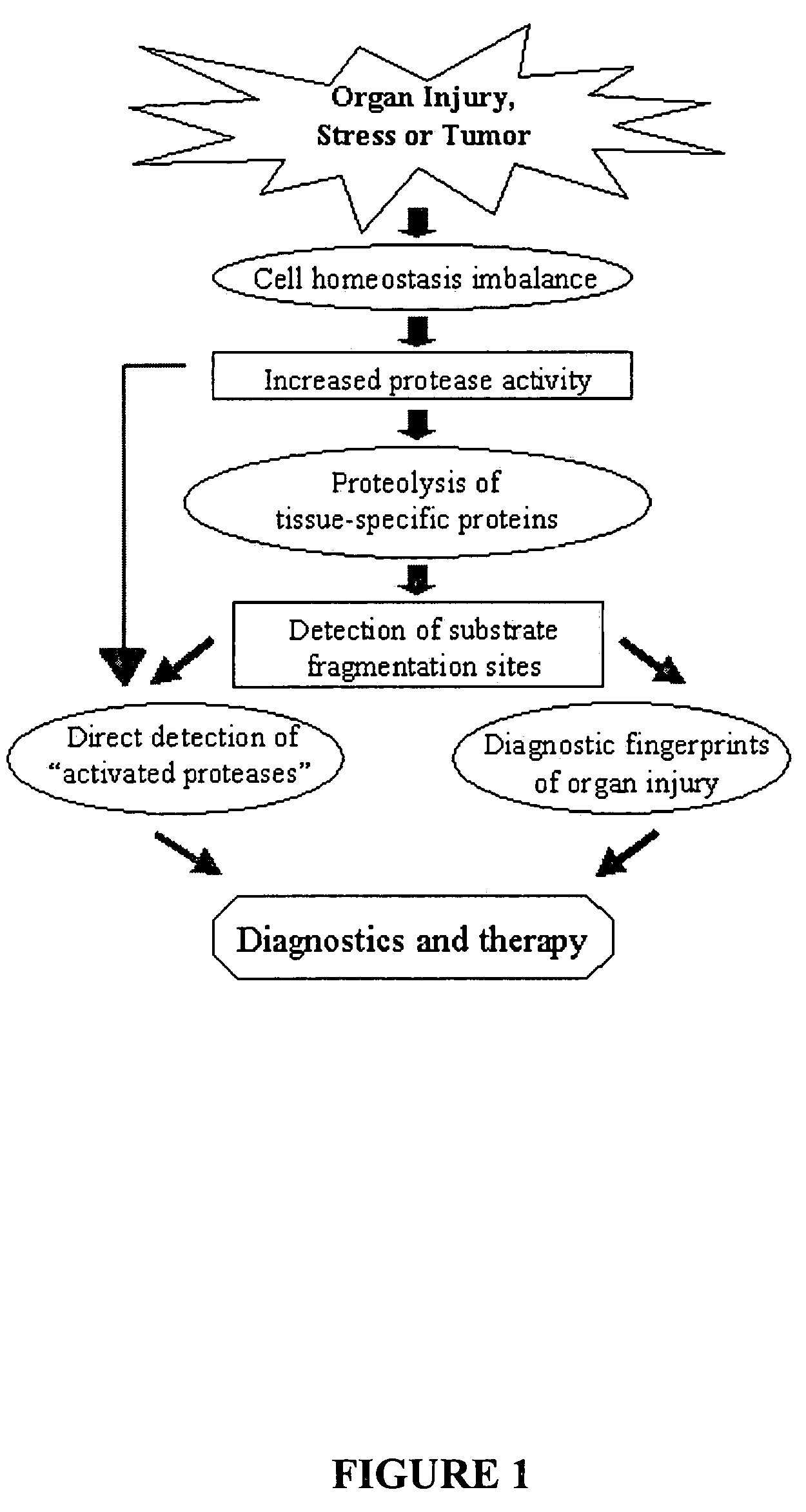
[0292]TBI was induced in rodents as described above. 48 h following TBI or sham operation on naïve rats, samples of CSF were collected and analyzed for presence of five novel synaptic protein markers; Synaptotagmin (top), and Synaptojanin-1 (bottom) (FIG. 5) were identified to be vulnerable to endogenous proteolytic attack, producing major breakdown products (BDPs) in the ipsilateral hippocampus. Ipsilateral cortical samples were also analyzed and they showed very similar patterns of proteolysis. These unique BDP's when accumulated in biofluids such as CSF and blood, can be detected by immunological techniques such as Western blots or ELISA and thus are excellent diagnostic marker for organ-specific (brain or spinal cord or peripheral nerve) injury or stress (FIG. 1).


PUM

PropertyMeasurementUnit
temperaturesaaaaaaaaaa

Abstract

The present invention identifies biomarkers that are diagnostic of nerve cell injury, organ injury, and / or neuronal disorders. Detection of different biomarkers of the invention are also diagnostic of the degree of severity of nerve injury, the cell(s) involved in the injury, and the subcellular localization of the injury.

Description

CROSS-REFERENCE TO RELATED APPLICATIONS[0001]The present application claims the priority of U.S. Provisional Patent application No. 60 / 562,819 filed Apr. 15, 2004; and entitled “PROTEOLYTIC MARKERS AS DIAGNOSTIC BIOMARKERS FOR CANCER, ORGAN INJURY AND MUSCLE REHABILITATION / EXERCISE OVERTRAINING” which is incorporated herein by reference in its entirety.[0002]The subject invention was made with government support under a research project supported by National Institutes of Health Grant #R01 NS39091, National Institutes of Health Grant #R01 NS40182, U.S. Army Grant #DAMD 17-99-1-9565, and U.S. Army Grant #DAMD 17-01-1-0765.FIELD OF THE INVENTION[0003]The invention provides for the reliable detection and identification of biomarkers that are uniquely produced in brain injury and / or stress, neuronal disorders, organ injury and / or stress, cancer and cancer treatment, and muscle rehabilitation / exercise overtraining and that are important for the diagnosis and prognosis and the monitoring ...

Claims


Application Information

Patent Timeline
Patent Type & Authority Patents(United States)
IPC IPC(8): G01N33/567G01N33/48C12Q1/37G01N33/68
CPCC12Q1/37G01N33/6896Y10T436/118339Y10T436/25375
Inventor WANG, KEVIN KA-WANGHAYES, RONALDLIU, MING CHENOLI, MONIKA
Owner UNIV OF FLORIDA RES FOUNDATION INC
Why Eureka
Try Eureka