Method and system for automatically identifying regions of trabecular bone tissue and cortical bone tissue of a target bone from a digital radiograph image

a digital radiograph image and target bone technology, applied in the field of image processing techniques, can solve the problems of inability or undesirable disadvantages of prior art approaches, inability to determine the specific density of cortical bone and trabecular bone, and limited application of qct in practi

Inactive Publication Date: 2009-05-26
COMPUMED
View PDF5 Cites 32 Cited by
  • Summary
  • Abstract
  • Description
  • Claims
  • Application Information

AI Technical Summary

Problems solved by technology

Unfortunately, prior art approaches are incapable or have undesirable disadvantages associated therewith.
Unfortunately, DXA only measures an integrated bone mineral density and is incapable for determining the specific density of the cortical bone and the trabecular bone.
However, the application of QCT is limited in practice due to high costs involved.
For example, a single QCT procedure can cost several hundred dollars.
There is also the added inconvenience and burden for the patient since the QCT procedure is not as readily available at hospitals and health care clinics as an X-ray machine due to QCT equipment's high installation and maintenance costs.
The actual procedu...

Method used


Image

  • Method and system for automatically identifying regions of trabecular bone tissue and cortical bone tissue of a target bone from a digital radiograph image
  • Method and system for automatically identifying regions of trabecular bone tissue and cortical bone tissue of a target bone from a digital radiograph image
  • Method and system for automatically identifying regions of trabecular bone tissue and cortical bone tissue of a target bone from a digital radiograph image

Examples


second embodiment

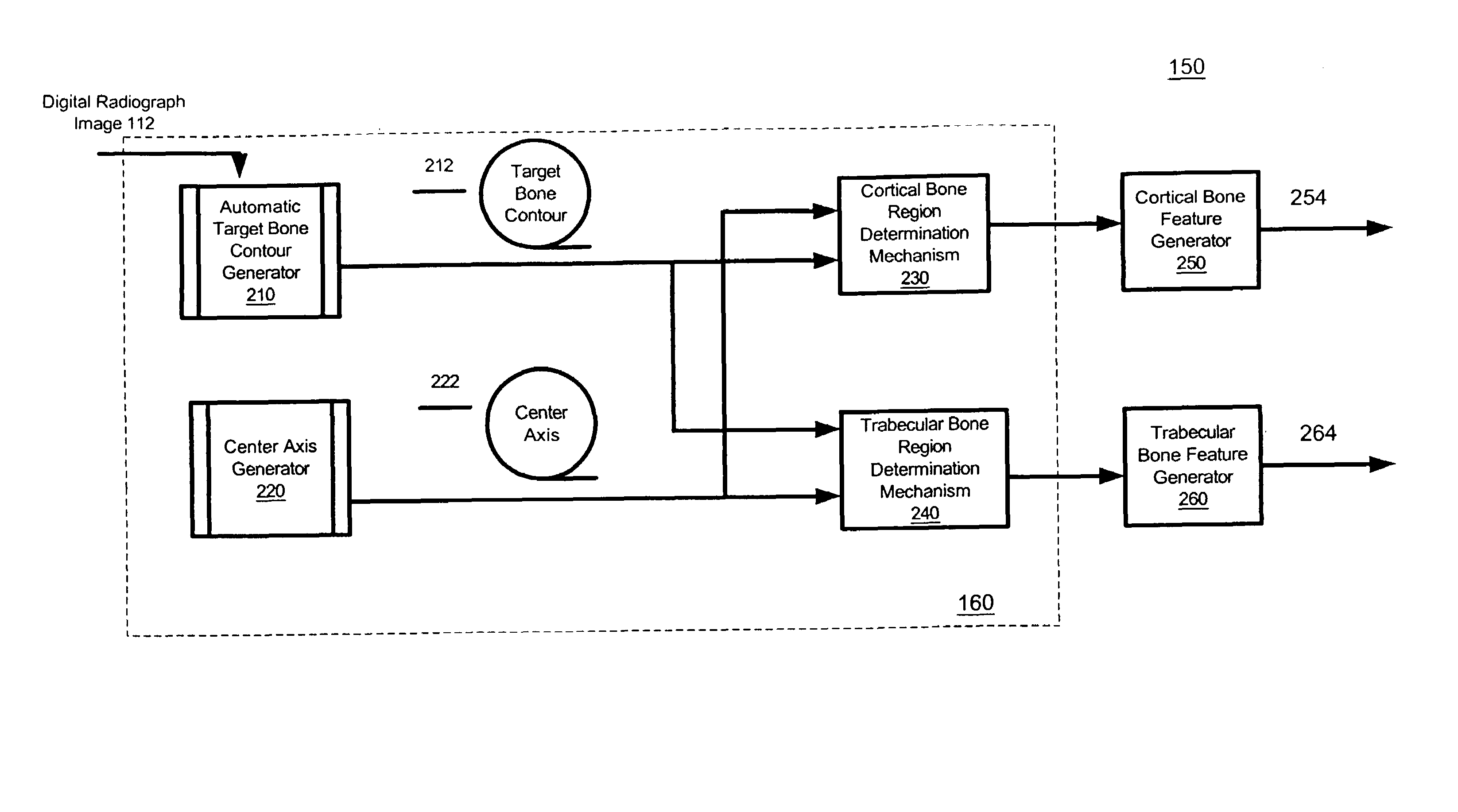
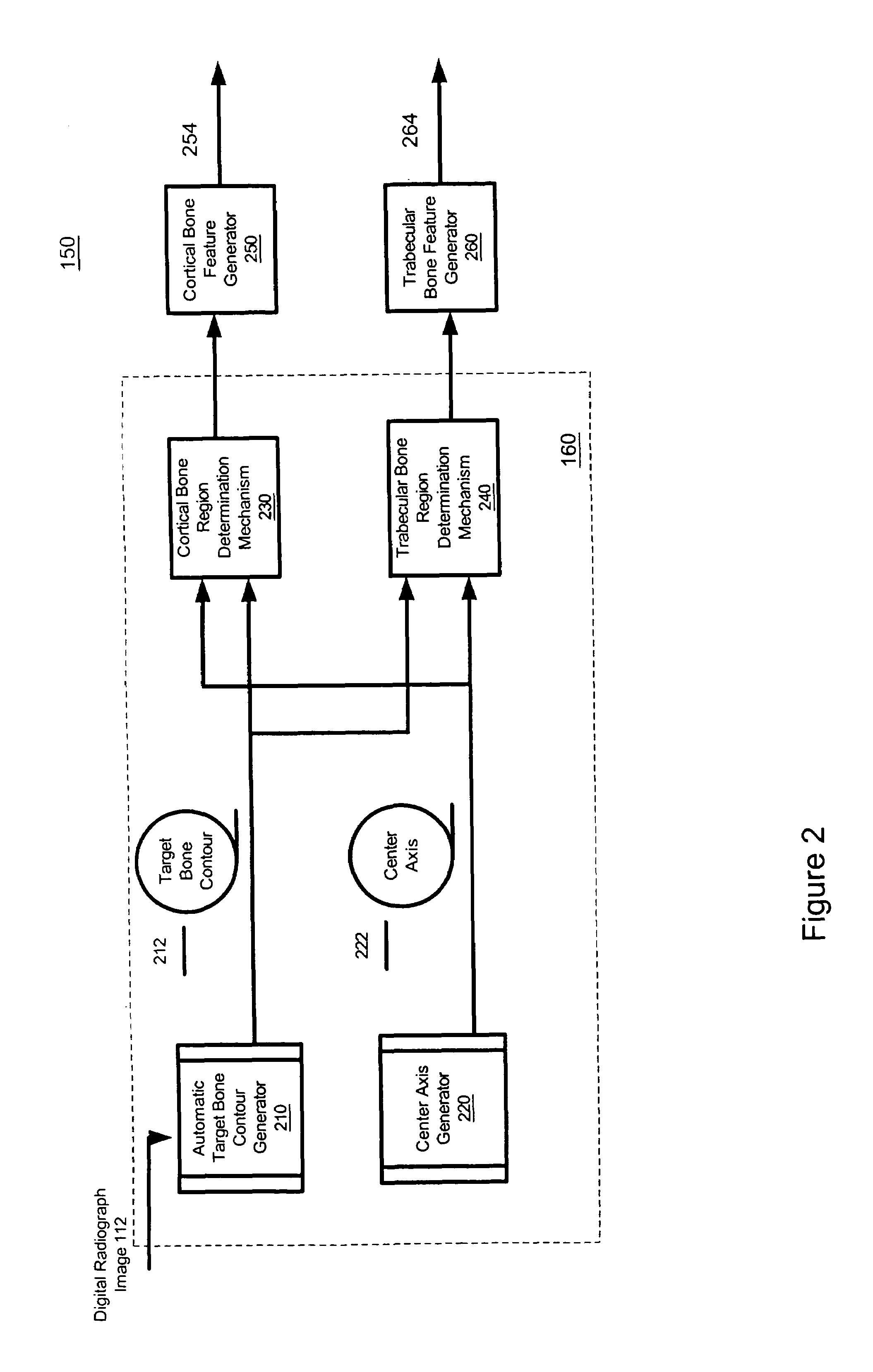
[0122 of Target Bone Feature Generation Mechanism that Employs a Two-Dimensional (2D) Bone Tissue Classifier

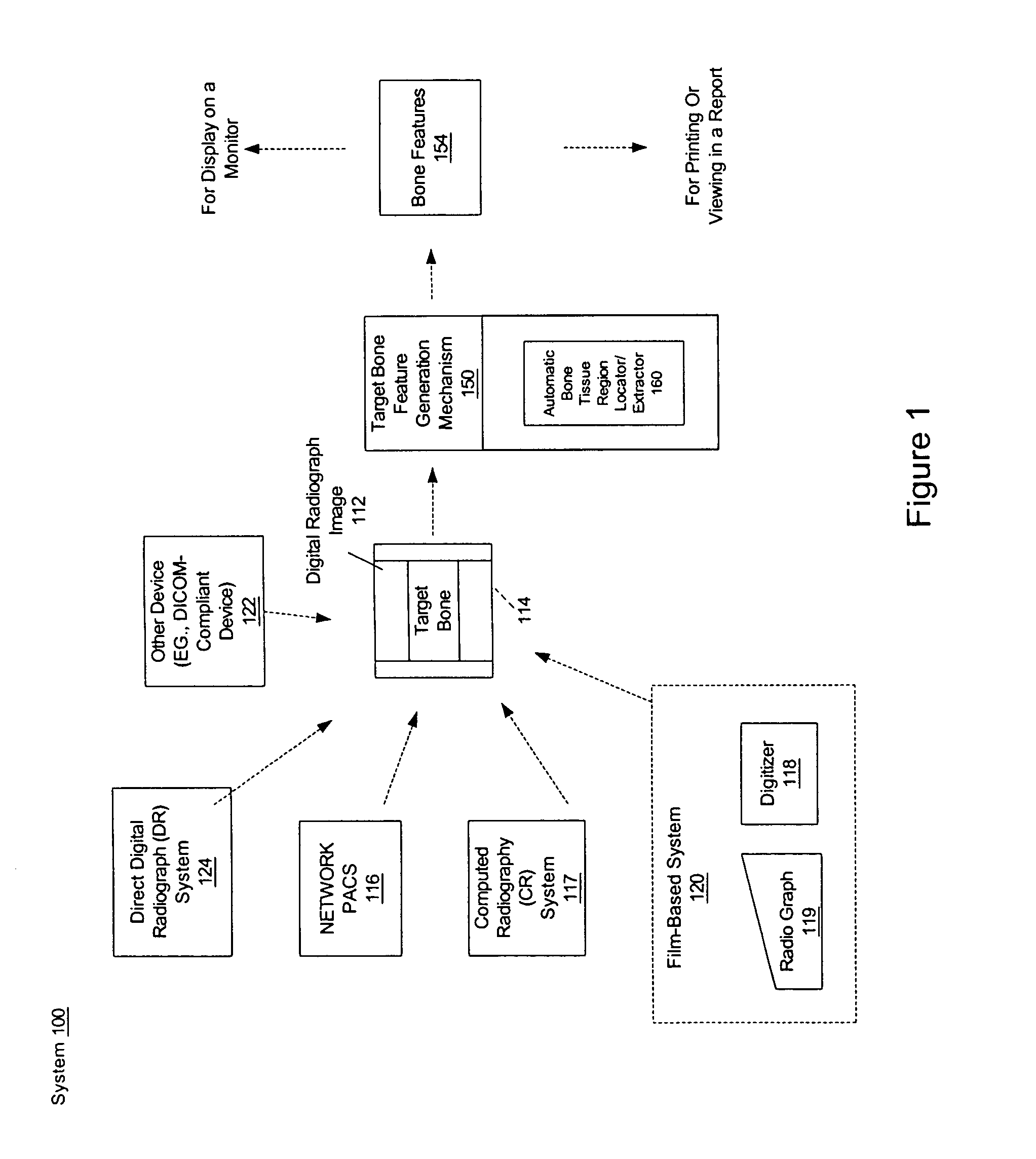
[0123]FIG. 19 is a block diagram illustrating in greater detail the target bone feature generation mechanism 150 of FIG. 1 in accordance with another embodiment of the present invention. The target bone feature generation mechanism 150 includes automatic bone tissue region locator / extractor 1960. The automatic region of bone tissue locator / extractor 1960 includes an automatic target bone contour generator 1910 for manually or automatically generating a target bone contour 1912 and for manually or automatically generating the middle or center axis 1914 of the target bone.

[0124]The automatic region of bone tissue locator / extractor 1960 includes an anchor point generator 1920 for automatically generating one or more anchor points 1922. In one embodiment, the anchor points include a neckline that is a line that is the minimum width of the target bone in the horizontal direction. T...


PUM


Abstract

System and method for automatically extracting at least one region of a first type of bone tissue from a target bone. A digital radiograph image with the target bone is received. Based on the digital radiograph image, at least one region of a first type of bone tissue is automatically identified or extracted. This region can then be utilized for different purposes, such as assessment, qualitative assessment, quantitative assessment, analysis, spatial domain analysis, frequency domain analysis, and calculation of one or more target bone features.

Description

[0001]This application claims the benefit of Provisional Patent Application Ser. No. 60 / 504,231 filed Sep. 19, 2003.FIELD OF THE INVENTION[0002]The present invention relates generally to image processing techniques applied to medical images, and more particularly, to a method and system for automatically identifying regions of trabecular bone tissue and cortical bone tissue of a target bone from a digital radiograph image.BACKGROUND OF THE INVENTION[0003]Radiographic Absorptiometry (RA) is utilized to measure volumetric bone mineral density (BMD) in the 2nd, 3rd, and 4th middle phalanges. The assignee of the present patent application has developed a technology referred to as the Osteogram™ technology to automatically generate the contour of the middle phalanges from a digital version of the 2-dimensional radiograph. The contour of the middle phalange may then be utilized to calculate the bone mineral density of the middle phalange. This BMD measure can then be utilized by health ca...

Claims


Application Information

Patent Timeline
IPC IPC(8): G06K9/00G06V10/26
CPCG06K9/34G06T7/0012G06T7/0083G06K2209/055G06T2207/10081G06T2207/10116G06T2207/30008Y10S128/922G06T7/12G06V10/26G06V2201/033
Inventor AL-DAYEH, LOUAIBI, XIAOLI
Owner COMPUMED
Why Eureka
Try Eureka