
Treatment of bioprosthetic tissues to mitigate post implantation calcification

a bioprosthetic tissue and calcification technology, applied in the field of medical methods/devices, can solve the problems of undesirable stiffening or degradation of bioprosthetic materials, side effects, and connective tissue proteins in these materials can become calcified, and achieve low acid stability for storage, promote long-term, and lessen the potential for untoward or undesirable reactions

Inactive Publication Date: 2009-08-25
EDWARDS LIFESCIENCES CORP
View PDF46 Cites 212 Cited by
  • Summary
  • Abstract
  • Description
  • Claims
  • Application Information

AI Technical Summary

Benefits of technology

[0020]The various embodiments of the method of mitigating post-implantation calcification of bioprosthetic tissues offer significant advantages over previous practices, as the desirable features of blocking the free amine groups or reducing aldehyde and acid functional groups lessens the potential for untoward or undesirable reactions between the fixed tissue and glutaraldehyde that is present in storage and / or sterilization solutions.
[0021]Also contemplated is the use of pH buffer formulations that contain antioxidants (e.g., ascorbic acid) that promote long-term, low acid stability for storage.

Problems solved by technology

One problem associated with the implantation of many bioprosthetic materials is that the connective tissue proteins (i.e., collagen and elastin) within these materials can become calcified following implantation within the body.
Such calcification can result in undesirable stiffening or degradation of the bioprosthesis.
However, factors thought to influence the rate of calcification include the patient's age, the existence of metabolic disorders (i.e., hypercalcemia, diabetes, etc.), dietary factors, the presence of infection, parenteral calcium administration, dehydration, in situ distortion of the bioprosthesis (e.g., mechanical stress), inadequate anticoagulation therapy during the initial period following surgical implantation and immunologic host-tissue responses.
Unreacted glutaraldehyde or glutaraldehyde adsorbed during storage can leach out into the body post-implantation and cause side effects, as glutaraldehyde is suspected to be cytotoxic.

Method used


Image

  • Treatment of bioprosthetic tissues to mitigate post implantation calcification
  • Treatment of bioprosthetic tissues to mitigate post implantation calcification

Examples


examples

[0100]It is understood that the examples and embodiments described herein are for illustrative purposes only and that various modifications or changes in light thereof will be suggested to persons skilled in the art and are to included within the spirit and purview of this application and are considered within the scope of the appended claims. All publications, patents, and patent applications cited herein are hereby incorporated by reference in their entirety for all purposes.

[0101]While the foregoing is a complete description of the preferred embodiments of the invention, various alternatives, modifications, and equivalents may be used. Moreover, it will be obvious that certain other modifications may be practiced within the scope of the appended claims.


PUM

PropertyMeasurementUnit
temperatureaaaaaaaaaa
timeaaaaaaaaaa

Abstract

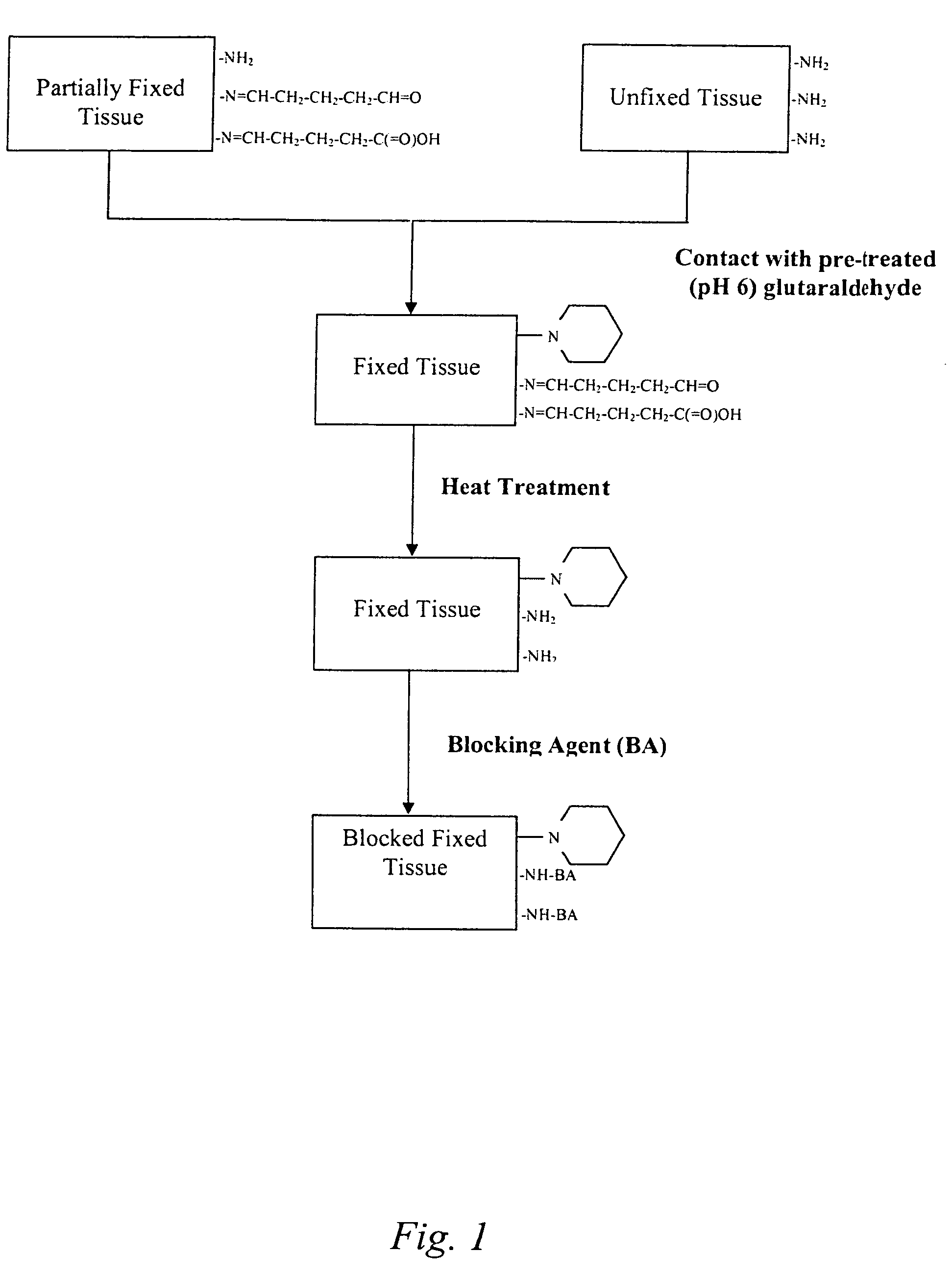
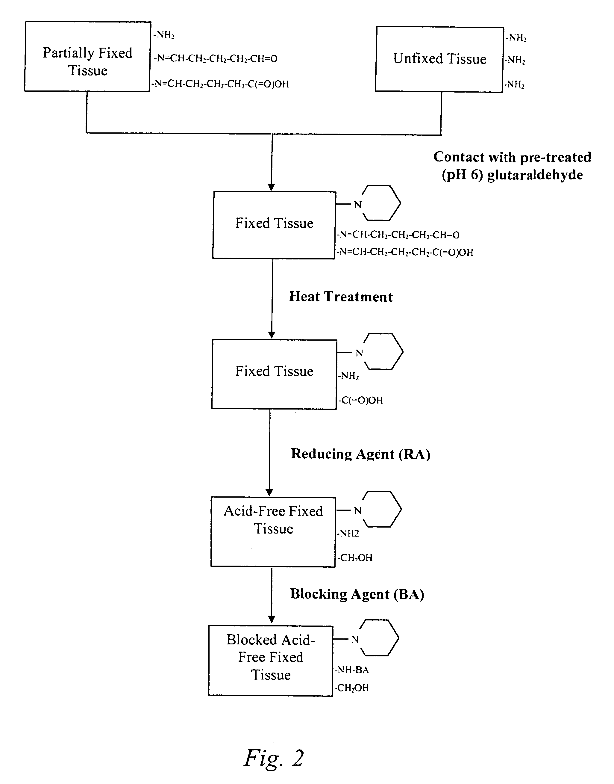
The present invention provides methods for treating tissue to inhibit post-implant calcification of a biological tissue. In one method of this invention, a tissue is immersed in or otherwise contacted with a pretreated glutaraldehyde solution, i.e., a heat-treated or pH-adjusted glutaraldehyde solution. The tissue may be partially fixed with glutaraldehyde prior to, after, or concurrently with the step of contacting the tissue with the pretreated gluteraldehyde. Contact with the pretreated gluteraldehyde produces free amine groups on the tissue, which are subsequently blocked by contacting the crosslinked tissue with a blocking agent. In another embodiment, a tissue is contacted with either a non-pretreated glutaraldehyde or a pH-adjusted glutaraldehyde solution for a period of time sufficient to crosslink the tissue. The crosslinked tissue is then treated with a reducing agent that reduces aldehyde and carboxylic acid groups on the fixed tissue.

Description

CROSS REFERENCE TO A RELATED PATENT APPLICATION[0001]Priority is herewith claimed under 35 U.S.C. §119(e) from co-pending Provisional Patent Application No. 60 / 665,618, filed Mar. 25, 2005, entitled “TREATMENT OF BIOPROSTHETIC TISSUES TO MITIGATE POST IMPLANTATION CALCIFICATION”. The disclosure of this Provisional Patent Application is incorporated by reference herein in its entirety.FIELD OF THE INVENTION[0002]This invention pertains generally to medical methods / devices and more particularly to a method for fixing (e.g., tanning or crosslinking) and sterilizing biological tissue to decrease the fixed tissue's propensity for post-implantation calcification and decrease the thrombogenicity of the fixed tissue.BACKGROUND OF THE INVENTION[0003]Implantable biological tissues can be formed of human tissues preserved by freezing (i.e., cryopreserving) the so called homograft tissues, or of animal tissues preserved by chemically fixing (i.e., tanning) the so called bioprosthesis (Carpentie...

Claims


Application Information

Patent Timeline
Patent Type & Authority Patents(United States)
IPC IPC(8): A61K31/11A61F13/00A61K31/115
CPCA61L27/3604A61L27/3683A61L2400/02
Inventor DOVE, JEFFREY S.
Owner EDWARDS LIFESCIENCES CORP
Learn More