Detection of influenza virus

a technology of influenza virus and detection method, applied in the field of detection of influenza virus, can solve the problems of local outbreaks of flu, limited use of flu vaccines, and high training requirements, and achieve the effect of reducing the difficulty of on-site or field testing

Inactive Publication Date: 2009-09-29
AVC ROYALTY FUND I
View PDF5 Cites 7 Cited by
  • Summary
  • Abstract
  • Description
  • Claims
  • Application Information

AI Technical Summary

Problems solved by technology

It occasionally causes local outbreaks of flu.
In addition, significant amounts of virion particles are commonly required to obtain a positive test result, limiting their use to a short window of time when virus shedding is at its highest levels.
Reverse-transcriptase PCR-based diagnostics (RT-PCR) has resulted in advances in capabilities, but is laborious and requires highly trained personnel making on-site or field-testing difficult.
Unfortunately, RT PCR is not easily adapted to high throughput screening of subjects in an epidemic setting or to field uses in an agricultural or point-of-care setting.
Additionally, the complexity, diversity and rapid emergence of new influenza strains has made diagnosis of high risk strains difficult, and therefore rapid response is at present nearly impossible.
For epidemiologists, diversity resulting from high mutation rates and genetic reassortment make it difficult to anticipate where new strains may originate and respond with the timely introduction of new diagnostic primers for PCR.

Method used


Image

  • Detection of influenza virus
  • Detection of influenza virus
  • Detection of influenza virus

Examples


example 1

NS1 Protein is Expressed in Human Clinical Specimens

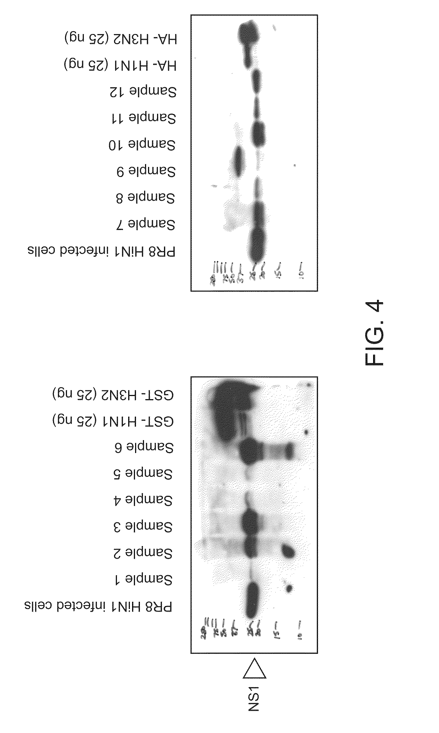
[0089]Human nasal secretions were examined for the presence and amount of NS1 from Influenza A. Human nasal aspirates were collected and stored in M4 viral transport media (Remel, Inc, Lenexa, Kans.) at −80° C. Stored material was thawed and run on 10% SDS-PAGE. Western blot analysis was performed with monoclonal antibodies to NS1, 3H3 and 1A10 (Arbor Vita Corporation, Sunnyvale, Calif.). The results for six samples are shown in FIG. 4. The results show that NS1 is present in large amounts in nasal secretions.

[0090]To investigate the timeline of when NS1 was produced and secreted by cells infected with influenza A virus, MDCK cells were infected with human influenza A / PR / 8 at a MOI of 0.1. Supernatant as well as cells were collected and lysed in 1% TRITON® X-100 and subjected to SDS-PAGE and western analysis with monoclonal antibody 3H3 which is pan-reactive to NS1. NS1 was detected in infected cells within 24 hours after infection...

example 2

NS1 Interacts with PDZ in Cells

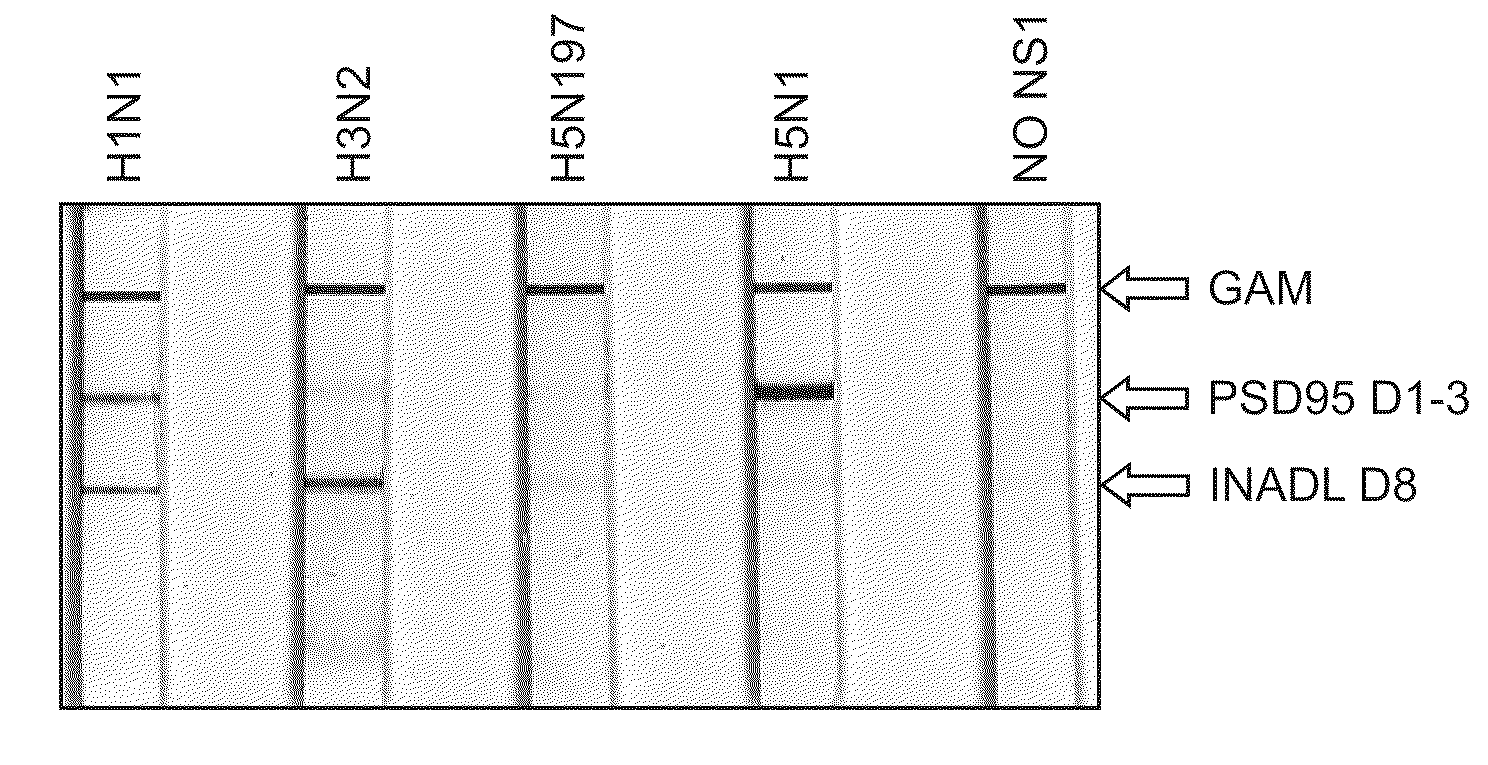
[0091]To verify that NS1 interacts with PDZ proteins in cells, a series of PDZ pull-down experiments were performed. 293 HEK cells were transfected with plasmids containing HA-NS1-H5N1B or with HA-NS1-H3N2. Lysates were prepared as described herein. Glutathione-SEPHAROSE -PDZ beads were prepared (10 ug of DLG1d1,2 10 ug of NeDLGd1,2, and 10 ug PSD95d1,2,3) and used to pulldown 150 ug of lysate from transfected 293ET cells as shown in FIGS. 6 and 7. Following an overnight incubation at 4° C. and multiple washes with HNTG buffer, a membrane was prepared with the pulldowns. The membrane was probed with F63-3G1 supernatant (1:5). All 3 of the PDZs tested successfully pulldown NS1 from cell expressing HA-H5N1B (see FIG. 6).

[0092]Similarly, glutathione-SEPHAROSE-PDZ beads were prepared (40 ug of INADLd8) and used to pulldown 150 ug of lysate from 293ET cells transfected with H3N2. Following an overnight incubation at 4° C. and multiple washes with PBS, a wes...

example 3

Monoclonal Antibodies to NS1

[0094]Monoclonal antibodies were prepared to specifically bind to subtype NS1 proteins (e.g., H5N1), NS1 PL classes (e.g., ESEV) and for pan-specificity (influenza A). The strategy for the generation of monoclonal antibodies to NS1 is as follows:[0095]1. GST and MBP fusion proteins of NS1 were generated. The cloning vectors were obtained from Pharmacia (GST) or New England Biolabs (MBP). The NS1 coding regions were synthesized using overlapping oligonucleotides by DNA 2.0 (Menlo Park, Calif.).[0096]2. Mice were immunized with MBP-NS1 fusion proteins at doses ranging from 10-100 ug per dose in CFA then IFA and PBS.[0097]3. Splenocytes and lymphocytes were harvested 3 days after the last boost with the corresponding GST-NS1 fusion protein and fused with FOX-NY myeloma cells according to Kohler and Milstein (Nature 1975).[0098]4. The hybridomas were screened first with MBP-NS1 in an ELISA. The positive wells were cloned and rescreened with a panel of MBP and...


PUM

PropertyMeasurementUnit
dissociation constantaaaaaaaaaa
dissociation constantsaaaaaaaaaa
concentrationaaaaaaaaaa

Abstract

The present application describes methods for detecting influenza A and / or influenza B and / or distinguishing between pathogenic and seasonal influenza A subtypes. Many of these preferred formats employ pan-specific antibodies (i.e., that react with all or at least multiple strains within an influenza type) to detect presence of influenza A and / or influenza B and PDZ domains in combination with panspecific antibodies to influenza A to distinguish pathogenic and seasonal influenza A subtypes.

Description

CROSS-REFERENCES TO RELATED APPLICATIONS[0001]The present application is a continuation-in-part of PCT / US06 / 41748 filed Oct. 21, 2006, incorporated by reference in its entirety for all purposes. The present application is also a continuation in part of U.S. Ser. No. 11 / 481,411, an application claiming benefit under 35 USC 119(e) of 60 / 792,274, filed Apr. 14, 2006, 60 / 765,292, filed Feb. 2, 2006, 60 / 726,377, filed Oct. 13, 2005; and 60 / 696,221, filed Jul. 1, 2005, each of which is incorporated by reference in its entirety for all purposes.BACKGROUND OF THE INVENTION[0002]Influenza is caused by an RNA virus of the orthomyxoviridae family. There are three types of these viruses and they cause three different types of influenza: type A, B and C. Influenza virus type A viruses infect mammals (humans, pigs, ferrets, horses) and birds. This is very important to mankind, as this is the type of virus that has caused worldwide pandemics. Influenza virus type B (also known simply as influenza ...

Claims


Application Information

Patent Timeline
Patent Type & Authority Patents(United States)
IPC IPC(8): C12Q1/70G01N33/00G01N33/53G01N33/542C12M1/34
CPCG01N33/56983G01N2500/02G01N2333/11Y02A50/30
Inventor LU, PETER S.BELMARES, MICHAEL P.GARMAN, DAVE
Owner AVC ROYALTY FUND I
Why Eureka
Try Eureka