
Regional anesthetic method and apparatus

a regional anesthetic and needle technology, applied in the field of regional anesthetics and epidural needles, can solve the problems of insufficient epidural anesthesia, inability to accurately place epidural needles, and inability to accurately diagnose epidural injuries, etc., to achieve accurate and safe placement of regional anesthetic needles, increase the number of sounds or pitches, and improve the effect of accuracy and safety

Active Publication Date: 2010-01-12
COOPER HEALTH SYST THE
View PDF19 Cites 13 Cited by
  • Summary
  • Abstract
  • Description
  • Claims
  • Application Information

AI Technical Summary

Benefits of technology

This approach provides safer and more accurate placement of regional anesthesia needles by offering real-time anatomical information, reducing the risk of complications and improving the precision of needle placement beyond tactile methods.

Problems solved by technology

The catheter is advanced only two to three centimeters into the epidural space in order to reduce the likelihood that it might exit though an intervertebral foramen, with resulting inadequate epidural anesthesia.
Because the person placing the needle must rely on tactile information, the incidence of errors such as dural punctures, wet taps during lumbar epidurals, and spinal chord injury during cervical or thoracic epidural placements is a serious problem.
While the apparatus and methods for administering regional anesthesia have proved successful over a long period of time, there are drawbacks to those approaches and methods.
Use of the Touhy needle therefore runs the risk that the practitioner, i.e., physician, operator, or other person trained to perform this procedure, might overshoot the epidural space and enter the subdural space between the dura mater and the arachnoid mater of the spine, or the subarachnoid space.
Such a mistake could result in extreme over-application of anesthesia with a possible high level of spinal anesthesia, necessitating endotracheal intubation of the trachea and mechanical ventilation of the patient.
Similar complications could also occur during the disconnection of the syringe from the Touhy needle and insertion of the catheter, as the patient might move, or the needle might not be held properly in position.
Such movement of the needle could result in the undesirable entry of the needle and / or catheter into the subdural or subarachnoid spaces.
In order to obtain better information during regional anesthesia needle placement, it has been proposed to use X-ray fluoroscopic techniques to place the needles, but in many cases it is not possible to use X-ray, for example in the case of pregnant women or at the bedside in the trauma intensive care unit (TICU).
The use of a 2D echo device is complicated by the need for the use of an acoustic gel, which can compromise sterility when used to assist with regional anesthetic needle placement.
Moreover, the 2D echo device is not useful for imaging structures inside the bony spinal canal because the structure of bone tissue scatters ultrasound randomly, such that no intelligible image can be obtained from sound reflected from tissue structures that are deep to bone.

Method used


Image

  • Regional anesthetic method and apparatus
  • Regional anesthetic method and apparatus
  • Regional anesthetic method and apparatus

Examples


Embodiment Construction

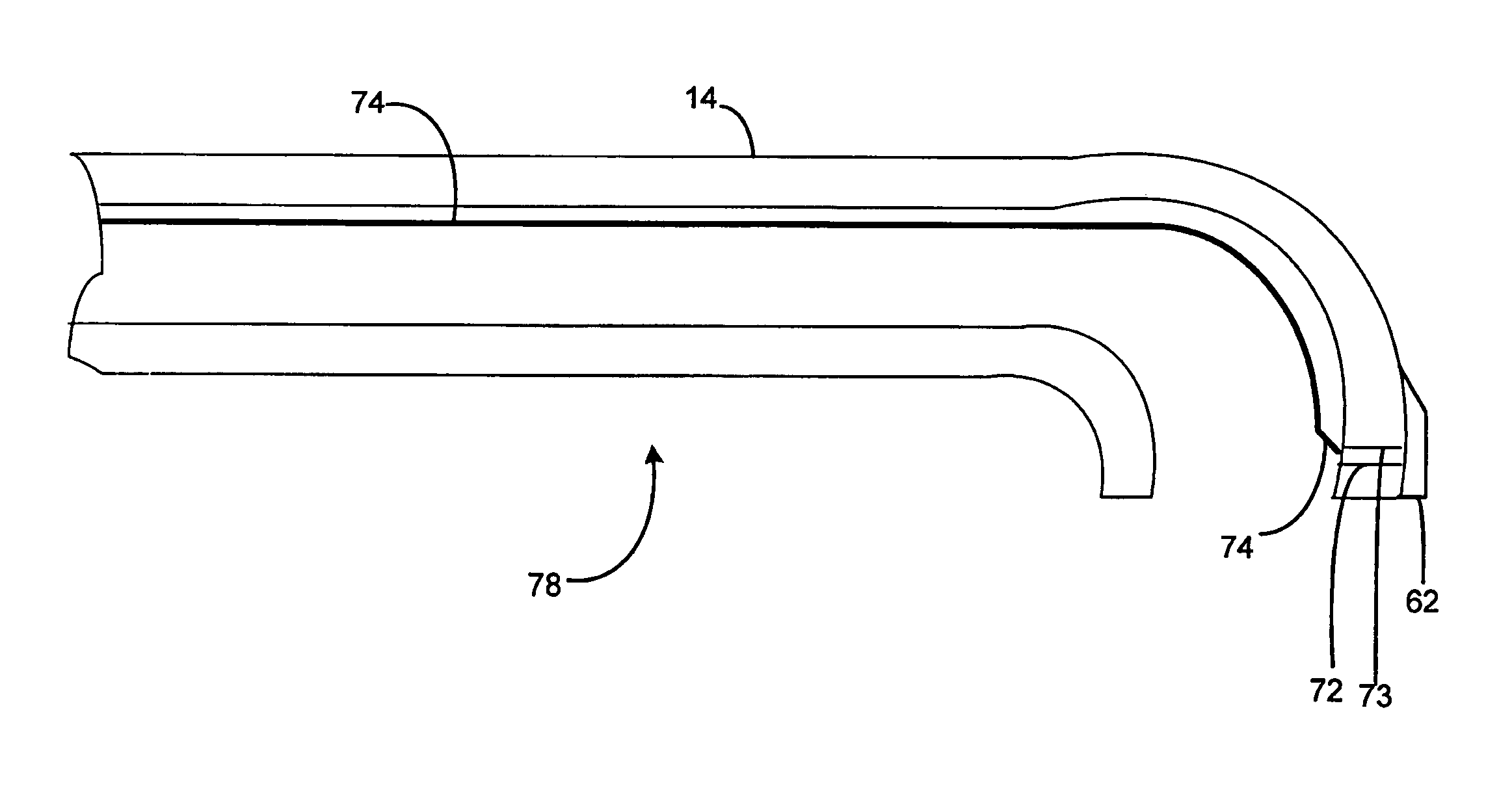
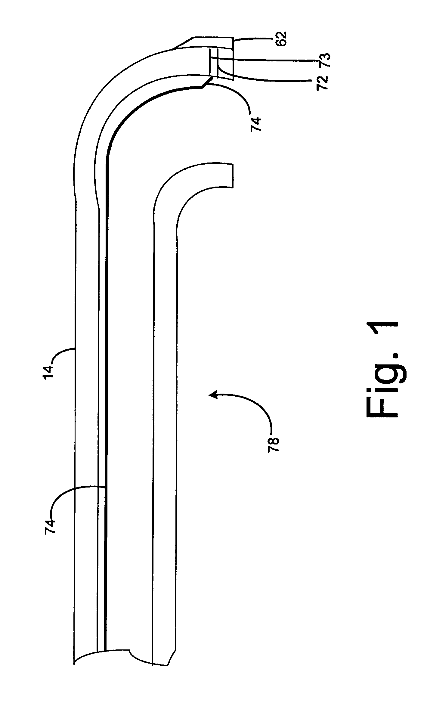
[0025]FIG. 1 shows an embodiment of a regional anesthetic needle 78 according to the invention wherein a conventional Touhy needle 14 is modified by providing a piezo-electric crystal 62 on the distal portion of the needle, connected by wires 72, 73 through the front of the needle, and the wire 74 continuing through the lumen of the needle. The wire 74 can be adhered to the inside wall forming the lumen. In some embodiments conductive material is used rather than wire, for example conductive polymers can be adhered to the inside wall of the lumen.

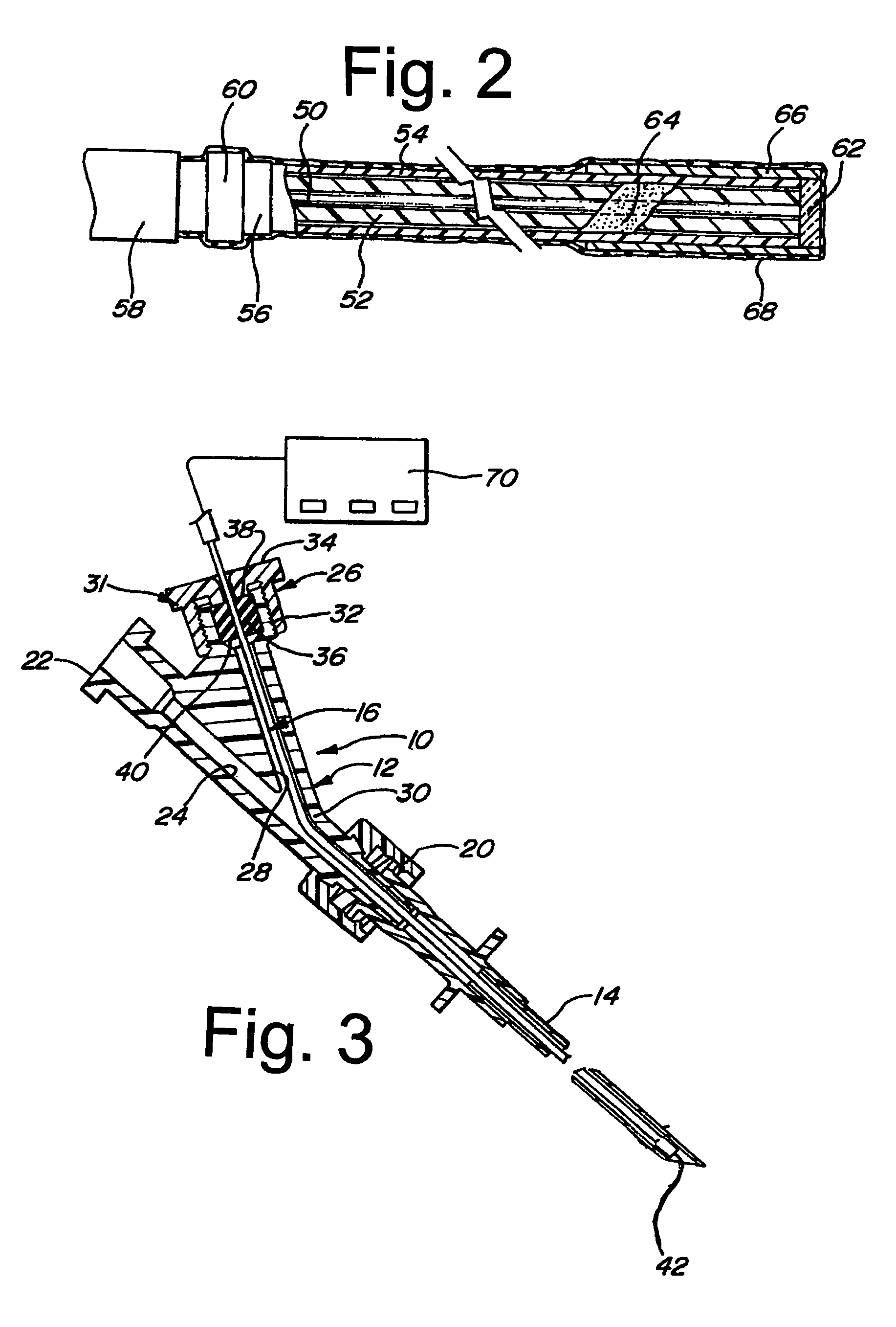
[0026]FIG. 2 shows an embodiment of stylet 16 having a transducer 62 at the distal end. A solid center conductor 50 is surrounded by a dielectric tube 52. The conductor in this embodiment is surrounded by a Teflon dielectric tube, which provides improved noise suppression during operation. Conductor 50 can be composed of copper, silver, or silver-plated copper. A conductive adhesive 54 is coextruded over dielectric tube 52 which is a thin l...


PUM


Abstract

A system and method for regional anesthesia using ultrasound to assist in locating the anesthetic needle are disclosed. One or more piezo-electric crystal transducers is placed either at the tip of the needle or near the tip of the needle on a stylet which is inserted into the lumen of the needle. The transducer(s) are pulsed with ultrasonic frequency, the reflected ultrasonic signals from structures are detected and converted into a digital signal and may be displayed in an oscilloscopic format to indicate anatomical structures forward to the needle and allow the medical professional to avoid errors and / or increase efficiency and accuracy. An array of transducers can be pulsed in a phased array fashion to generate a 2D image, or a single transducer can be pulsed to generate a 1D image.

Description

FIELD OF THE INVENTION[0001]This invention relates generally to the field of regional anesthesia, epidural needles and methods of placement of epidural needles and other regional anesthesia needles.BACKGROUND OF THE INVENTION[0002]Regional anesthesia involves the introduction of local anesthetics, with the intention of blocking the nerve supply to a specific part of the body so the patient cannot feel pain in that area when a surgical operation is performed, for pain relief during onset of labor or during labor, or for chronic pain. Regional anesthesia is used in both epidural and subdural or spinal procedures, and can involve plexus blocks and blocks of peripheral nerves.[0003]Epidural anesthesia, one form of regional anesthesia, has gained popularity over the years as being an effective manner of blocking pain without requiring entry to the dura mater of the spinal cord (i.e., a spinal anesthesia). In fact, epidural anesthesia is often the anesthesia of choice in child birth. The ...

Claims


Application Information

Patent Timeline
Patent Type & Authority Patents(United States)
IPC IPC(8): A61B8/14
CPCA61B8/12A61B8/4461A61B17/3401A61B8/42A61B5/4824A61B2017/3413
Inventor HIRSH, ROBERT
Owner COOPER HEALTH SYST THE
Learn More