Apparatus and method for magnetic resonance angiography utilizing flow pulses and phase-encoding pulses in a same direction

a technology of magnetic resonance angiography and flow pulses, applied in the field of magnetic resonance imaging, can solve the problems of inability to impress flow pulses with optimal time integral values, limited temporal space which can be spared for flow pulses in the pulse sequence, etc., and achieve the effect of high blood flow extractability

Active Publication Date: 2010-01-12
TOSHIBA MEDICAL SYST CORP
View PDF13 Cites 7 Cited by
  • Summary
  • Abstract
  • Description
  • Claims
  • Application Information

AI Technical Summary

Benefits of technology

[0009]An object of the present invention is to realize

Problems solved by technology

Since, however, the flow pulses are impressed on the same axis as that of a readout gradient magnetic field for frequency encoding, a temporal space which can b

Method used


Image

  • Apparatus and method for magnetic resonance angiography utilizing flow pulses and phase-encoding pulses in a same direction
  • Apparatus and method for magnetic resonance angiography utilizing flow pulses and phase-encoding pulses in a same direction
  • Apparatus and method for magnetic resonance angiography utilizing flow pulses and phase-encoding pulses in a same direction

Examples


Embodiment Construction

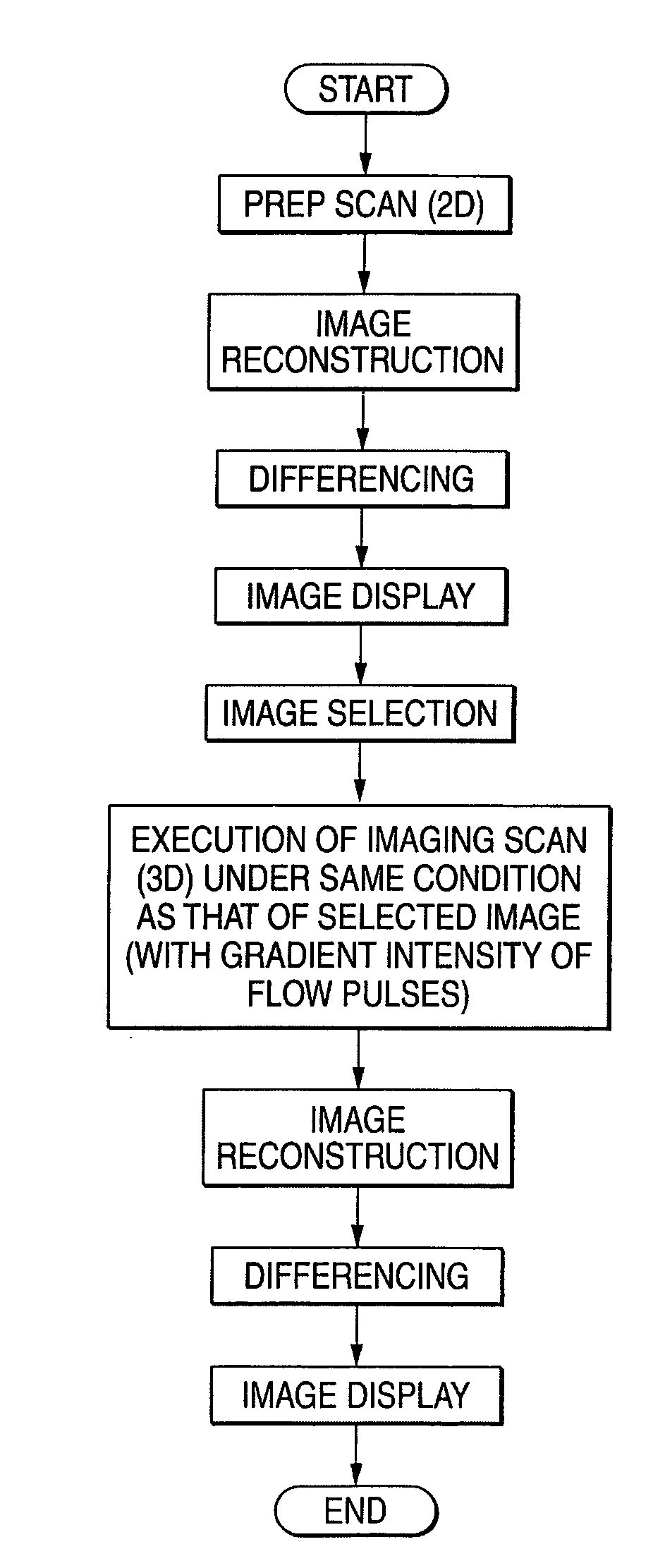
[0031]Now, an embodiment according to the present invention will be described with reference to the drawings.

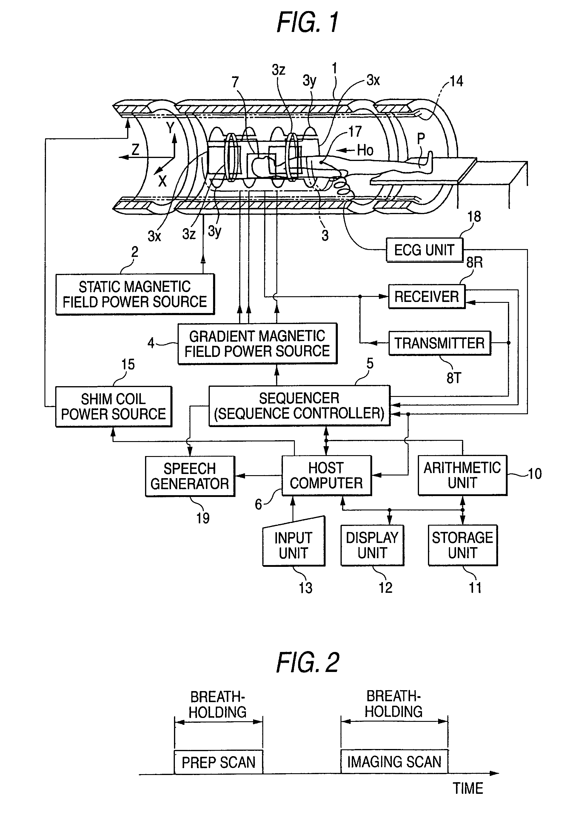
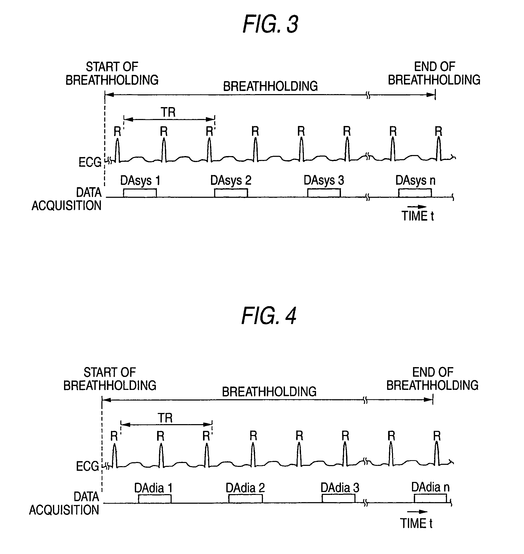
[0032]FIG. 1 shows the schematic construction of a magnetic resonance imaging apparatus according to this embodiment. The magnetic resonance imaging apparatus includes a patient couch section on which a patient P being a subject is laid, a static magnetic field generation section which generates a static magnetic field, a gradient magnetic field generation section which serves to add positional information to the static magnetic field, a transmission / reception section which transmits / receives an RF (radio frequency) signal, a control / calculation section which takes charge of the control of the whole system and image reconstruction, an electrocardiographic measurement section which measures an ECG (electrocardiogram) signal being a signal representative of the cardiac phase of the patient P, and a breathholding command section which commands the patient P to suspend respiratio...


PUM


Abstract

A magnetic resonance imaging apparatus includes an RF coil unit which generates RF pulses toward a subject, and which receives an MR signal from the subject. Gradient magnetic field coils generate a gradient magnetic field for slice selection, a gradient magnetic field for phase encoding and a gradient magnetic field for frequency encoding, respectively. An arithmetic unit generates image data on the basis of the MR signal, and a sequence controller controls phase encoding gradient magnetic field coils in order to generate flow pulses for dephasing or rephasing the MR spin of blood flow within the subject, in the same direction as that of the phase encoding gradient magnetic field.

Description

CROSS-REFERENCE TO RELATED APPLICATIONS[0001]This application is based upon and claims the benefit of priority from the prior Japanese Patent Application No. 2002-295190, filed Oct. 8, 2002, the entire contents of which are incorporated herein by reference.BACKGROUND OF THE INVENTION[0002]1. Field of the Invention[0003]The present invention relates to a magnetic resonance imaging apparatus and method which are furnished with MR angiography capable of separating an artery and a vein without using any contrast medium.[0004]2. Description of the Related Art[0005]Magnetic resonance imaging is an imaging method wherein the atomic nucleus spin of a subject located in a static magnetic field is magnetically excited by the radio frequency signal of its Larmor frequency, and wherein an image is reconstructed from a magnetic resonance signal generated with the excitation. One imaging method which has recently come into the limelight in the field of the magnetic resonance imaging, is MR angiog...

Claims


Application Information

Patent Timeline
IPC IPC(8): A61B5/05A61B5/055G01N24/08G01R33/48G01R33/563
CPCG01R33/56308G01R33/56316G01N24/08
Inventor MIYAZAKI, MITSUESUGIURA, SATOSHI
Owner TOSHIBA MEDICAL SYST CORP
Why Eureka
Try Eureka