Imaging apparatus

Active Publication Date: 2010-03-09
ESAOTE
View PDF21 Cites 14 Cited by
  • Summary
  • Abstract
  • Description
  • Claims
  • Application Information

AI Technical Summary

Benefits of technology

[0046]The advantages of this invention are self-evident from the above, and consist in that a stable structure is provided for supporting a diagnostic and/or therapeutic tool, e.g. a syringe or a needle and a ultrasound probe, which may assume and maintain more easily a defined position relative to the body under examination or a part thereof (total body mode and dedicated mode). By this arrangement, the injection may be performed in a much more accurate manner and with no risk for the patient, especially when compared to manual displacement of the syringe or needle. Thanks to the inventive apparatus, both the anatomy of the region under examination and the image of the needle may be simultaneously display

Problems solved by technology

The perfusion of said contrast agents shall be as accurate as possible and requires a critical time selection, which may not be easy, especially due to the anatomic peculiarities of the shoulder which, as is known, is a very complex anatomic region, especially when said operations are carried out in a wholly manual manner.
Any error in the selection of the injection point and/or of the needle orientation, or a poor synchr

Method used


Image

  • Imaging apparatus
  • Imaging apparatus
  • Imaging apparatus

Examples


Embodiment Construction

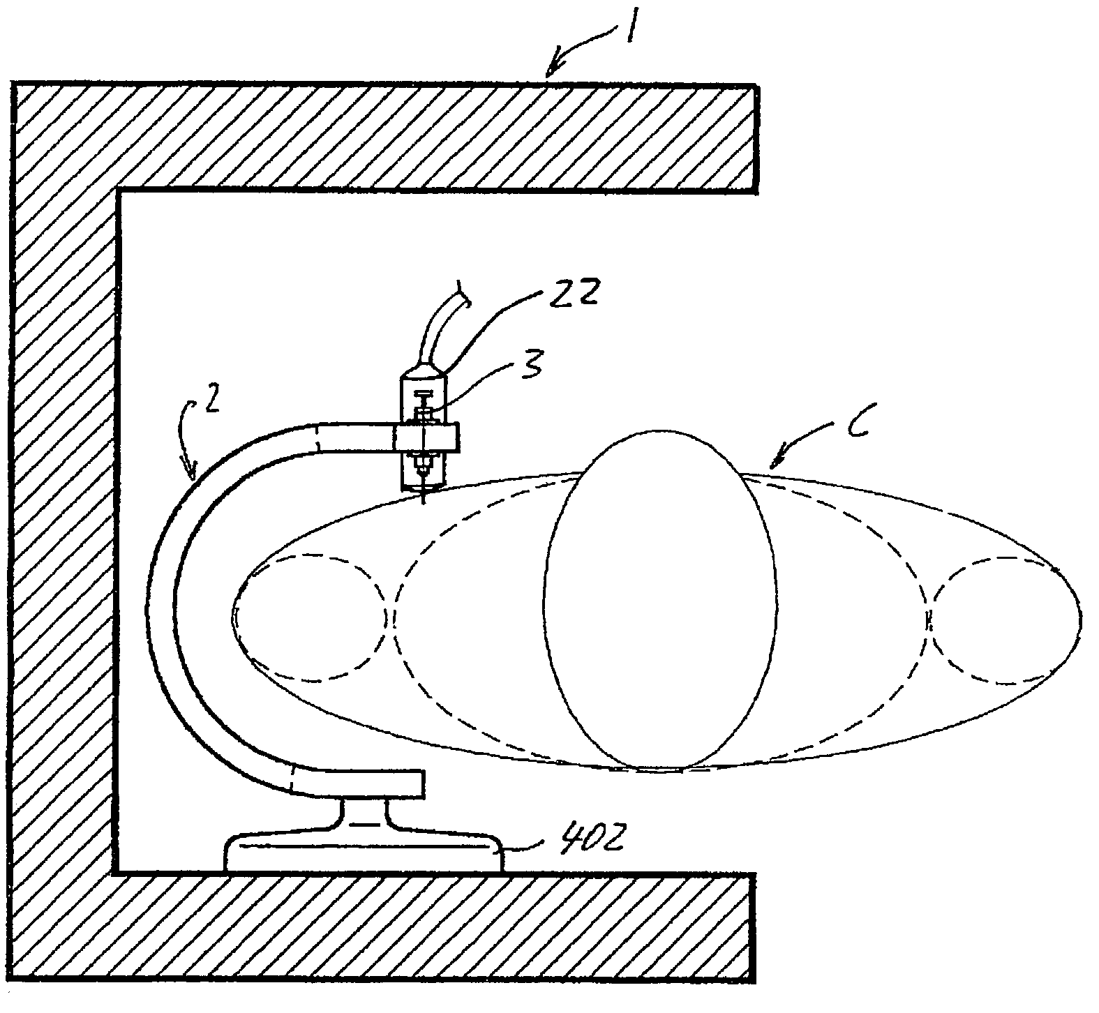
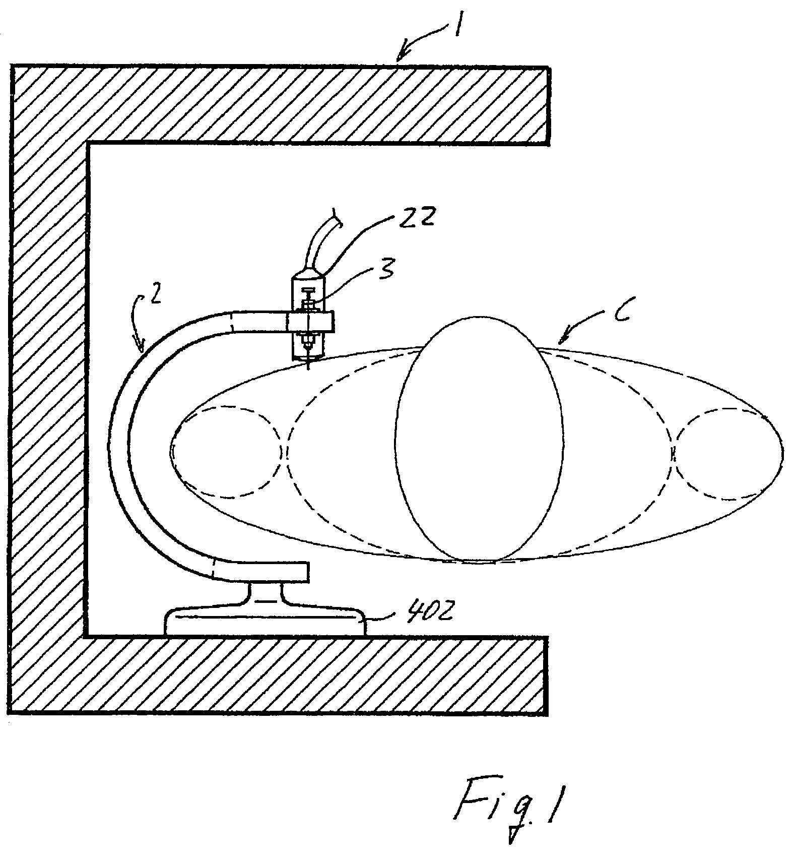
[0059]Referring to FIG. 1, an embodiment of a dedicated combined MRI imaging and ultrasound imaging apparatus of this invention is shown. The example illustrated and described and particularly related to the examination of the shoulder is not to be considered a limitation of the present invention which may be applied to whatever anatomical district or whatever kind of body or part under examination. As is known, the above mentioned dedicated type of apparatus has the advantage of a relatively low cost and of a considerable comfort, versatility and ease of use and installation, particularly when compared with larger apparatuses. This apparatus for imaging a body C under examination or a part thereof comprises a magnetic structure 1 having at least two opposite poles which define an intermediate cavity, between which a static magnetic field is generated in a predetermined imaging volume of said cavity. The cavity may be accessed from one or more openings of the magnetic structure 1. T...


PUM


Abstract

A combined imaging apparatus having a magnetic resonance imaging apparatus with at least one transmitting coil for exciting the matter of a body under examination or of a part thereof and at least one coil for receiving the signals transmitted by the body under examination or a part thereof, as well as an electronic unit for processing the received signals to create a diagnostic image and including a device for supporting and/or guiding at least one diagnostic and/or therapeutic tool. The combined apparatus further including an echographic imaging apparatus.

Description

BACKGROUND OF THE INVENTION[0001]1. Field of the Invention[0002]The invention relates to a imaging apparatus comprising a magnetic resonance imaging (MRI) apparatus, with at least one transmitting coil for exciting the matter of a body under examination or of a part thereof and at least one coil for receiving the signals transmitted by the body under examination or a part thereof, as well as electronic means for processing said received signals to create an image and which apparatus has further means for supporting an guiding a surgical or therapeutic tool.[0003]2. Description of Related Art[0004]As is known, MRI examinations are often combined with the use of various tools, which may consist of diagnostic assistance instruments or of therapeutic instruments. This may be the case, for instance, of shoulder dedicated MRI apparatuses, wherein contrast agents are often to be injected in the region under examination. The perfusion of said contrast agents shall be as accurate as possible...

Claims


Application Information

Patent Timeline
IPC IPC(8): A61B5/05A61B5/055A61B8/00G01R33/28
CPCG01R33/28A61B5/055A61B5/4528A61B8/00A61B8/4416A61B5/0035
Inventor SATRAGNO, LUIGIBIGLIERI, EUGENIO
Owner ESAOTE
Why Eureka
Try Eureka