Apparatus and method for the analysis of retinal vessels

a retinal vessel and apparatus technology, applied in the field of apparatus and method for the analysis of retinal vessels, can solve the problems of unsatisfactory reproducibility and high artery-to-vein ratio in individual examinations, and achieve the effects of improving the reproducibility of individual determined artery-to-vein ratios, reducing measurement uncertainty, and increasing individual validity of determined values

Active Publication Date: 2010-03-16
IMEDOS INTELLIGENTE OPTISCHE SYST DER MEDIZIN & MESSTECHNIK GMBH
View PDF4 Cites 71 Cited by
  • Summary
  • Abstract
  • Description
  • Claims
  • Application Information

AI Technical Summary

Benefits of technology

[0012]Accordingly, it is the primary object of the invention to improve the reproducibility of individually determined artery-to-vein ratios and to reduce the measurement uncertainty in determining the artery-to-vein ratio in order to substantially increase the individual validity of the determined values for vessel diagnosis.
[0017]It can also be advantageous when the means for determining the artery-to-vein ratio have an image evaluating device by which the recorded images can be validated visually or automatically. Further, it is possible that an image adjustment is carried out by means of an image correction unit for correcting eye movements.
[0021]Further, the systematic measurement error of the individual measurement caused by changes in the imaging scale from one recording to another is randomized and substantially reduced by the invention. Above all, this is a matter of reducing the measurement uncertainty in determining the central equivalents.

Problems solved by technology

However, the methods are very uncertain and are usable only conditionally for individual diagnoses because, as a result of the random systematic errors which vary between individuals, the scatter of the individual central equivalents and of the artery-to-vein ratios in individual examinations is very high and leads to unsatisfactory reproducibility.

Method used


Image

  • Apparatus and method for the analysis of retinal vessels
  • Apparatus and method for the analysis of retinal vessels

Examples


Embodiment Construction

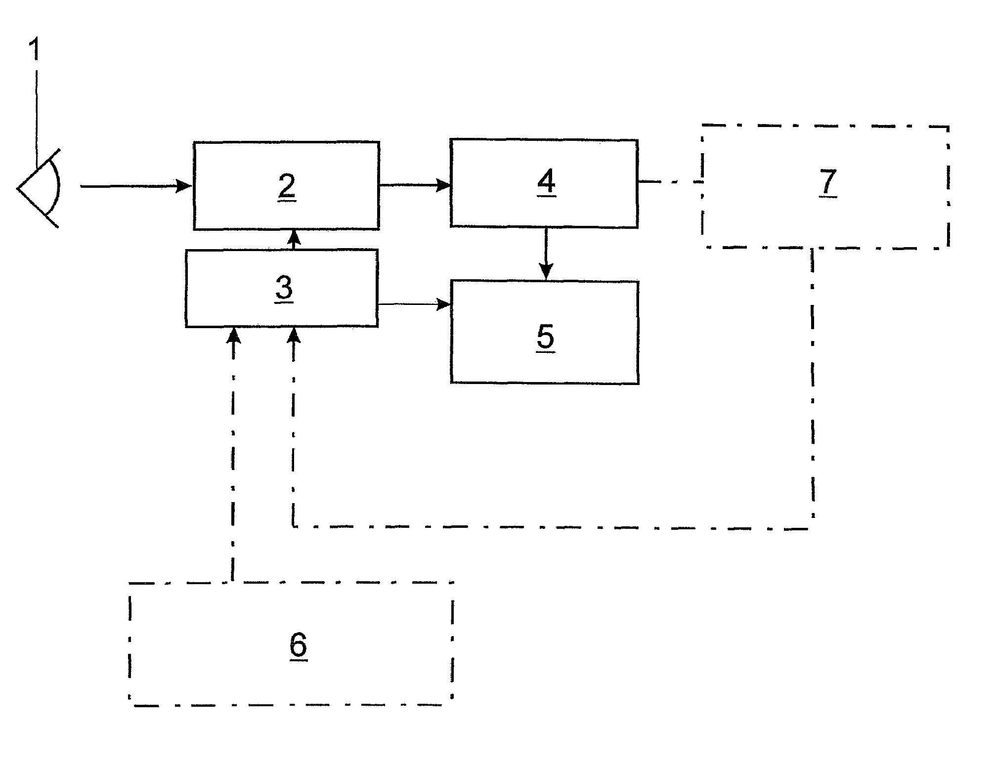
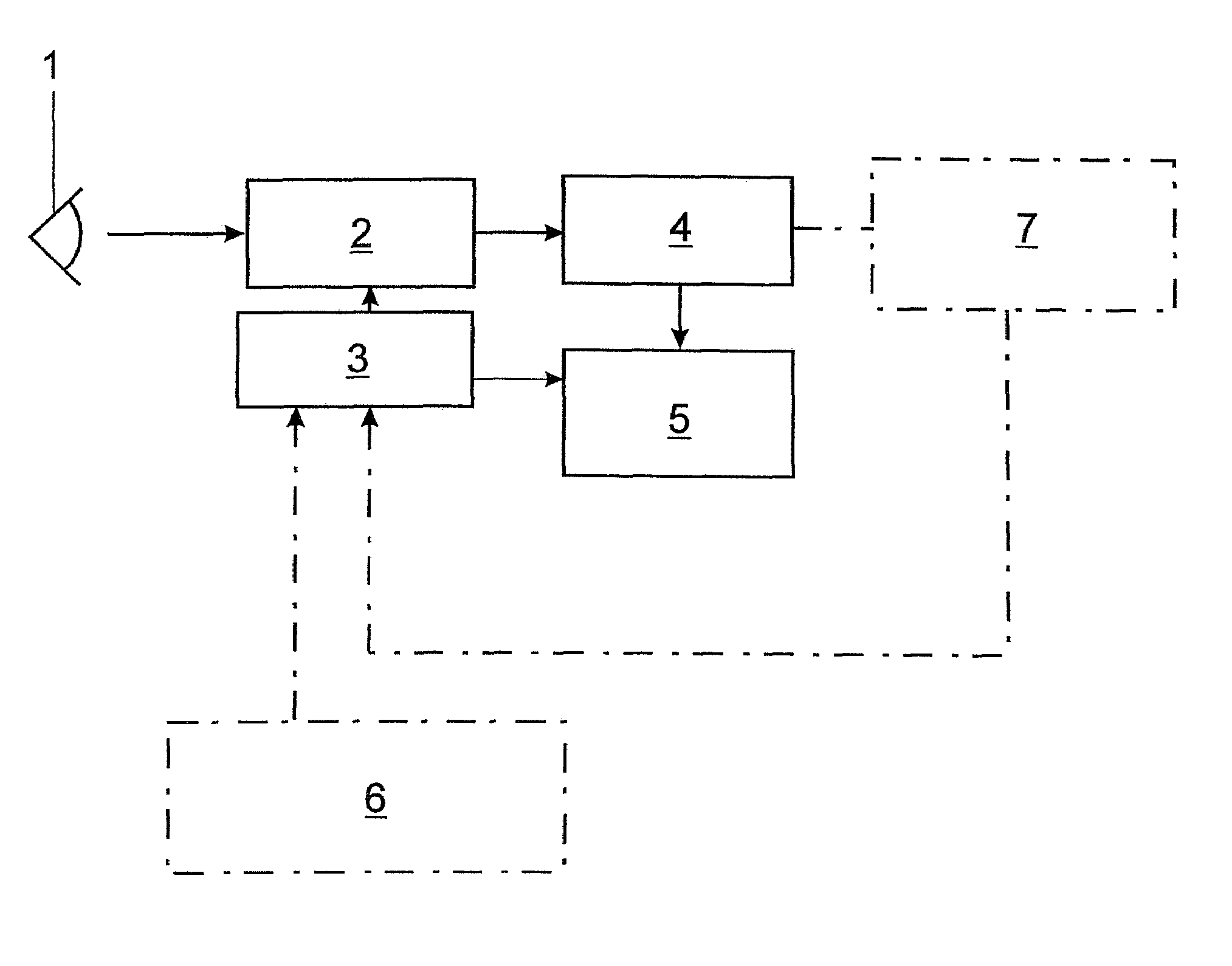
[0043]A nonmydriatic or mydriatic retinal camera 2 with digital imaging or a digitizing system for fundus photography is provided for examining the eye 1 of a patient and is connected to an image sequence control unit 3 for controlling the sequence of image recordings of the ocular fundus. The images which are recorded by the method according to the invention are stored at least temporarily with image designation and time of recording in an image sequence storage 4 from which an image sequence evaluating device 5 takes images for evaluation to determine a mean AVR value.

[0044]Of course, the apparatus comprises input media and output media for dialog mode and for displaying and outputting results, e.g., a keyboard, mouse, screen and printer, which are not shown in the drawing.

[0045]The first embodiment example is directed to the determination of AVR values based on an individual image sequence, by which is meant snapshots of the ocular fundus which are taken at fixed time intervals b...


PUM


Abstract

The object of an apparatus and a method for the analysis of retinal vessels is to improve the reproducibility of individually determined artery-to-vein ratios and to reduce the measurement uncertainty in determining the artery-to-vein ratio in order to substantially increase the individual validity of the determined values for vessel diagnosis. At least two images are recorded successively as an image sequence in a predetermined timed sequence adapted to the vasomotricity of the vessels and are evaluated such that a mean artery-to-vein ratio is formed from artery-to-vein ratios that are determined on the basis of the at least two images.

Description

CROSS-REFERENCE TO RELATED APPLICATIONS[0001]This application claims priority of German Application No. 10 2006 018.445.9, filed Apr. 18, 2006, the complete disclosure of which is hereby incorporated by reference.BACKGROUND OF THE INVENTION[0002]a) Field of the Invention[0003]The invention is directed to an apparatus for the analysis of retinal vessels which contains a retinal camera for recording images of the ocular fundus, at least one image storage, means for detecting arterial and venous vessels in the images, means for determining the vessel diameters of the detected arterial and venous vessels, and means for determining artery-to-vein ratios (AVRs) from the determined vessel diameters.[0004]b) Description of the Related Art[0005]The invention is further directed to a method for the analysis of retinal vessels through the evaluation of digital fundus images in which a ring-shaped measurement zone containing vessel portions which proceed from a central vessel, which are to be d...

Claims


Application Information

Patent Timeline
Patent Type & Authority Patents(United States)
IPC IPC(8): G03B9/28G03B1/48
CPCA61B3/12
Inventor VILSER, WALTHARDKASSNER, CHRISTINE
Owner IMEDOS INTELLIGENTE OPTISCHE SYST DER MEDIZIN & MESSTECHNIK GMBH
Why Eureka
Try Eureka