

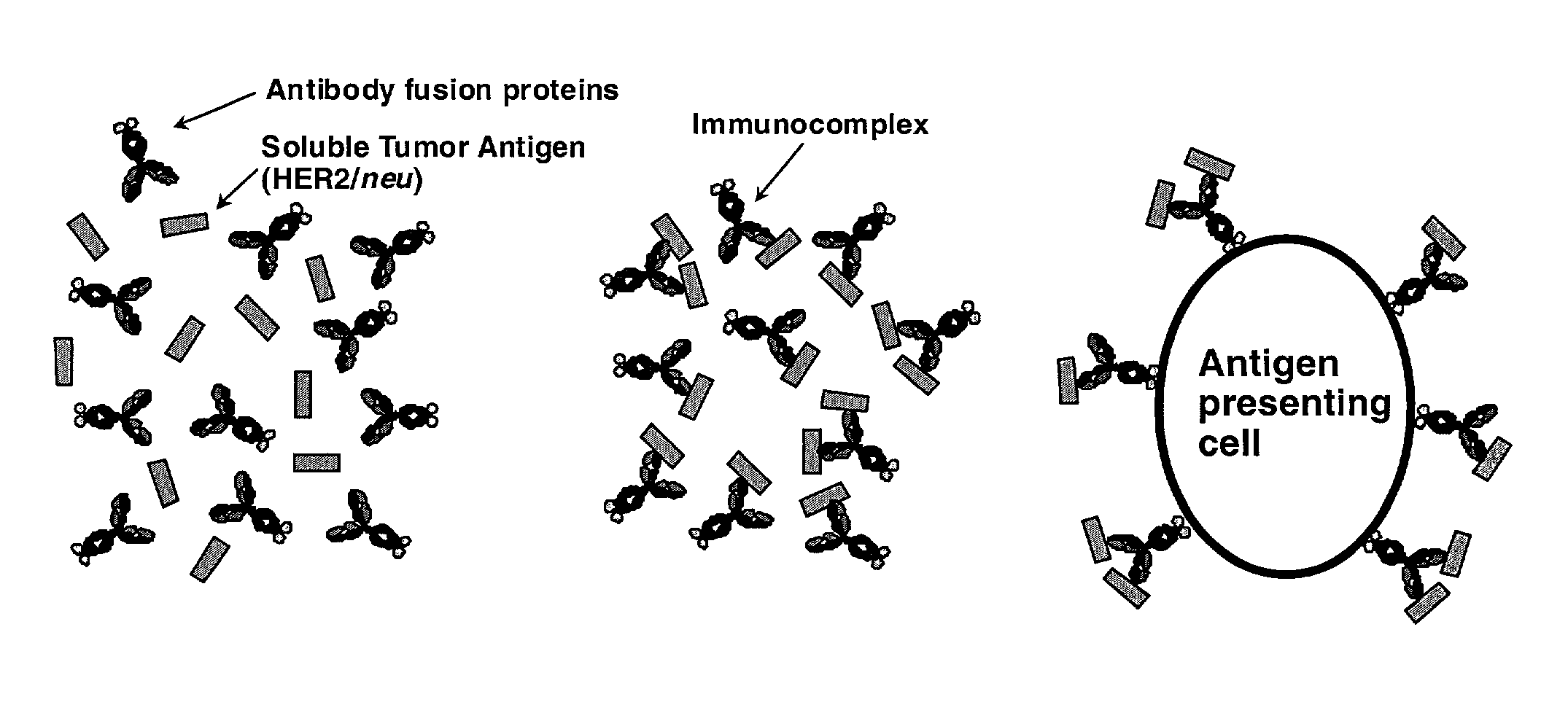
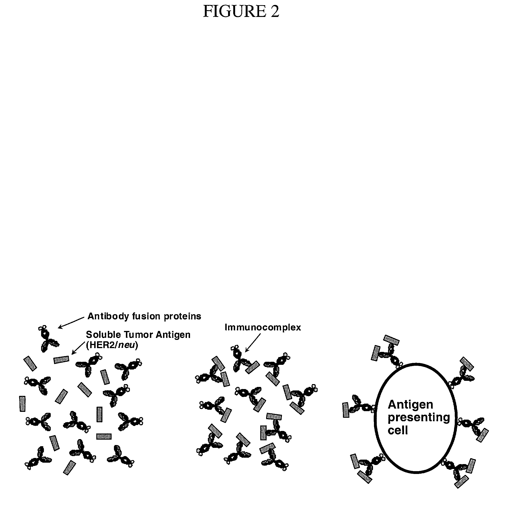
Antibody fusion proteins: effective adjuvants of protein vaccination
- Summary
- Abstract
- Description
- Claims
- Application Information
AI Technical Summary
Benefits of technology
Problems solved by technology
Method used
Image

Examples
examples
[0137]The following examples utilize the HER2 / neu antigen / antibody and various cytokines (e.g., IL-2, IL-12, GMCSF) as well as the protein A antigen from Staphylococcus aureus. However, once again, it is to be emphasized that the methods of the current invention (e.g., use of antibody-immunostimulant fusions as adjuvants of antigenic delivery) are applicable to many different combinations of antibodies and immunostimulants, etc., and are useful in the treatment of myriad diseases / conditions (i.e., not just for the treatment of HER2 / neu presenting tumors or staphylococcal infections).
Example I
Anti-HER2 / Neu Antibody Fusion Proteins as Effective Enhancers of Extracellular Domain HER2 / Neu Protein Vaccination
[0138]The molecule HER2 / neu is overexpressed in a number of human cancers (e.g., breast, ovarian, prostate and lung cancers) and is associated with poor prognosis. As described above, some DNA and peptide based vaccines which target HER2 / neu have elicited significant protection again...
example i
Results for Example I
ECDHER2 Vaccination and Anti-Tumor Activity
[0155]BALB / c mice were vaccinated subcutaneously on week 0 and week 5 with either PBS, ECDHER2 alone, ECDHER2 plus IgG3 or ECDHER2 plus either IgG3-(GMCSF), IgG3-(IL-2) or IgG3-(IL-12) (as described above). No apparent side effects were observed throughout the duration of the vaccination. Eight weeks after the initial vaccination, 106 TUBO cells were injected subcutaneously into the left flank of vaccinated mice. At 7 days post-challenge, measurable tumors were present in all mice vaccinated with PBS, ECDHER2 alone and ECDHER2 plus IgG3, while two out of eight mice in the ECDHER2 plus IgG3-(GMCSF) or ECDHER2 plus IgG3-(IL-12) group of vaccinated mice and five out of seven mice in the ECDHER2 plus IgG3-(IL-2) vaccinated mice showed no tumors. See, FIG. 3a. Tumors grew uniformly and progressively in all PBS treated mice whereas mice vaccinated with ECDHER2 alone and ECDHER2 plus IgG3 showed dispersions in the sizes of tum...
example ii
Use of Antibody-Immunostimulant Fusion Proteins to Enhance Immune Response Against Staphylococcus aureus Virulence Factor Protein A
Protein A and Antibody-Immunostimulant Fusion Proteins
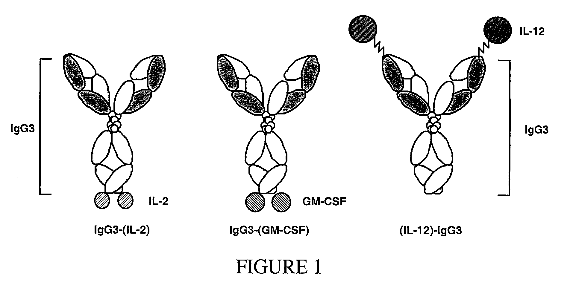
[0171]As outlined above, antibody-immunostimulant (e.g., cytokine) fusion proteins specific for the extracellular domain of the human tumor associated antigen HER2 / neu (ECDHER2) were constructed and their action characterized. Such fusion proteins were composed of human IgG3 (containing the variable region of Trastuzumab (Herceptin, Genentech, San Francisco, Calif.)) which was genetically fused to the immunostimulatory cytokines interleukin-2 (IL-2), interleukin-12 (IL-12), or granulocyte-macrophage colony stimulator factor (GMCSF). See, Penichet, M. L. and Morrison, S. L. 2001, “Antibody-cytokine fusion proteins for the therapy of cancer”J Immunol Methods 248: 91-101; Peng, L. S., et al. 1999, “A single-chain IL-12 IgG3 antibody fusion protein retains antibody specificity and IL-12 bioactivity and de...
PUM
| Property | Measurement | Unit |
|---|---|---|
| Volume | aaaaa | aaaaa |
| Volume | aaaaa | aaaaa |
| Volume | aaaaa | aaaaa |
Abstract
Description
Claims
Application Information