
Method and apparatus for medical intervention procedure planning and location and navigation of an intervention tool

a technology of medical intervention and procedure planning, applied in the field of imaging system, can solve problems such as failure to place lead, affect the electrical conduction system of the heart, and ineffective contraction of the left ventricl

Inactive Publication Date: 2010-08-17
GENERAL ELECTRIC CO +1
View PDF79 Cites 198 Cited by
  • Summary
  • Abstract
  • Description
  • Claims
  • Application Information

AI Technical Summary

Benefits of technology

"The patent describes an imaging system for use in medical intervention procedures planning. The system includes a medical scanner for generating cardiac image data, a data acquisition system for acquiring the data, an image generation system for generating a viewable image from the data, a database for storing information from the acquisition and image generation systems, an operator interface system for managing the scanner, data acquisition, image generation, and database, and a post-processing system for analyzing the cardiac image data and displaying it in a viewable image. The system can be used for planning procedures such as bi-ventricular pacing, atrial fibrillation, and atrial flutter. The patent also describes a computer system for managing and displaying the probe information and the stored information from an interventional procedure planning session. The system can also use the pre-stored 3D images to guide interventional tools in real-time. Overall, the patent provides a technical solution for improving the accuracy and precision of medical intervention procedures."

Problems solved by technology

Some patients with CHF also experience left bundle branch block (LBBB), which negatively impacts the electrical conduction system of the heart.
In patients with CHF and LBBB, delayed left ventricular ejection results from delayed ventricular depolarization, and in the presence of LBBB, ventricular contraction is asymmetrical, which causes ineffective contraction of the left ventricle.
Interventional bi-ventricular pacing therapy may involve a lengthy procedure, may result in unsuccessful lead placement in the CS due to the CS anatomy, or the lead itself may dislodge from the CS.
In most cases, these situations are identified only at the time of the interventional procedure, resulting in abandonment of the procedure or the scheduling of a second procedure where, using a surgical incision, the LV lead is placed epicardially.

Method used


Image

  • Method and apparatus for medical intervention procedure planning and location and navigation of an intervention tool
  • Method and apparatus for medical intervention procedure planning and location and navigation of an intervention tool
  • Method and apparatus for medical intervention procedure planning and location and navigation of an intervention tool

Examples


Embodiment Construction

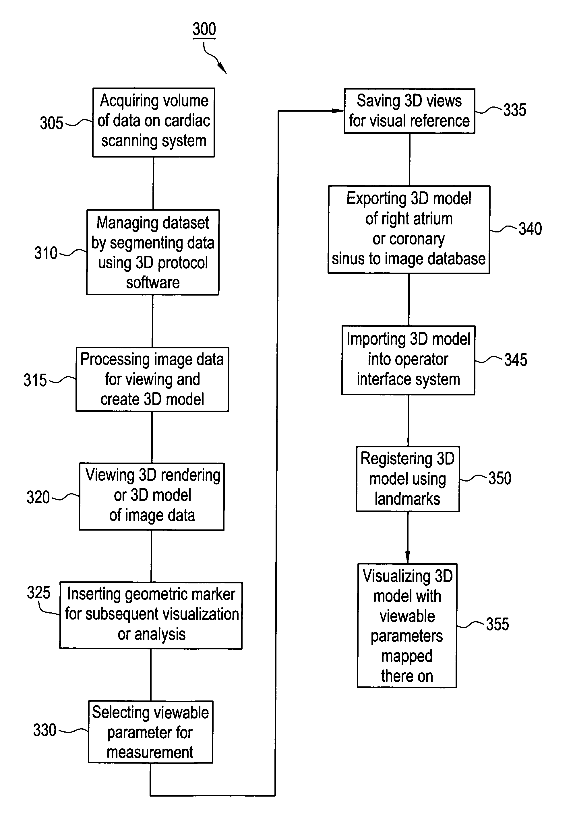
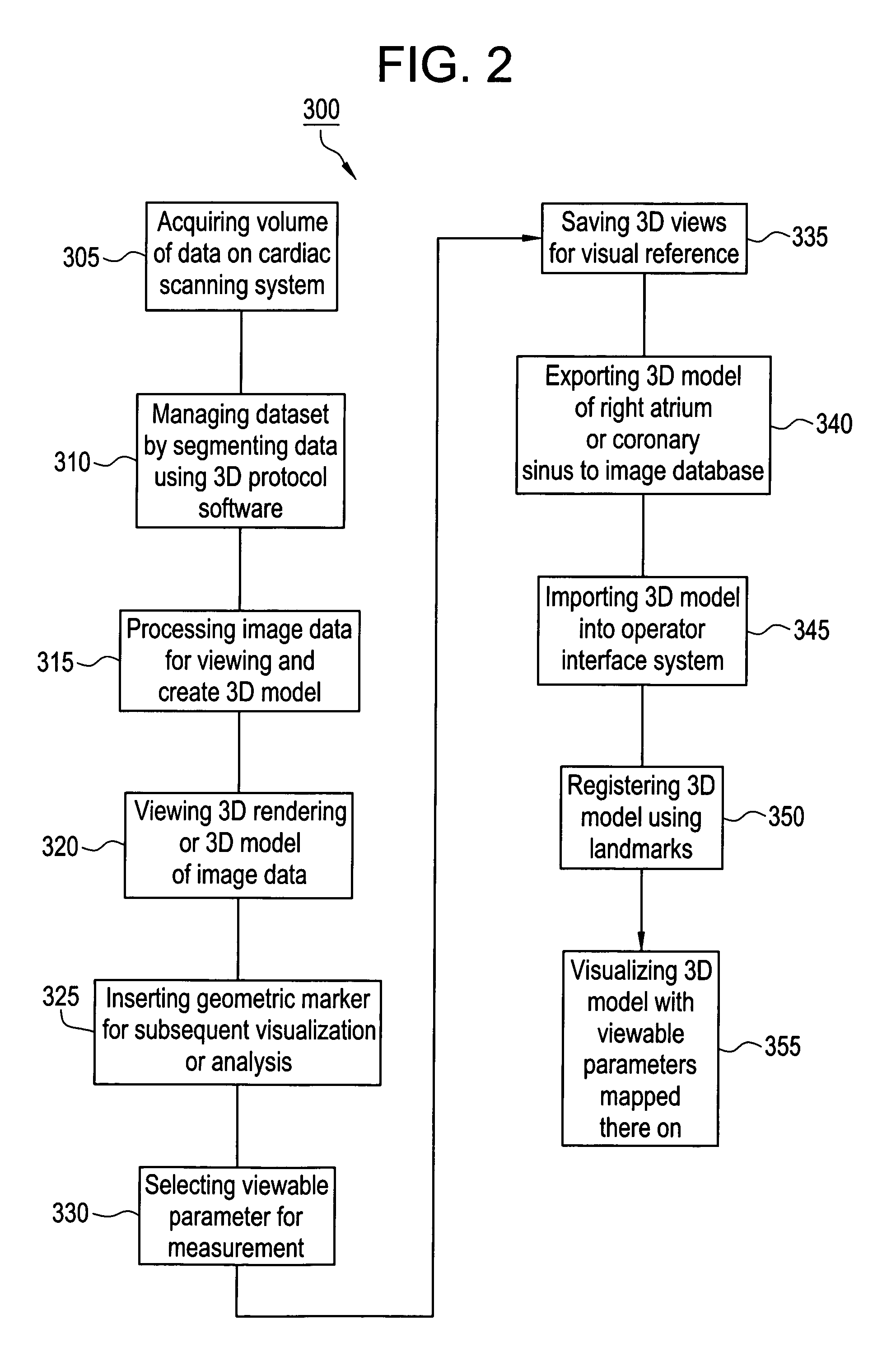
[0020]A detailed description of an embodiment of the invention is presented herein by way of exemplification and not limitation with reference to FIGS. 1-3.

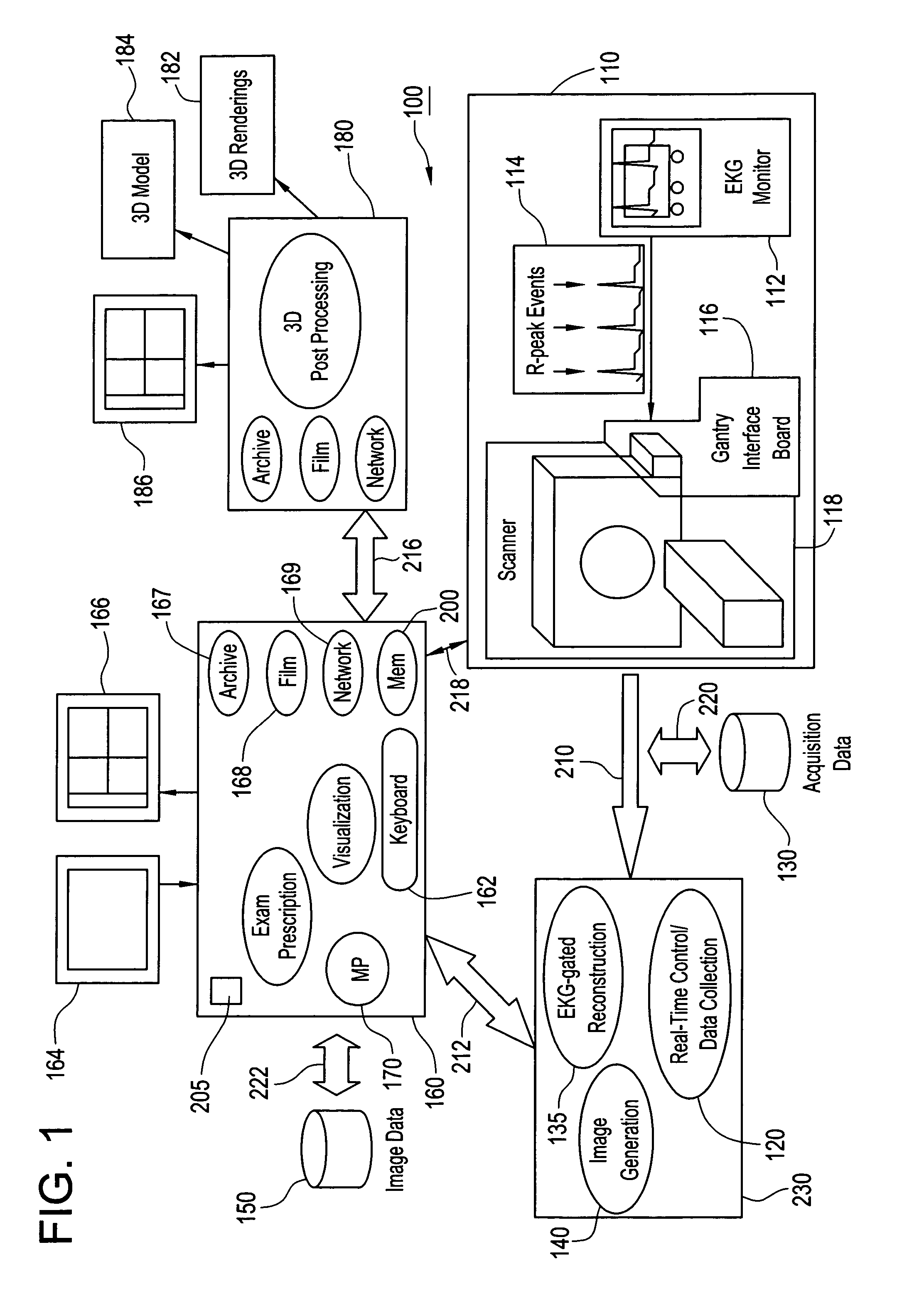
[0021]FIG. 1 depicts a generalized schematic of an imaging system 100 for use in medical intervention procedure planning, such as, for example, bi-ventricular procedure planning, atrial fibrillation procedure planning, or atrial flutter procedure planning. The imaging system 100 includes: a medical scanner system 110 for generating cardiac image data, such as, for example, image data of the left atrium, the left ventricle, the right atrium and the coronary sinus, a data acquisition system 120 for acquiring the cardiac image data from medical scanner system 110, an acquisition database 130 for storing the cardiac image data from data acquisition system 120, an image generation system 140 for generating a viewable image from the cardiac image data stored in acquisition database 130, an image database 150 for storing the viewable im...


PUM


Abstract

A system and method for a medical intervention procedure within a cardiac chamber having an imaging system to obtain image data of the cardiac chamber and to create a 3D model from that image data, an interventional system to register the 3D model with a real-time image of the cardiac chamber and to display the 3D model, and an interventional tool positioned in the cardiac chamber to be displayed upon the interventional system and to be navigated in real-time over the registered 3D model. Preferably, the method and system also includes a storage medium to store the 3D model and wherein the interventional system receives the stored 3D model to register with the real-time image of the cardiac chamber.

Description

CROSS REFERENCE TO RELATED APPLICATIONS[0001]The present application is a continuation-in-part of assigned U.S. patent application, Ser. No. 10 / 065,595 filed Nov. 1, 2002, the contents of which are incorporated herein by reference thereto which claims the benefit of U.S. Provisional Application Ser. No. 60 / 385,749, filed Jun. 4, 2002, which is incorporated by reference in its entirety. The present application also claims the benefit of U.S. Provisional Application No. 60 / 494,607, filed Aug. 12, 2003, the contents of which are incorporated by reference herein in their entirety.BACKGROUND OF THE INVENTION[0002]This invention relates generally to an imaging system, and more particularly to a method and apparatus for use of the imaging system in medical intervention procedure planning and location and navigation of an intervention tool.[0003]Medical diagnostic and imaging systems are present in modern health care facilities. Such systems provide invaluable tools for identifying, diagnos...

Claims


Application Information

Patent Timeline
Patent Type & Authority Patents(United States)
IPC IPC(8): A61B5/00A61B5/055A61B5/352A61B6/03A61B6/12A61B8/00A61B8/12A61B18/04A61B19/00A61M25/00G01T1/161
CPCA61B5/0456A61B19/52A61B5/7207A61B19/50A61B6/504A61B2019/5289A61B6/487A61B90/36A61B2090/364A61B34/10A61B5/352
Inventor VASS, MELISSASRA, JASBIR S.
Owner GENERAL ELECTRIC CO
Learn More