
Kits for diagnosis and monitoring of pathogenic infection by analysis of cell-free pathogenic nucleic acids in urine

a technology of pathogenic nucleic acids and urine, which is applied in the direction of enzymology, organic chemistry, transferases, etc., can solve the problems of difficult storage of unaltered biological samples, requiring special equipment and qualified personnel, and requiring the consideration of biological samples as at risk

Inactive Publication Date: 2010-09-28
INST NAT PER LE MALATTIE INFETTIVE LAZZARO SPALLANZANI IRCCS
View PDF19 Cites 96 Cited by
  • Summary
  • Abstract
  • Description
  • Claims
  • Application Information

AI Technical Summary

Benefits of technology

"The patent describes new methods for diagnosing or monitoring infectious diseases caused by bacterial or parasitic pathogens by detecting and quantifying pathogen-related transrenal nucleic acid sequences. These methods can be applied to urine samples, with or without DNA isolation, and can include steps consisting of the fractionation of the sample and the pretreatment of the sample with a denaturing agent. The analysis of the nucleic acids can be performed through various techniques such as hybridization, polymerase chain reaction, and single-strand conformation polymorphism. The invention provides a sensitive method for detecting the initial phases of an infection, which can have significant clinical implications. The patent also describes a kit for the diagnosis of DNA of the pathogenic agent in urine."

Problems solved by technology

In the first system, the principal disadvantage is that the biological sample must be considered to be at risk for the transmission of the pathogenic agent, whereas in the latter case, there is no possibility of discriminating between past and current infections.
However, they may require the presence of special equipment and qualified personnel.
Furthermore, the biological samples—in the case of plasma, blood, or cell cultures—are difficult to store unaltered, except under controlled temperature conditions.
The application of this system to bacteriological infections is not clear, because it has not been well studied and because prokaryotic DNA package is different from that of eukaryotic DNA.
In the case of malaria, a diagnostic system that can function during the initial periods of the infection is not yet available.
In fact, at present, using current diagnostic systems, it is still difficult to distinguish between the presence of malaria in the initial period and viral hepatitis.

Method used


Image

  • Kits for diagnosis and monitoring of pathogenic infection by analysis of cell-free pathogenic nucleic acids in urine
  • Kits for diagnosis and monitoring of pathogenic infection by analysis of cell-free pathogenic nucleic acids in urine
  • Kits for diagnosis and monitoring of pathogenic infection by analysis of cell-free pathogenic nucleic acids in urine

Examples


example 1

Stabilization and Preparation of the Samples

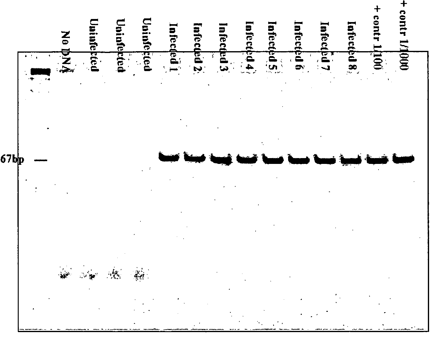
[0045]The first step in this method is the preparation of the DNA present in the urine sample. This method was described previously in studies of the detection of transrenal DNA in urine. All of the preparation takes place at room temperature (on the order of 24° C.).[0046]A quantity consisting of approximately 50 to 60 ml of urine is collected from the patient in sterile containers.[0047]Within 30 minutes after collection of the urine, 0.5M EDTA and 0.5M Tris-HCl (at a pH of 8.5) are added until a final concentration of 10 mM is reached. The purpose is to inhibit possible nuclease enzymes. Specifically, the EDTA is added to inhibit nucleases that are dependent on divalent ions, and the Tris-HCl is added so that a pH value can be reached that inhibits acid nucleases. This stabilizes the specimen, in the sense that it stabilizes the DNA fragments.[0048]The samples are then divided into aliquot portions of 5 ml and stored at a temperature of...

example 2

Isolation and Purification of the Nucleic Acids

[0049]Because the idea is to analyze DNA fragments whose length is less than 1000 nucleotides, and preferably less than 500, and more preferably less than 300, and yet more preferably less than 200, and even yet more preferably between 100 and 200 nucleotides, the commercially available DNA isolation kits cannot be used, because they are based on the isolation of DNA having a higher molecular weight.

All of the steps in the following method take place at room temperature (20 to 25° C.). As mentioned above, the sample may consist of aliquot parts of urine, or else of the urine supernatant or of the sediment.[0050]2 volumes of GITC [guanidine isothiocyanate] (at a concentration of 6M) were added to 1 volume consisting of 5 ml of sample and mixed well.[0051]The DNA whose length is less than 1000 base pairs of nucleic acids, and preferably less than 500, and more preferably less than 300, and yet more preferably less than 200, and even yet m...

example 3

Preparation for PCR

The PCR primers are usually constructed for amplicons measuring 60 to 120 nt or 250 to 400 nt. In this case, it was necessary to find primers that could also function for sequences consisting of 150 to 250 nt.

The FastPCR method (biocenter.helsinki.fi / bi / bare-1_html / oligos.htm) was used to design the primer sequences based on the complete human genome sequence.

[0052]The Primer 3 method (frodo.wi.mit.edu / cgi-bin / primer3 / primer3_www.cgi) was utilized to select the nested (semi-nested) primer.

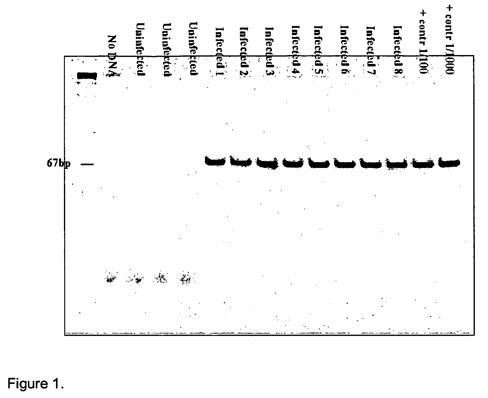
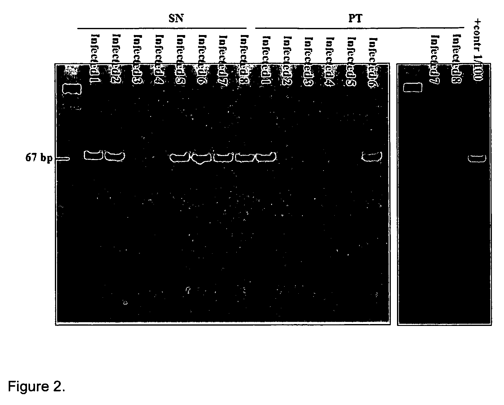
[0053]This method was used to create tuberculosis primers. The primer that was constructed for tuberculosis was reciprocal to the sequence IS 6110, which is present only in the Mycobacterium tuberculosis genome, as indicated below:

[0054]

IDN 1 F-785: ACCAGCACCTAACCGGCTGTGG(SEQ ID NO: 1)andIDN 2 R-913: CATCGTGGAAGCGACCCGCCAG.(SEQ ID NO: 2)(Product: 129 nt)

Instead, for the nested primer, the following sequence was selected:

[0055]

IDN 1 F-785: ACCAGCACCTAACCGGCTGTGG(SEQ ID NO: 1)andID...


PUM

PropertyMeasurementUnit
Tmaaaaaaaaaa
Tmaaaaaaaaaa
temperatureaaaaaaaaaa

Abstract

The present invention relates to a method for diagnosing and / or monitoring a bacterial or parasitic infection by detection and quantification of the transrenal nucleic acids, derived from bacterial pathogenic agents or from parasites, in urine. The detection method optionally includes the isolation and the purification of the nucleic acids from urine by methods known in the art including pairing with molecular probes that are specific for the pathogenic agents, PCR hybridization, PCR, nested PCR, SSCP, LCR, and SDA. Diagnostic kits based on these detection methods are also claimed.

Description

RELATED APPLICATIONS[0001]This application claims priority to Italian patent application RM2005000068, filed Feb. 17, 2005, bearing attorney docket number 6627PTIT, which is incorporated herein by reference in its entirety.FIELD OF THE INVENTION[0002]Disclosed is a system for the diagnosis of infections caused by pathogenic agents, exemplified by tuberculosis and malaria, based on the molecular analysis of the transrenal nucleic acids (Tr-NA) detected in urine.BACKGROUND OF THE INVENTION[0003]Early analysis and the possibility of rapidly genotyping the pathogenic agent are among the principal objectives of research in the diagnostic field.[0004]At present, the development of new diagnostic assays should take into consideration the following factors: compatibility to high-throughput screening methods; a higher degree of diagnostic sensitivity in the preliminary phase of the infection; and, particularly in developing countries, the ease of handling of biological samples, for more wide...

Claims


Application Information

Patent Timeline
Patent Type & Authority Patents(United States)
IPC IPC(8): C07H21/04A61K9/14C12N9/12
CPCY10S435/81Y02A50/30C12Q1/689C12Q2600/16C12Q2600/156C12Q1/686
Inventor MELKONYAN, HOVSEPCANNAS, ANGELATOMEI, LOUIS DAVIDUMANSKY, SAMUIL R.
Owner INST NAT PER LE MALATTIE INFETTIVE LAZZARO SPALLANZANI IRCCS
Learn More