Diagnostic device and method

a diagnostic device and a technology for biotechnology, applied in the field of clinical diagnostics and biotechnology, can solve the problems of inability to perform diagnostics using blood-containing samples, inability to perform diagnostics, and inability to meet the needs of patients,

Inactive Publication Date: 2011-01-18
QUALIGEN INC
View PDF14 Cites 22 Cited by
  • Summary
  • Abstract
  • Description
  • Claims
  • Application Information

AI Technical Summary

Problems solved by technology

This large variation coupled with other factors such as storage conditions, coagulation, and the fragility of red blood cells, produces considerable technical problems in performing diagnostics using blood-containing samples.
Centrifugation of blood is, however, also problematic.
For example, centrifuges are expensive, involve multiple steps, are often unavailable at points of care such as bedside, schools or at home, and usually require electrical power for operation.
But these advances often create entirely new problems.
For example, filters tend to retain significant amounts of plasma, and analytes present in low concentrations are frequently difficult to detect in the serum derived from small volumes of blood.
Existing filters also tend to clog, and have undesirably slow flow rates.
But choices here are fairly limited.
Filters are relatively simple to produce and use, but tend to cause excessive hemolysis of red blood cells.
Capillary action, however, is generally too weak to effect rapid separation of large volumes.
One of the major limitations of applying known magnetic separation to blood separation is that multiple anti-ligands are required to remove all of the various types of cells and sub-cellular particles.
Furthermore, lack or absence of ligands on the cells due to pathological conditions, genetic diseases or genetic variations or life cycle of cells generally reduce the efficiency with which the anti-ligands bind with the target cells.
The problems with known magnetic separation devices are exacerbated with increasing sample volumes, especially sample volumes over one milliliter.
Since many diagnostic applications require serum volumes of up to one milliliter to satisfy the requirements of multiple tests or batteries of tests, magnetic separation has not been particularly useful.
Moreover, assays such as glucose or hemoglobin tests are highly susceptible to interference caused by biological or chemical substances in the sample, including proteins, bilirubin, and drugs.

Method used


Image

  • Diagnostic device and method
  • Diagnostic device and method
  • Diagnostic device and method

Examples


experiment set 1

[0102

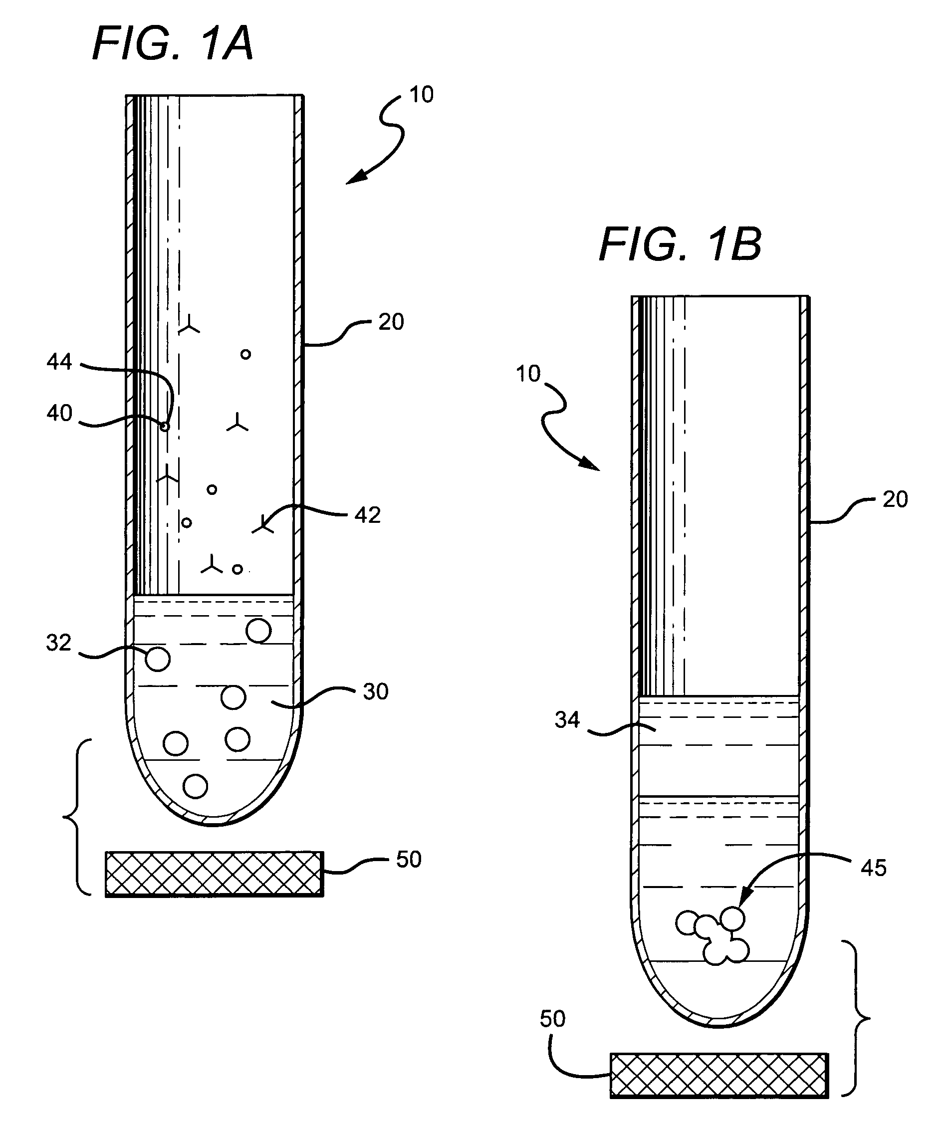
[0103]In a first series of experiments, precipitation of red blood cells was performed using mouse anti-red blood cell antibodies, paramagnetic beads coated with goat anti-mouse antibodies and 0.5 ml of anti-coagulated whole blood. Heparinized, EDTA or citrated whole blood was mixed with 10 μl undiluted mouse anti-red blood cell antibody solution (Red Out™; Robbins Scientific Corp.) for 2 minutes; next 10 μl of a solution (isotonic PBS, pH 7.4) containing paramagnetic beads (Cortex Biochem Inc., MagaCell™ or MagaBeads™; 30 mg / mL; or Pierce MagnaBind™) coated with goat anti-mouse antibodies was added and mixed gently for 2 minutes. The bottom of the tube was placed on a magnet (permanent iron magnet; approximately 0.2 Tesla). Precipitation of the cellular network started instantly, and was substantially finished after about 2 minutes. Plasma was collected by aspiration from the top of the vessel.

[0104]Significantly, methods and apparatus described herein have been found to separ...

experiment set 2

[0106

[0107]In a second series of experiments, precipitation of red blood cells on a microscope slide was performed using whole blood, mouse anti-red blood cell antibodies and paramagnetic beads coated with goat anti-mouse antibodies. To 0.2 ml fresh whole blood, 5:1 of an anti-red blood cell antibody containing solution (Red Out™) was added, mixed and incubated for 2 min at room temperature. Then, 5:L of a solution containing paramagnetic beads coated with goat anti-mouse antibodies (MagaCell™ or MagaBeads™ or MagaBind™) was added, mixed, and after another 2 minutes, two disk magnets were positioned at opposite ends of the slide. After about 1.5 minutes, a clear plasma containing zone was formed between the two magnets and this was retrieved with a pipette without disturbing the laterally-fixed cell-containing network.

experiment set 3

[0108

[0109]Flat envelopes sealed on three of four sides were prepared from transparency acetate sheets (for example, the 3M Inc. product, 3MCG3460), or plastic sheet protectors (e.g., Avery-Dennison PV119E), or from small “ZipLock” or ITW Inc. MiniGrip™ bags (2.5×3 cm; 2.0 mil). One-half to 1 mL of anti-coagulated whole blood was injected into the bag; 10 uL of Red Out™ (see above) was added and mixed with blood by gently rocking the container. After 2 minutes, the anti-mouse coated magnetic beads (MagaCell™) were added, mixed and the open side of the bag sealed. Two minutes later the bag was placed horizontally on a rectangular permanent iron magnet (approximately 0.2 Tesla). The magnetic particles and attached cellular networked moved adjacent to the magnet, leaving a clear layer of plasma as supernatant. The bag was then rotated into an upright position while still on the magnet, opened, and plasma aspirated using a pipette. It was also discovered that envelopes or bags (containi...


PUM

PropertyMeasurementUnit
wavelengthaaaaaaaaaa
volumesaaaaaaaaaa

Abstract

A method of separating a cell-containing sample into a substantially cell-depleted portion, and a cell-containing portion comprising at least one of a stem cell, a lymphocyte, and a leukocyte comprises a step in which the sample is received in a vessel with at least one flexible wall. In another step, an additive and particles are added to the sample, wherein the additive substantially binds to the at least one of the stem cell, lymphocyte, and leukocyte, and the particles and wherein the particles substantially bind to the at least one of the stem cell, lymphocyte, and leukocyte, and the additive, thereby producing a cell-containing network. In a further step, the network is separated from the substantially cell-depleted portion by applying a magnetic force.

Description

[0001]This application is a divisional application of U.S. patent application with the Ser. No. 10 / 371,837 filed Feb. 20, 2003, now abandoned, which is continuation-in-part of U.S. patent application with the Ser. No. 09 / 949,314, which was filed Sep. 7, 2001, now U.S. Pat. No. 6,821,790, and which is a continuation-in-part of U.S. patent application with the Ser. No. 09 / 514,686, filed Feb. 28, 2000, now U.S. Pat. No. 6,291,249, issued Sep. 18, 2001, and claiming the benefit of U.S. patent application Ser. No. 09 / 261,068 filed Mar. 2, 1999, all of which are incorporated herein by reference herein.FIELD OF THE INVENTION[0002]The field of the invention is clinical diagnostics and biotechnology.BACKGROUND OF THE INVENTION[0003]In vitro diagnostic tests to identify and treat diseases have become common tools in hospitals, homes and physician's offices. Biological fluids such as blood, urine or cerebrospinal fluids, which may at times contain blood, are the most frequently employed biolog...

Claims


Application Information

Patent Timeline
Patent Type & Authority Patents(United States)
IPC IPC(8): C12M1/34G01N33/53C12M3/00C12N11/18G01N1/34G01N33/49
CPCB01L3/5021B01L3/505G01N33/491B01L2300/0681G01N2001/4016Y10S436/807Y10T436/25375Y10T436/25125Y10T436/255Y10T436/113332Y10T436/2575B03C1/01B03C1/288B03C1/30B03C2201/18B03C2201/26
Inventor WYATT, JAMESSCOTT, MATTHEWMAHANT, VIJAY K
Owner QUALIGEN INC
Why Eureka
Try Eureka