

Image-based methods for measuring global nuclear patterns as epigenetic markers of cell differentiation
a global nuclear pattern and epigenetic marker technology, applied in image enhancement, image data processing, instruments, etc., can solve the problem of not by itself indicating nuclear organization
- Summary
- Abstract
- Description
- Claims
- Application Information
AI Technical Summary
Benefits of technology
Problems solved by technology
Method used
Image

Examples
example 1
DNA Positional Persistence
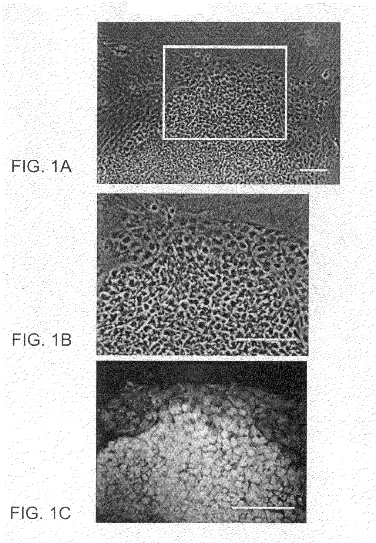
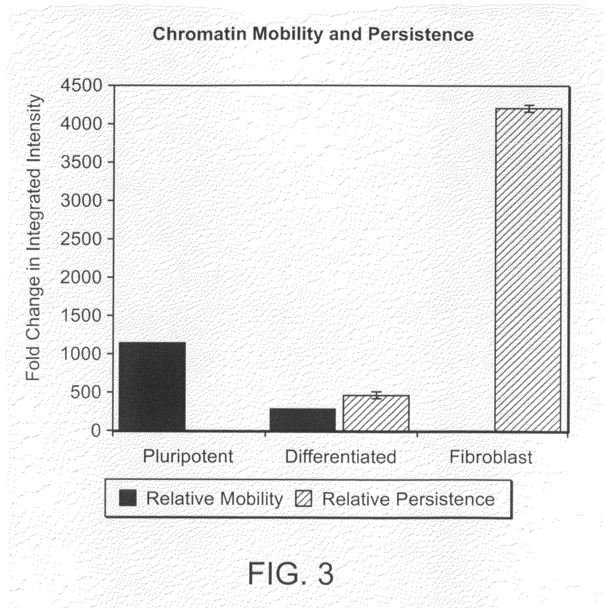
[0356]Chromatin movement within the nucleus was measured by live cell, time-lapse confocal microscopy of chromatin labeled with the vital DNA dye, Syto-16. Time lapse confocal images of Syto-16-loaded pluripotent cells hESC, 2 day differentiated hESC and CF-1 mouse fibroblasts (5 cells per condition) were analyzed for the integrated intensity (area x intensity) of DNA domains that changed between 30 min time intervals (mobile DNA areas) or that were persistent during a 1 hr interval (persistent areas pluripotent cells. Whole cell movement was excluded by analyzing in the frame of reference of the center of the nucleus. Movement was shown by the integrated intensity of chromatin spots that were fixed in position (relative to the center of mass of the nucleus) for one, two or three images at 30 min intervals. Movement in pluripotent cells decreased 4-fold after 2 days of differentiation and was more than 1000-fold greater than in fibroblasts. Areas of persist...
example 2
Reduced Speed but Increased Cohesion of Chromatin During Differentiation
[0357]Chromatin movement was further analyzed in hESC transfected with the fluorescent histone GFP-H2B (FIG. 4). Single confocal slices at 0.4 μm spacing (FIG. 4A) show density variations, including low-density voids (arrow in FIG. 4A, 0.4 μm).
[0358]The movement of cell-specific bright chromatin densities was analyzed by overlaying images at 0, 5 and 10 min, assigned to separate channels (FIGS. 4B-F) and by vector analysis where movement of bright spots was traced with a line at 1 min intervals and arrows were placed at the last position of the spot (FIG. 4G). Different kinds of nuclear movement were detected including internal mixing of chromatin, indicated by movement of bright chromatin domains without envelope deformation (pluripotent hESC, FIG. 4B), deformation of the envelope and chromatin (pluripotent hESC, FIG. 4C), and multidirectional streaming of chromatin (2 day differentiated hESC, FIG. 4D). Long-ra...
example 3
Relative Mechanical Deformation of Stem Cells and Somatic Cell Nuclei
[0359]Nuclear organization and structure correlate with mechanical stiffness of stem cell nuclei. Single hESC were isolated by brief treatment with Ca2+ / Mg2+ free HBSS with 1 mM EDTA and deposited in standard DSR media containing Hoechst 33342, Oregon 488 Wheat Germ Agglutinin and propidium iodide. Propidium iodide-positive cells were excluded, as were cells with clear cytoplasm, since this may indicate transient lysis during isolation. Cells were aspirated into pipettes (6-11 μm diameter) at fixed hydrostatic pressure for 1 min until steady state aspiration length was achieved. The steady state length of cell and nuclei dimpling into the pipette was measured. As cells differentiate for 2 and 6 days, nuclei become more solid and are not aspirated into the pipettes. A relative deformability was calculated by comparing the distance aspirated cells moved into the pipette. Cytoplasmic and nuclear aspirated length decre...
PUM
| Property | Measurement | Unit |
|---|---|---|
| half time | aaaaa | aaaaa |
| half time | aaaaa | aaaaa |
| half time | aaaaa | aaaaa |
Abstract
Description
Claims
Application Information