Method and apparatus for analysis of a sample of cells

a cell and method technology, applied in the field of cell and method and apparatus for analysis, can solve the problems of unsatisfactory approach in many aspects, time-consuming and hence cost-intensive, interference with light, etc., and achieve the effect of simplifying and complementing the analysis method

Active Publication Date: 2011-05-24
PHASE HOLOGRAPHIC IMAGING PHI
View PDF4 Cites 10 Cited by
  • Summary
  • Abstract
  • Description
  • Claims
  • Application Information

AI Technical Summary

Benefits of technology

[0019]The present invention has the advantages over the prior art that in combination with a resolution comparable to conventional optical microscopes it offers new possibilities for detecting phase changes and variations in the shape of undyed cellular samples comprising living and / or dead cells over time without removing the sample from the cell culture vessel. It is also possible to make the analyzes some time afterward and numerically or even automatically set the proper focal depth. This will simplify and complement the analyzing methods of today.

Problems solved by technology

This approach is unsatisfactory in many aspects, since it may kill a specimen whose life processes are under study.
It is also often very time consuming and hence costly such as in determining the number of cells in a cell culture vessel.
A laser illuminates the object and the scattered light interferes with the light of a reference source originating from the same laser.
The problem with prior art is to be able to analyse the characteristics of a large amount of cells and to determine the number of living or / and dead cells in a sample.

Method used


Image

  • Method and apparatus for analysis of a sample of cells
  • Method and apparatus for analysis of a sample of cells
  • Method and apparatus for analysis of a sample of cells

Examples


Embodiment Construction

[0028]The following description focuses on an embodiment of the present invention applicable to a device and a method for analyzing a sample of cells without influencing the cells by the use of digital holographic microscopy.

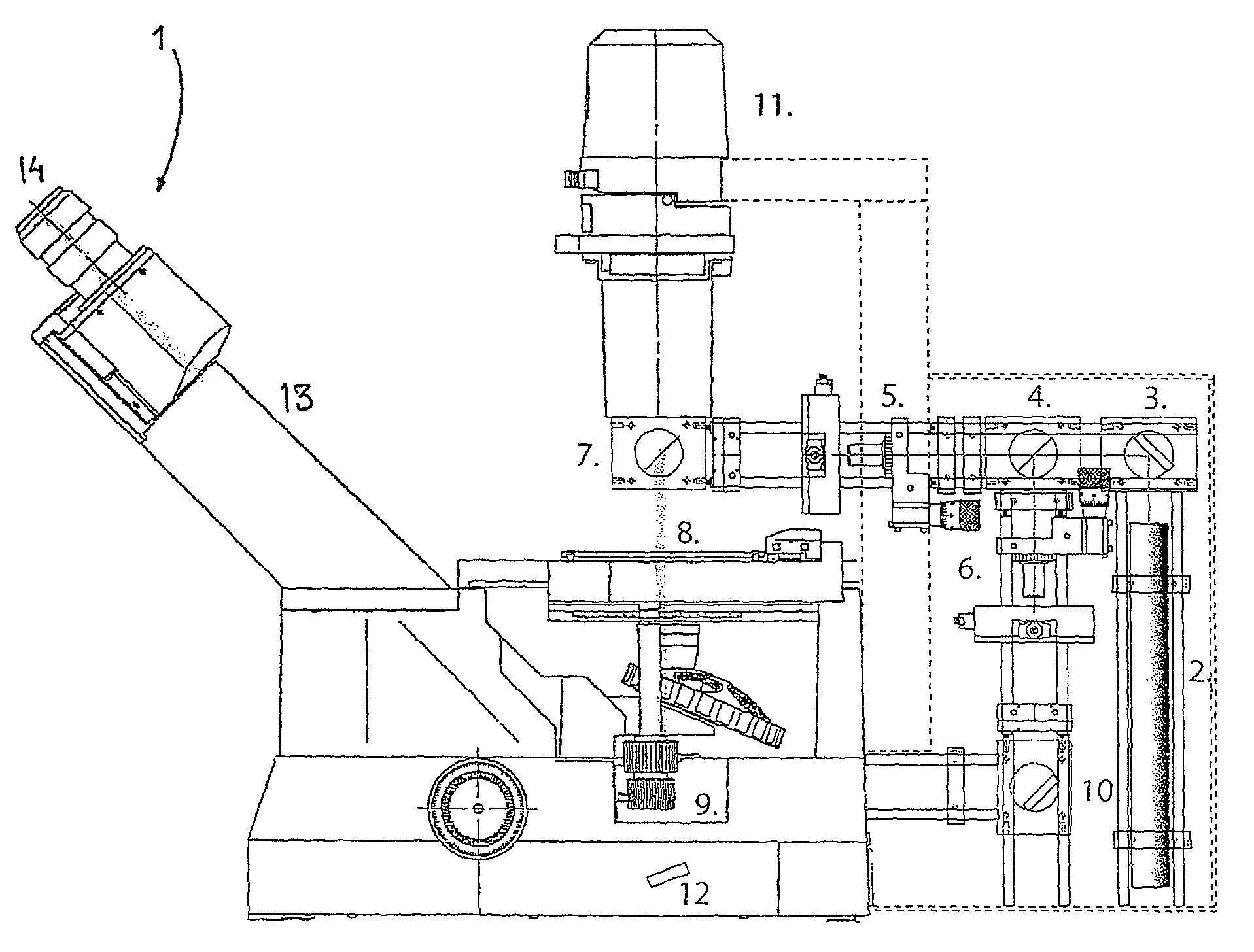
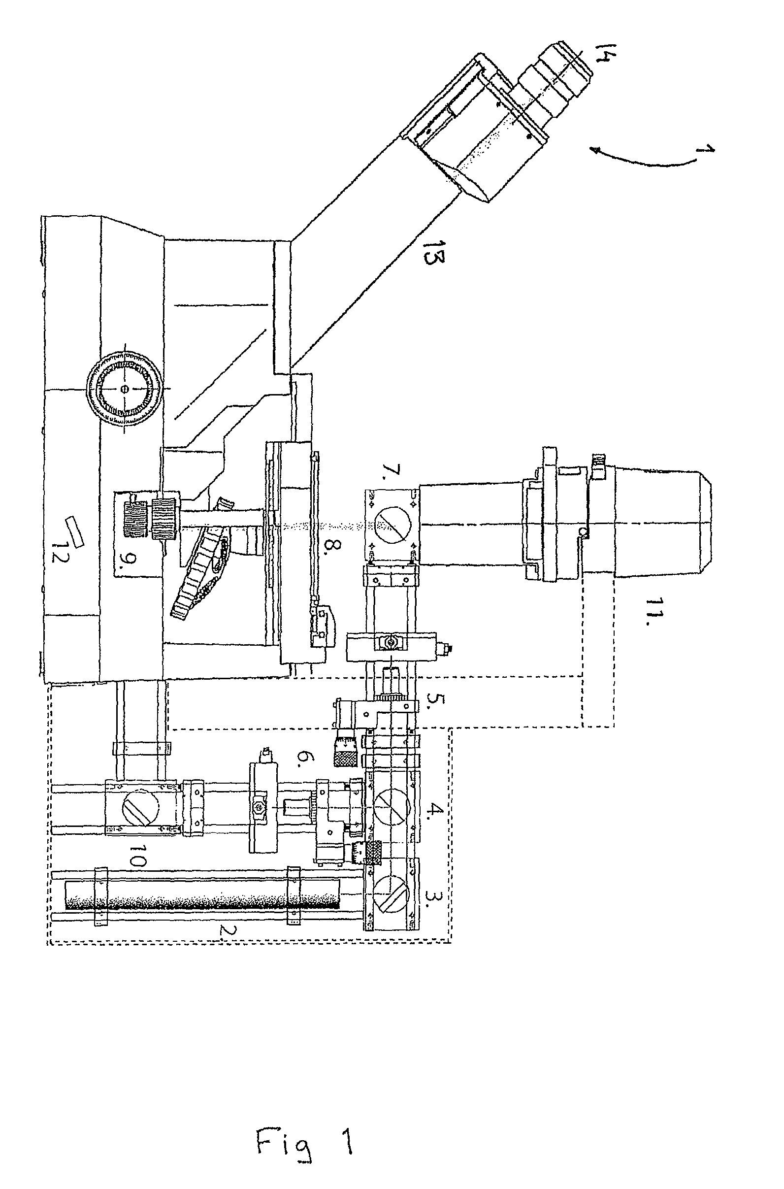
[0029]FIG. 1 illustrates an embodiment comprising a phase contrast microscope 1. The embodiment further comprises a laser 2, such as a He—Ne laser, emitting light at a wavelength of 633 nm.

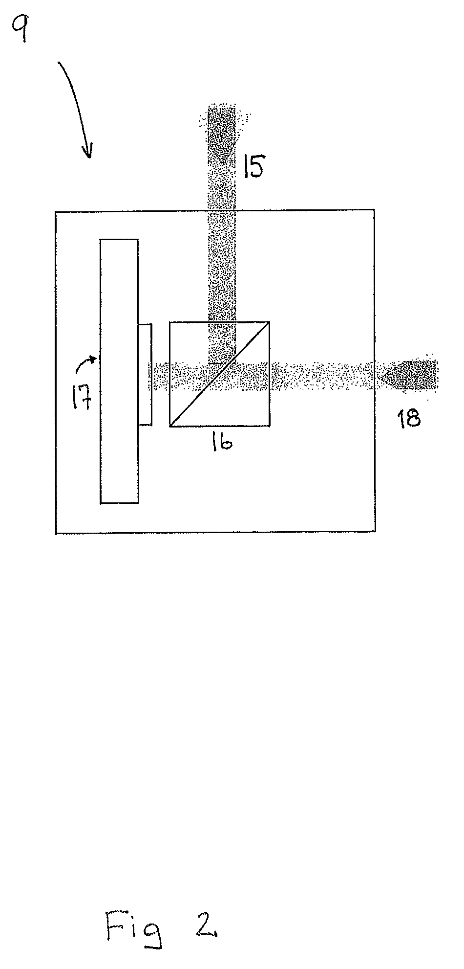
[0030]The light beam from the laser 2 is directed via a mirror 3 towards a first beam splitter 4 dividing the beam into an object beam and a reference beam. The object beam passes through a spatial filter 5, towards a second beam splitter 7. A sample 8 is arranged so that the object beam passes through the sample before reaching a beam collector device 9. The reference beam passes through a spatial filter 6 and is then reflected by a mirror 10, which diverts the reference beam towards the beam collector device 9. As shown in FIG. 2, the reference beam 18 and the object beam 15...


PUM


Abstract

A non-destructive method and device for analyzing a sample comprising transparent living and / or dead cells, by 5 means of a digital holographic microscope, where the sample (8) is exposed to light from a laser (2). The light that travels through the cells in the sample will experience a difference in the optical path length compared to the surrounding media and the wave front that emerges from the 10 cells will thus be phase shifted. This distortion can be detected in the digital hologram, which is reconstructed from the interference pattern detected by a digital sensor (17), such as a CCD or a CMOS, as phase differences or phase shifts and thereby creating a digital hologram. The 15 phase shift of each element of the hologram is then used for analyzing the characteristics of the cells in the sample.

Description

AREA OF INVENTION[0001]The present invention relates in general to the field of cell analysis. More particular the invention relates to a method and device for non-destructive analysis and characterization of a sample comprising living or / and dead cells in a cell culture vessel by use of digital holographic microscopy. This is to measure the proliferation and viability of the cells in the sample.PRIOR ART[0002]To analyse cells, which are almost transparent, they are often dyed or prepared in a certain way to become visual. This approach is unsatisfactory in many aspects, since it may kill a specimen whose life processes are under study. It is also often very time consuming and hence costly such as in determining the number of cells in a cell culture vessel.[0003]Thus, there is a need for a new method for analysing samples without influencing the sample.[0004]Digital holographic (DH) microscopes use the principle of holography to image an object. A laser illuminates the object and th...

Claims


Application Information

Patent Timeline
Patent Type & Authority Patents(United States)
IPC IPC(8): G01B9/021
CPCG02B21/14G02B21/36G03H1/0005G03H1/0443G03H1/0866G03H2210/62G03H2001/0428G03H2001/0447G03H2001/045G03H2210/12G03H2001/005G03H1/041
Inventor GUSTAFSSON, MATSSEBESTA, MIKAEL
Owner PHASE HOLOGRAPHIC IMAGING PHI
Why Eureka
Try Eureka