
Assessment of oocyte competence

a morphological assessment and oocyte technology, applied in the field of oocyte competence assessment, can solve the problems of difficult application of classical cytogenetic techniques to polar bodies, chromosome abnormalities are almost always lethal to the developing, and the method of identifying healthy oocytes and embryos based on morphological assessment is not very successful

Inactive Publication Date: 2011-09-27
YALE UNIV
View PDF2 Cites 2 Cited by
  • Summary
  • Abstract
  • Description
  • Claims
  • Application Information

AI Technical Summary

Benefits of technology

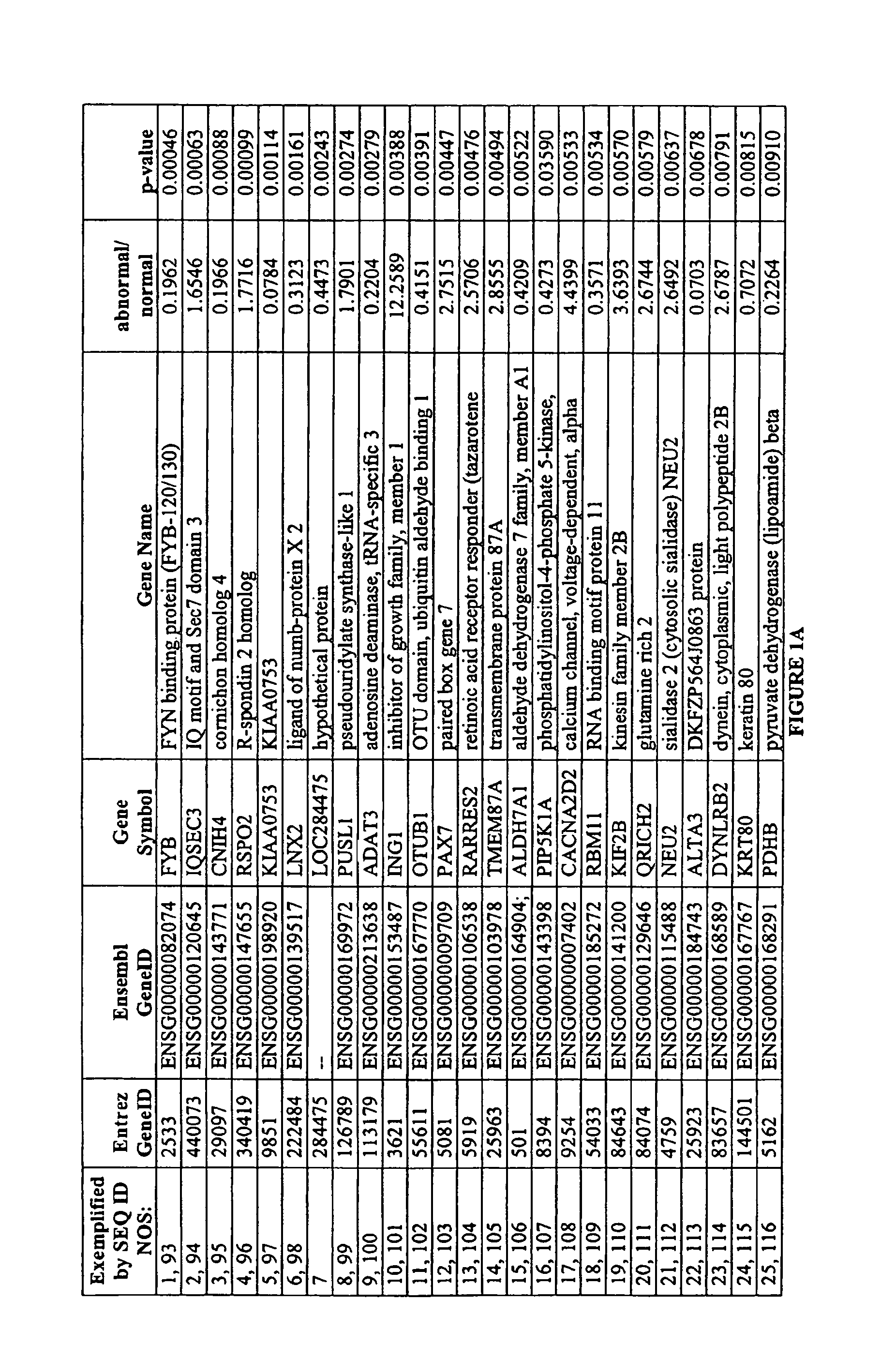
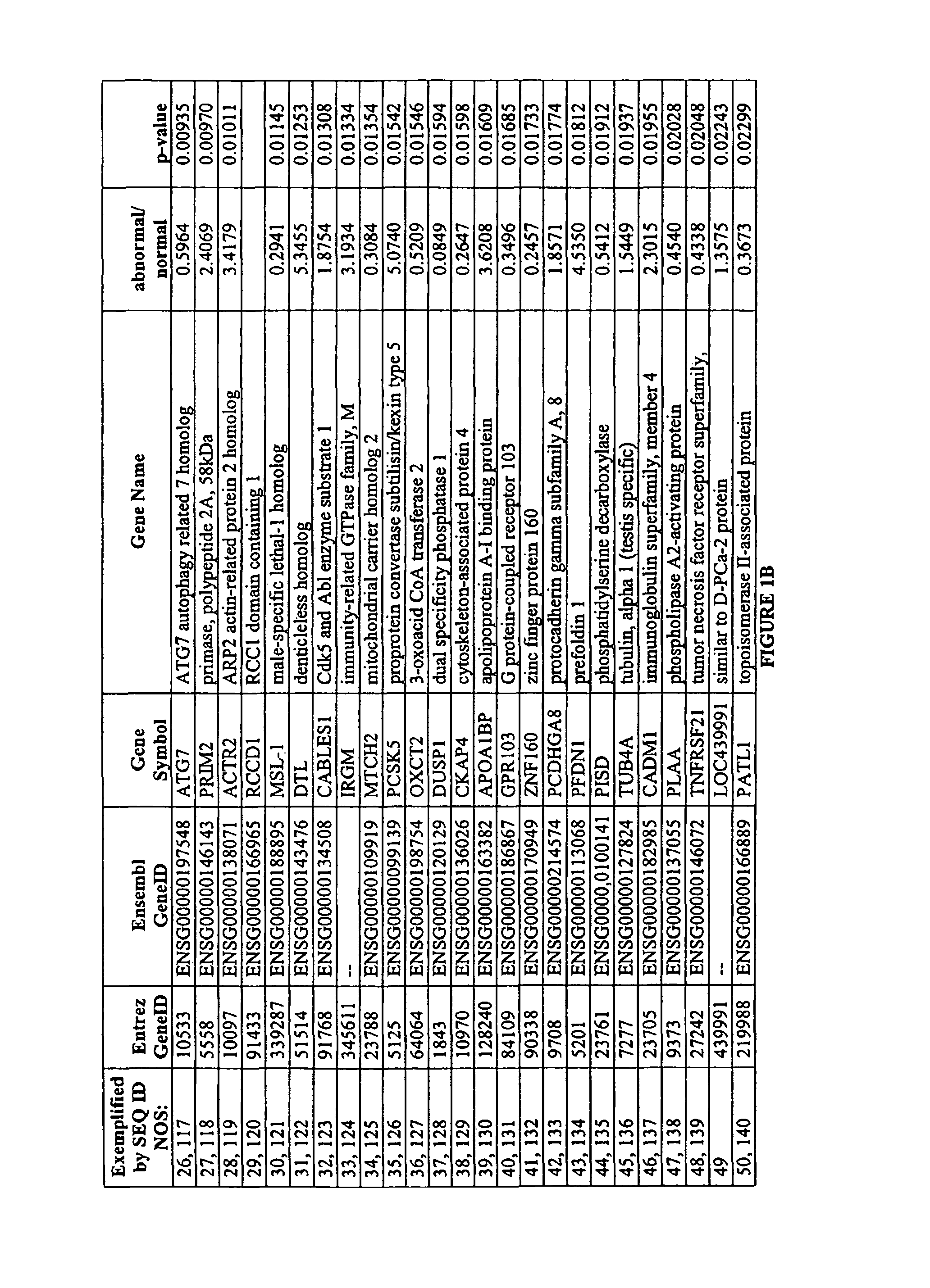
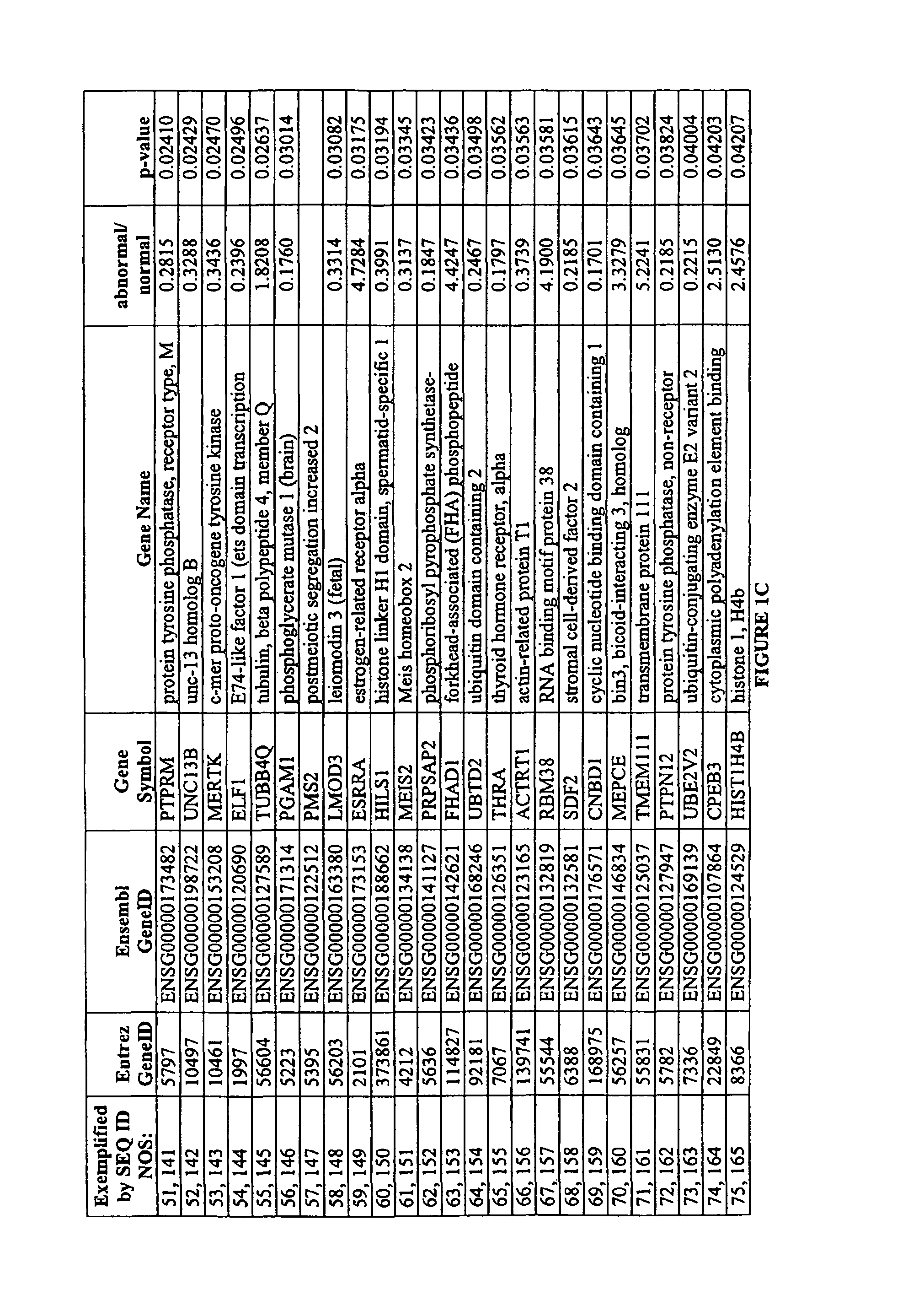
The present invention provides a method for evaluating the competence of a mammalian oocyte for fertilization and implantation. This is done by measuring the level of marker expression in the oocyte and comparing it to a control or reference standard. The sample can be derived from the oocyte, follicular fluid, cumulus cell, or culture medium. The level of marker expression is determined using nucleic acid microarray, Northern blot, reverse transcription PCR, Western blot, enzyme-linked immunosorbent assay, protein microarray, or FACS analysis. The invention also includes an array of nucleic acid probes immobilized on a solid support.

Problems solved by technology

These chromosome abnormalities are almost always lethal to the developing embryo and their high prevalence is responsible for many failed in vitro fertilization (IVF) treatments.
A great challenge for physicians is identifying which embryos are the most likely to result in a pregnancy and ensure that these embryos are among the limited number selected for transfer to the uterus.
Methods for identifying healthy oocytes and embryos based upon morphological assessments have not been very successful (for review, see Patrizio et al., 2007, Repro BioMedicine Online 15:346-353).
But, classical cytogenetic techniques are difficult to apply to polar bodies, due to problems of obtaining high quality chromosome spreads.
However, this method examines only less than half of the chromosomes.
In addition, the removal of a cell is an invasive procedure that may damage the embryo.
CGH has the major advantage that every chromosome is analyzed, rather than the limited subset assessed using FISH, but it is a time-consuming and labor-intensive method that is difficult to perform within the limited time available for preimplantation testing.
Despite the accuracy and sensitivity of real-time PCR, the method is limited by the restricted number of genes that can be assessed for each oocyte or embryo, generally less than 10 genes.

Method used


Image

  • Assessment of oocyte competence
  • Assessment of oocyte competence
  • Assessment of oocyte competence

Examples


experimental examples

[0095]The invention is further described in detail by reference to the following experimental examples. These examples are provided for purposes of illustration only, and are not intended to be limiting unless otherwise specified. Thus, the invention should in no way be construed as being limited to the following examples, but rather, should be construed to encompass any and all variations which become evident as a result of the teaching provided herein.

example 1

Identification of Markers Differently Expressed in Oocytes

[0096]The processing of oocytes was conducted in a dedicated DNA-free clean-room environment. A total of 27 oocytes, including seven single oocytes (three chromosomally normal and four chromosomally abnormal) and four pooled samples each consisting of five pooled oocytes of unknown chromosomal status were analyzed.

[0097]Oocytes were collected in sterile, RNase-free conditions and processed rapidly in order to minimize changes in marker expression. The zona pellucida was removed to ensure the exclusion of all cumulus cells from the sample and the polar body was separated from the oocyte. The oocyte was transferred to a microcentrifuge tube and then immediately frozen, while the polar body was thoroughly washed to remove any DNA contaminants before transfer to a separate microcentrifuge tube.

[0098]The polar body DNA was released by lysing the cell. Polar bodies were washed in four 10 uL droplets of phosphate-buffered saline—0.1...

example 2

Identification of Markers Differently Expressed in Cumulus Cells

[0112]The processing of cumulus cells was conducted in a dedicated DNA-free clean-room environment. A total of six cumulus cells (three chromosomally normal and three chromosomally abnormal) were analyzed.

[0113]Cumulus cells were collected in sterile, RNase-free conditions. The cumulus cells were separated from the oocyte mechanically and processed rapidly in order to minimize changes in marker expression. The zona pellucida was removed from the corresponding oocyte and the polar body was separated. The oocyte was transferred to a microcentrifuge tube and then immediately frozen, while the polar body was thoroughly washed to remove any DNA contaminants before transfer to a separate microcentrifuge tube.

[0114]The polar body DNA was released by lysing the cell. Polar bodies were washed in four 10 uL droplets of phosphate-buffered saline—0.1% polyvinyl alcohol, transferred to a microfuge tube containing 2 uL of proteinase ...


PUM

PropertyMeasurementUnit
temperatureaaaaaaaaaa

Abstract

Methods are provided for evaluating an oocyte for fertilization and implantation. For example, methods are provided for determining whether an oocyte expresses, or does not express, one or more of a group of markers identified as differently expressed between chromosomally normal and chromosomally abnormal oocytes. Also provided, for example, are methods for determining whether a cumulus cell expresses, or does not express, one or more of a group of markers identified as differently expressed between cumulus cells associated with chromosomally normal oocytes and cumulus cells associated with chromosomally abnormal oocytes. Methods are provided for the detection of marker expression of differentially expressed genes at the RNA level, as well as at the protein level.

Description

CROSS-REFERENCE TO RELATED APPLICATIONS[0001]This is a National Stage application of PCT International Application No. PCT / US2007 / 023216, filed Nov. 2, 2007, which in turn claims the benefit pursuant to 35 U.S.C. §119(e) of U.S. Provisional Application No. 60 / 856,157, filed on Nov. 2, 2006, which is hereby incorporated by reference in its entirety herein.REFERENCE TO SEQUENCE LISTING[0002]The Sequence Listing submitted on compact disk is hereby incorporated by reference. The file on the disk is named 047162-5031-00WO.txt. The file is 1657 kb and the date of creation is Nov. 2, 2007.BACKGROUND OF THE INVENTION[0003]Chromosome anomaly affects over 20% of oocytes obtained from women in their early thirties and this prevalence more than doubles as women enter their forties. These chromosome abnormalities are almost always lethal to the developing embryo and their high prevalence is responsible for many failed in vitro fertilization (IVF) treatments. Consequently the identification of ch...

Claims


Application Information

Patent Timeline
Patent Type & Authority Patents(United States)
IPC IPC(8): C12Q1/68
CPCC12Q1/6883C12Q1/6876C12Q2600/158
Inventor WELLS, DAGANPATRIZIO, PASQUALE
Owner YALE UNIV
Learn More