Multi-echo magnetic resonance imaging method and system

a magnetic resonance imaging and multi-echo technology, applied in the field of multi-echo magnetic resonance imaging method and system, can solve the problems of insufficient resolution for other applications, early destruction of magnetization preparations, and certain problems, and achieve the effect of faster and more accurate perfusion maps

Inactive Publication Date: 2011-11-15
NEW YORK UNIV
View PDF5 Cites 12 Cited by
  • Summary
  • Abstract
  • Description
  • Claims
  • Application Information

AI Technical Summary

Benefits of technology

[0014]Exemplary embodiments of the present invention may combine the advantages of the multi-echo gradient recalled EPI imaging and parallel imaging with the concept of the temporal resolution being governed by the time taken to reach the center of k-space. This combination can permits the snapshot imaging to be performed with high temporal resolution, and does not require the use of a sliding window reconstruction. Accordingly, the temporal resolution achieved in this exemplary manner can be closer to the actual resolution, rather than average. Since such resolution can be based on a Fourier approach to image acquisition, the image reconstruction is easier as compared to spiral imaging. In addition, exemplary embodiments of the present invention may be used to acquire high temporal resolution images using polarized gas or hyperpolarized gas. Such use may include without limitation lung imaging with hyperpolarized helium. For example, according to one exemplary embodiment of the present invention, cine imaging application where single-shot (e.g., the entire image data may be acquired in a single acquisition) can be utilized to improve temporal resolution. Further, the exemplary embodiments of the present invention may be used for perfusion imaging, fast T1 mapping or similar magnetization-prepared imaging techniques.
[0022]At least one of the exemplary embodiments of the present invention can provide faster and more accurate perfusion maps.

Problems solved by technology

This resolution may be insufficient for other applications, such as a study of extremely rapid mechanical cardiac functions, for example, valve motion, mechanical activation maps, and evaluation of mechanical dyssynchrony, which is a key indicator for early stages of systolic contraction and diastolic expansion.
Single echo SSFP (e.g., balanced or unbalanced SSFP) techniques allow for a temporal resolution of approximately 2.5 milliseconds, but suffer from certain problems.
For example, single echo SSFP techniques require application of a large number of radio frequency (“RF”) pulses into the patient's body, which is undesirable.
Also, for sequences that involve magnetization preparation (e.g., magnetization tagging), application of a high number of RF pulses causes an early destruction of the magnetization preparations (i.e., the tags fade faster).
This potentially limits the analysis of prepared datasets (i.e., tagged datasets) to a small fraction of the cardiac cycle.
The reconstruction based on this combination can be a lengthy process because each echo in the train of echoes (or echo-train) contributes to the same image, or cardiac phase, and it may not be easy to parallelize the reconstruction of the data acquired using this combination.
However, this approach can only yield temporal resolution of up to 60 msec (e.g., when used with single-shot spirals), with a corresponding spatial resolution of 4 mm.
While echo-planar imaging (EPI) is one of the renowned examples of fast snapshot imaging, this technique may suffer from various artifacts, such as susceptibility, and T2 or T2* signal attenuation effects, which reduce its practical usefulness to select regions of the body, and to low spatial resolutions.
Due to the short T2* of 3He (approximately 12 ms), EPI is not a practical solution for clinically useful spatial resolutions (3 mm or less).
Beside the situation that spiral imaging may be nontrivial to reconstruct, the sliding window approach can lead to an averaging of the data over multiple time points, making it difficult to determine the true temporal resolution of the data.
However, this segmented approach can make it difficult to acquire data with the type of temporal resolution that is achieved with EPI.
However, spiral imaging can be cumbersome to implement and the sliding window reconstruction leads to time averaging of the signal.
One of the major problems encountered in making accurate maps of T1 is the long time generally needed.
If a conventional two-dimensional (2D) inversion-recovery spin-echo sequence is used, data acqisition for each slice can take a significant time (e.g., several hours).

Method used


Image

  • Multi-echo magnetic resonance imaging method and system
  • Multi-echo magnetic resonance imaging method and system
  • Multi-echo magnetic resonance imaging method and system

Examples


first exemplary embodiments

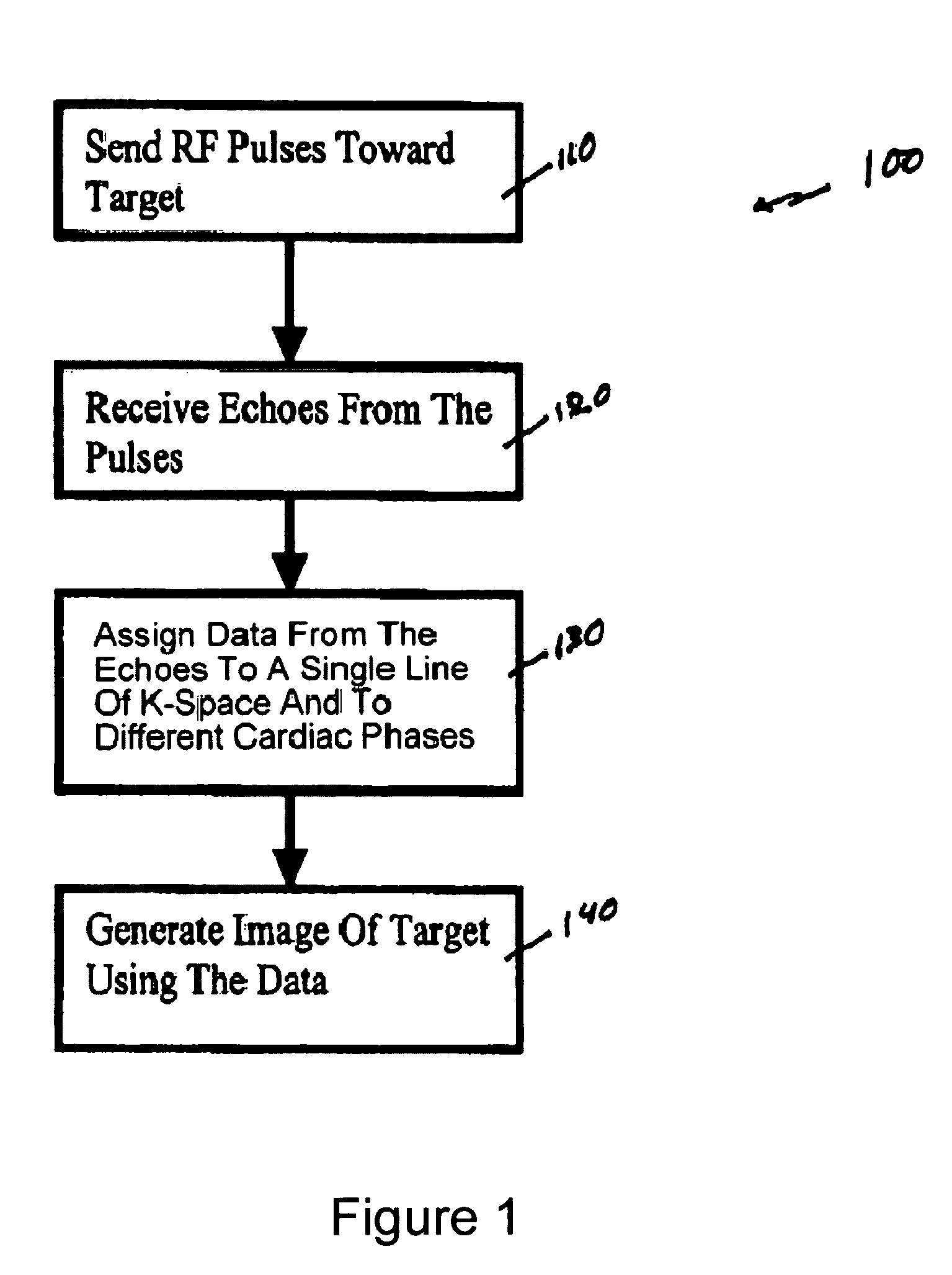
[0050]FIG. 1 shows a flow diagram of one exemplary embodiment of a phase-train imaging (“PTI”) method 100 for generating magnetic resonance images according to the present invention. FIG. 2 shows an exemplary embodiment of an MRI apparatus 10 according to the present invention which may be used to implement the method 100 described below with respect to FIG. 1.

[0051]For example, as shown in FIGS. 1 and 2, a series of radio frequency (“RF”) pulses 82 can be transmitted in step 110 by an RF transmitter 50 of the MRI apparatus 10 toward a target 80. In one exemplary embodiment, the exemplary method 100 can be applied to cardiac MRI techniques, and the target 80 may be patient's cardiac region. The series of RF pulses 80 can generate echoes 84 from the target 80, which may be received (in step 120) by an RF receiver 60 of the MRI apparatus 10. In one exemplary embodiment, a single device, e.g., a transceiver, can be used as both an RF receiver 60 and an RF transmitter 50. Because the mu...

second exemplary embodiments

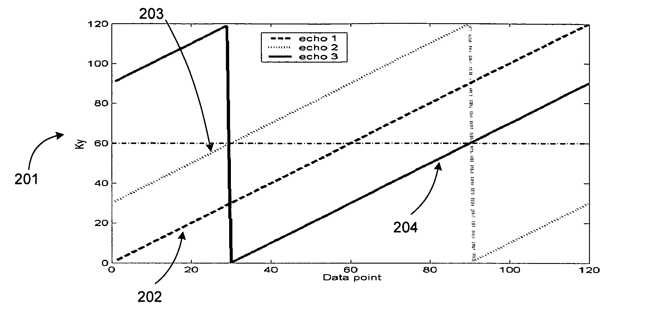
[0056]The MESSFP approach called Phase Train Imaging (PTI) (e.g., see V. M. Pai et al. “Phase train approach for very high temporal resolution cardiac imaging,” JCMR 2005; 7(1):98-99) can permit the acquisition of extremely high temporal resolution cine datasets in relatively short breath-hold durations. In this exemplary approach, unlike conventional MESSFP approaches, each set of echoes following an RF excitation pulse can acquire the same k-space line, and may be assigned to a different cardiac phase. Since all the echoes acquire the same k-space line for different cardiac phases, there may likely be no “complex” phase discontinuity in the “complex” phase-encode direction associated with this approach; since multiple echoes can be acquired, the number of RF pulses may be correspondingly smaller than that for single echo sequences, and effects of magnetization preparation pulses such as tagging can persist for a relatively longer duration. In addition, PTI is inherently more effic...

third exemplary embodiments

[0061]Exemplary Approach to Encode in all 3 Directions

[0062]For example, an echo train of 3 echoes can enable a first moment nulling in the Readout direction (beside the inherent zero-th moment nulling) at the end of each TR (repetition time), thus maintaining the conditions for trueFISP imaging (e.g., zero-th and first moments are generally nulled in the Slice Select and “complex” phase-encode directions in this approach). However, the 3-echo implementation of PCPTI can be primarily provided to encode the flow in-plane. In addition, because the implementation may have a long TR, it can potentially give rise to off-resonance banding artifacts if encoding for smaller velocities is attempted. An alternate approach can be to use a 2-echo implementation. One exemplary approach to achieving first moment nulling at the end of each TR with a 2-echo sequence can be to either implement a bipolar gradient following the second readout gradient, or to use a flyback gradient between the two read...


PUM


Abstract

Exemplary method, systems and arrangements can be provided for creating a high-resolution magnetic resonance image (“MRI”) or obtaining other information of a target. For example, radio-frequency (“RF”) pulses can be transmitted toward the target by a RF transmitter of a MRI apparatus. In response, multiple echoes corresponding to each pulse may be received from the target. Data from each of the echoes can be assigned to a single line of k-space of a distinct image, and stored in memory of the apparatus. An image of the target, velocity data and / or acceleration data associated with a target can be generated as a function of the data. In one exemplary embodiment, the data from different echoes can be assigned to the same k-space line and to different cardiac phases. The exemplary embodiments of the present disclosure can be utilized for the heart or for any other anatomical organ or region of interest.

Description

CROSS-REFERENCE TO RELATED APPLICATION(S)[0001]This application is based upon and claims the benefit of priority from U.S. Patent Application Ser. No. 60 / 643,798, filed Jan. 14, 2005, the entire disclosure of which is incorporated herein by reference.FIELD OF INVENTION[0002]The present invention relates to magnetic resonance imaging (“MRI”). More particularly, the present invention relates to method and system for generating MRI data using a multi-echo approach. The present invention also relates to methods and systems for increased temporal resolution or increased combinations of temporal and spatial resolution. The present invention further relates to methods and systems which are capable of imaging or characterizing flow dynamics with high temporal resolution including, without limitation, flow conditions in the heart or any organ of the body. The present invention still further relates to methods and systems which are capable of producing T1 maps, perfusion imaging or imaging an...

Claims


Application Information

Patent Timeline
Patent Type & Authority Patents(United States)
IPC IPC(8): A61B5/05
CPCG01R33/563A61B5/055G01R33/561
Inventor PAI, VINAY M.
Owner NEW YORK UNIV
Why Eureka
Try Eureka