Automated microscope for blood cell analysis

a blood cell and automatic technology, applied in the field of automatic microscopic analysis of stained blood cells, can solve the problems of inability to detect the presence of a single color, etc., and achieve the effect of low cos

Active Publication Date: 2011-11-29
MEDICA CORP
View PDF17 Cites 18 Cited by
  • Summary
  • Abstract
  • Description
  • Claims
  • Application Information

AI Technical Summary

Benefits of technology

[0008]The present invention uses new approaches to create a novel automated blood cell analysis system. An exemplary embodiment of the present invention provides a much improved, low cost, computer controlled microscope system for this analysis of blood cells. This system allows the tedious parts of the blood analysis to be done unattended, allowing the technician to focus on interpretation of the results.
[0009]In one aspect, microscope slides with a blood smear and cover glass are further prepared by coating with a thin film of oil of about 20μ thickness, which provides high quality digital images under high powered magnification that are virtually indistinguishable from the results from the traditional immersion technique without all the attending messy problems of the oil blob.

Problems solved by technology

All this focusing and stage movement is usually done manually and takes a significant amount of time.
Positional and alignment errors can be introduced during these manipulations.
Counting of cells is a mindless, tedious task, given to distraction and human error.
In sum, present day manual methods are time consuming, tedious, costly and can lead to errors.
Unfortunately, these automated systems are built around conventional microscopes, which are not designed for extended use in high volume applications.
Also focus, X-Y transport and objective lens mechanisms designed for manual use exhibit backlash effects making difficult reliable and reproducible positioning, which is necessary for high quality imaging operations.
Additionally, the adaptations of conventional microscopes do not address the problems of ambient jarring and temperature changes, which can translate to large positional errors.

Method used


Image

  • Automated microscope for blood cell analysis
  • Automated microscope for blood cell analysis
  • Automated microscope for blood cell analysis

Examples


Embodiment Construction

)

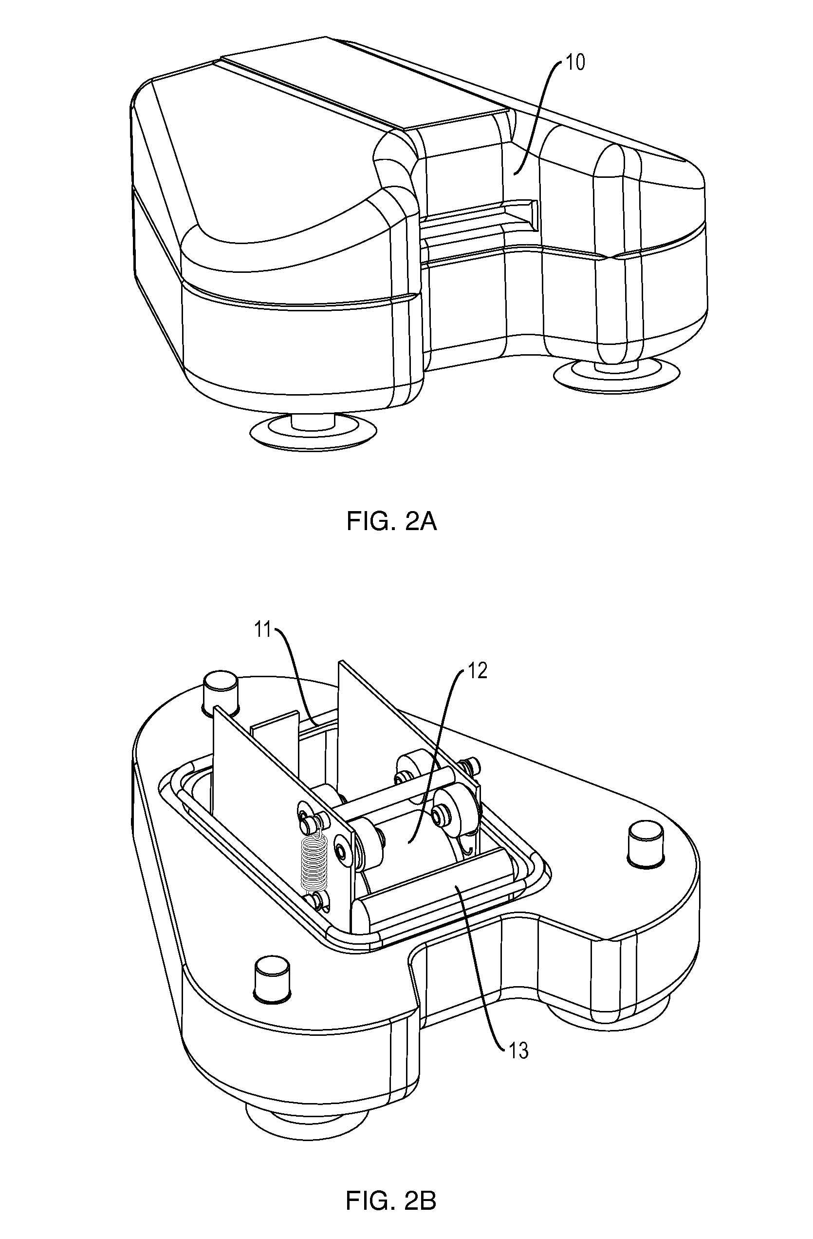
[0046]FIG. 2, the oiling device 5 consists of a slot 10 for access, a well 11 containing microscope immersion oil, an applicator 12 to coat the slide and a wiper 13 to remove excess oil.

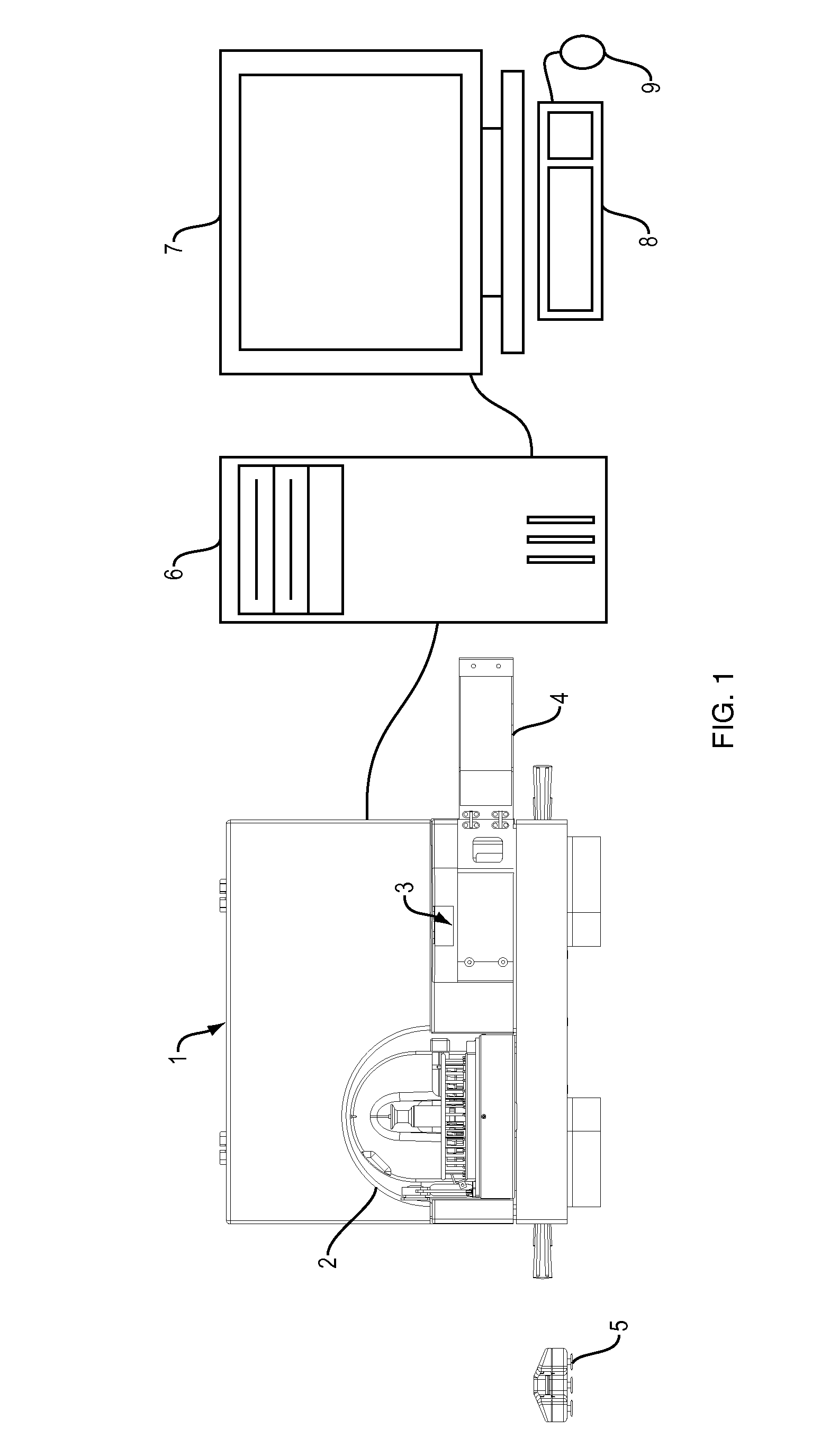
[0047]FIG. 1, the apparatus comprises a computer controlled microscope subsystem incorporated in a housing 1. The microscope subsystem communicates with the computer 6 via a cable connected to USB ports on each. The housing has portals for a slide carousel 14, a slot 3 for STAT samples and an access panel 4. A computer subsystem comprises a computer 6 with a monitor 7 and other peripherals for storage, display and user input, such as a keyboard 8 and mouse 9.

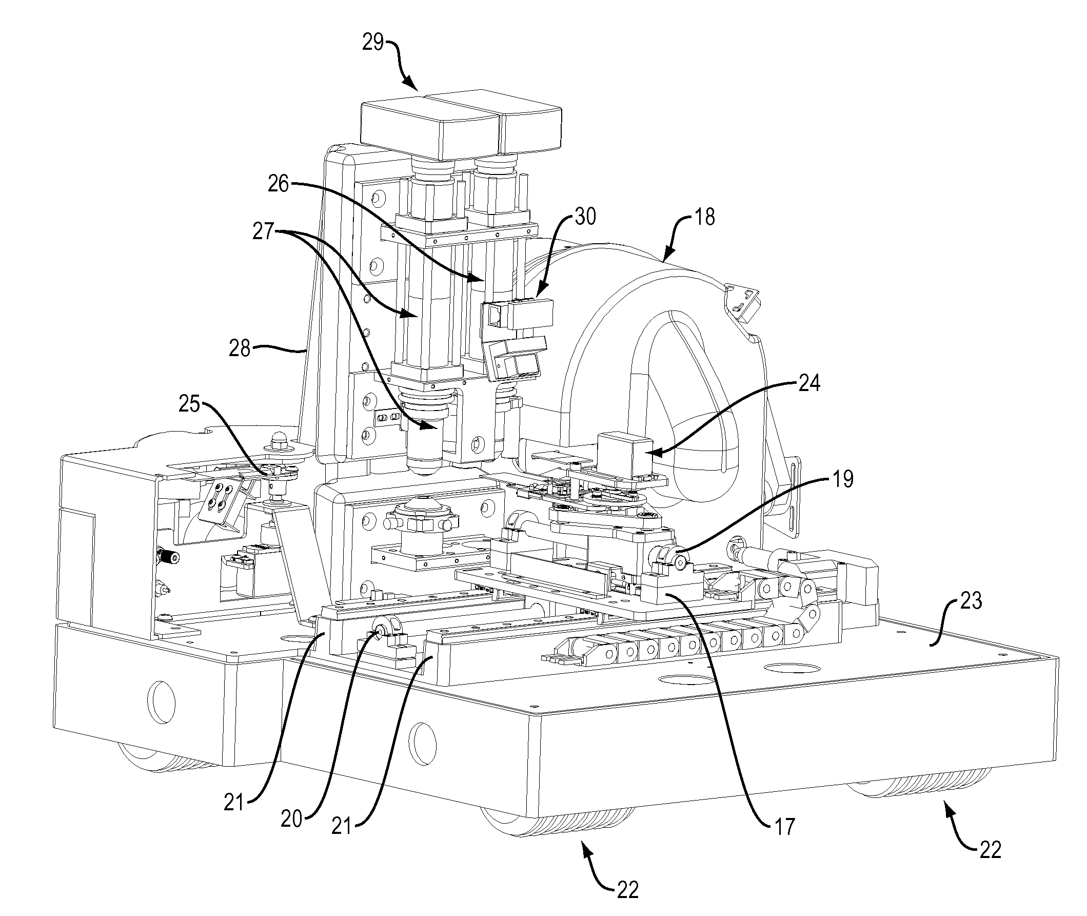
[0048]FIG. 5, the microscope subsystem has computer controlled, linear induction motors for movement in both the X 31 and Y 32 directions. The X-direction motor 31 travels along a center rail 20 and two linear bearings 21. The Y-direction motor 32 also travels along a center rail 19 and one linear bearing 37, attached to the X-direction motor. T...


PUM

PropertyMeasurementUnit
widthsaaaaaaaaaa
widthaaaaaaaaaa
sizeaaaaaaaaaa

Abstract

An apparatus and method of automated blood cell analysis uses technologies from other systems to create a new, robust, improved type of automated microscope, which uses electronic motors and a closed loop control system to minimize ambient factors, such as jarring and temperature changes. Pre-stained blood smear slides are first coated with a thin film of oil and are loaded into a carousel from which they can individually be analyzed. The slides are moved under a low magnification microscope; an optimal area of examination is determined; a focal plane map is calculated for that area, and the positions of white blood cell candidates are computed. The slide is then moved under a high power microscope where a refined focal plane map is computed and the individual white blood cell candidates are imaged. The cells are preclassified and the images are made available for analysis by the technician. Additionally, samples of red blood cells, equivalent to what the technician would do manually, are imaged and presented to the technician for evaluation. The technician may make notes on those cells, both red and white blood cells and archive them.

Description

CROSS REFERENCE TO RELATED APPLICATIONS[0001]Not ApplicableFEDERALLY SPONSORED RESEARCH[0002]Not ApplicableSEQUENCE LISTING OR PROGRAM[0003]Not ApplicableFIELD OF THE INVENTION[0004]The invention relates to an apparatus for automatic microscopic analysis of stained blood cells. The invention employs means of motion in the X, Y and Z directions that is computer controlled, reproducible and low friction, coupled with software for focusing, finding an appropriate area on a blood smear in which to perform the test, finding cell candidates at low power within that area, capturing digital images of those cells at high power, counting the cells and grouping them according to their color, size and morphology. The base of the apparatus is a low cost polymer concrete material that is stable to temperature changes and is energy absorbent. Additionally, the apparatus is mounted upon polymeric shock absorbers. The apparatus is unreactive to ambient temperature changes and vibrations.BACKGROUND O...

Claims


Application Information

Patent Timeline
Patent Type & Authority Patents(United States)
IPC IPC(8): G01N1/00
CPCG01N1/312G02B21/34G02B21/362G02B21/26G01N35/0099G01N2001/317Y10T436/25Y10T436/2575
Inventor VAN RYPER, WILLIAM JOHNCOTE, TYLERSMALL, STEVEN I.SHENG, AMY P.MA, IVAN HEE YIUJONES, RONALD
Owner MEDICA CORP
Why Eureka
Try Eureka