
Reducing imaging-scan times for MRI systems

a technology of mri and imaging scans, applied in the field of mri, can solve problems such as image artifacts and prevent accurate diagnosis, and achieve the effects of high accuracy, high-temporal efficiency, and exceeding sar limits

Active Publication Date: 2011-12-13
THE TRUSTEES OF THE UNIV OF PENNSYLVANIA
View PDF7 Cites 27 Cited by
  • Summary
  • Abstract
  • Description
  • Claims
  • Application Information

AI Technical Summary

Benefits of technology

[0010]To solve these and other problems, the present invention described herein, introduces a method of, and system for, rapid MRI imaging-scanning that provides 2D or 3D coverage, high precision, and high-temporal efficiency without exceeding SAR limits.
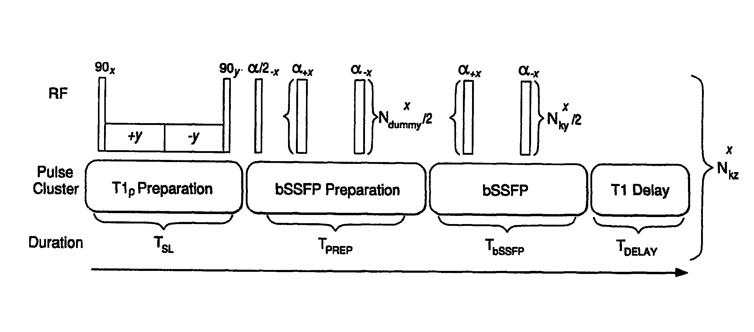
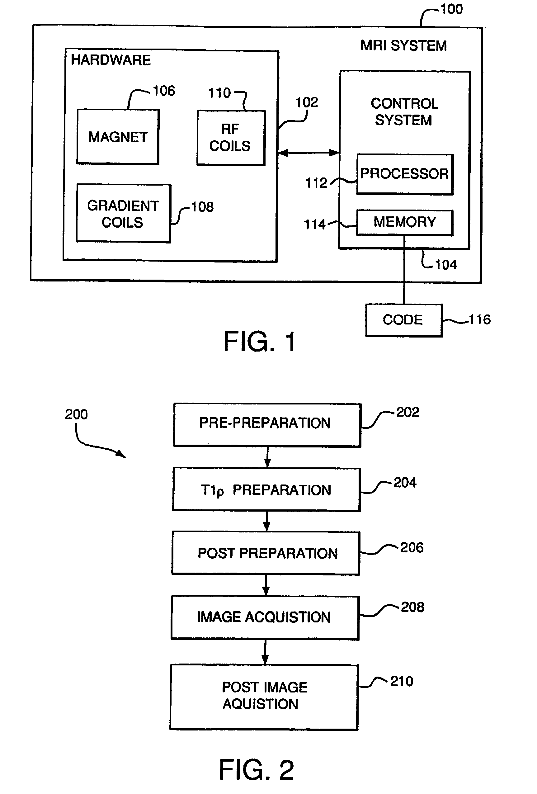
[0012]In one embodiment of the invention, the T1ρ pulse sequence includes five stages: a pre-preparation, T1ρ preparation, post-preparation, and image acquisition stages, and post image acquisition period which facilitates clinical imaging.
[0014]In another embodiment, methods to reduce T1ρ image blurring and incorrect measurement of T1ρ relaxation times or enhance signal to noise, are performed by a post-processing filter, a variable-flip-angle acquisition method, and / or a Half-Fourier acquisition method. Additionally, scan time can still be further shortened by reducing the T1ρ relaxation delay as part of the post image acquisition period, while compensating for magnetization saturation.

Problems solved by technology

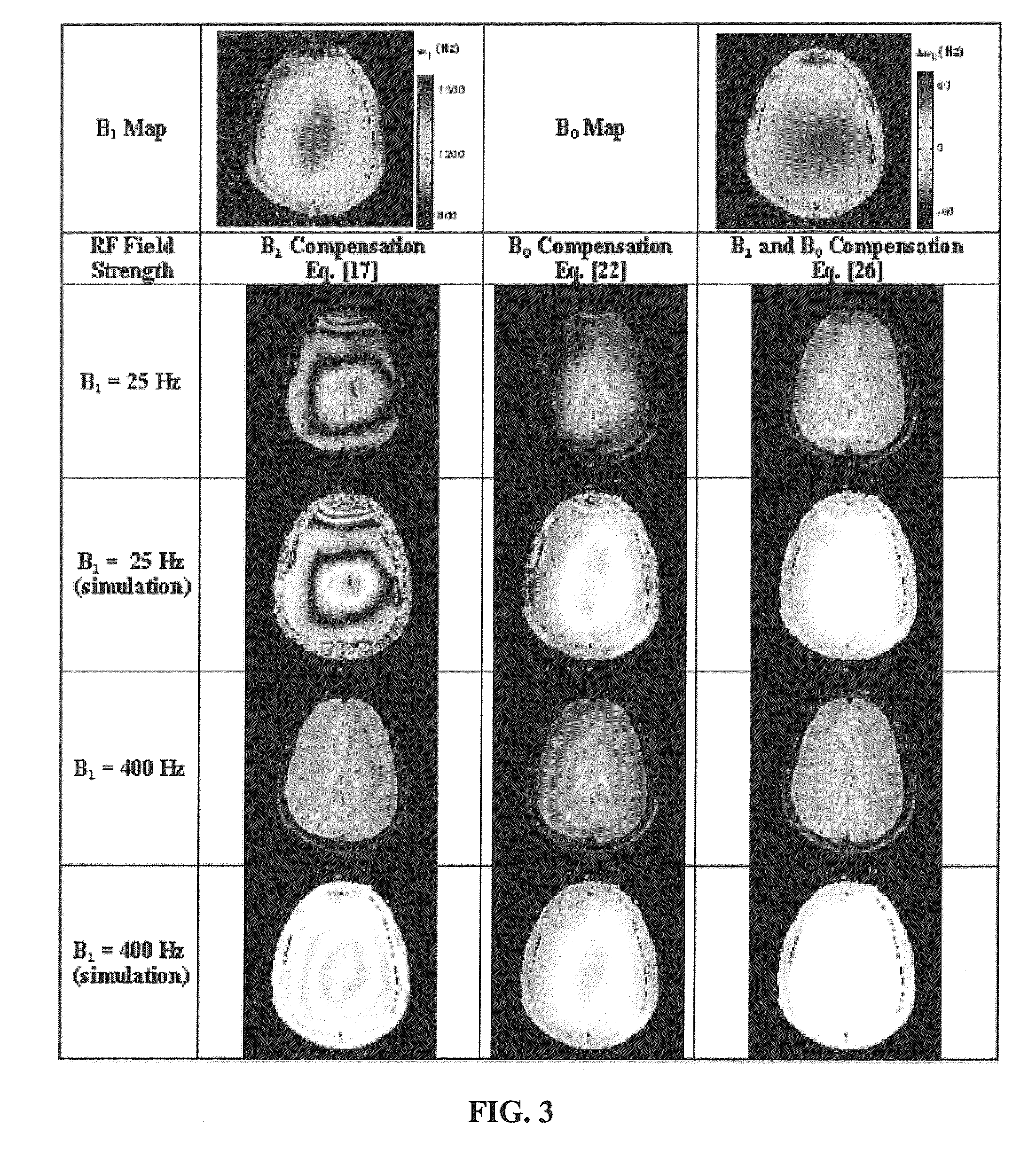
This is valuable, since magnetic field inhomogenieties can cause image artifacts and prevent accurate diagnoses.

Method used


Image

  • Reducing imaging-scan times for MRI systems
  • Reducing imaging-scan times for MRI systems
  • Reducing imaging-scan times for MRI systems

Examples


example

[0070]Materials And Methods: A MRI “phantom” and two healthy male volunteers were imaged on a 1.5 T Sonata Siemens clinical MRI scanner (Siemens Medical Solutions, Erlangen, Germany) using an eight-channel knee coil (MRI Devices Corp., Muskego, Wis.). The phantom consisted of gel of 2% (w / v) agarose in phosphate-buffered saline (Sigma-Aldrich, St. Louis, Mo.) doped with 0.2 mM MnCl2 to reduce T1. For this particular study, only healthy subjects were studied without any clinically meaningful acute or chronic medical problems.

[0071]Estimation of T1ρ in agarose phantoms: The ability of pulse sequence to estimate accurate T1ρ values was evaluated using two agarose bottle phantoms. A series of T1ρ-weighted images were acquired with the pulse sequence at seven spin-lock durations (TSL) (1, 5, 10, 20, 30, 35, and 40 mseconds). Other imaging parameters were TE=2.5 mseconds, short TR=5 mseconds, FOV=180 mm, 256×128 matrix size with 4 mm-thick sections and spin-locking frequency at 400 Hz. Th...


PUM


Abstract

Provided are methods and systems for rapid MRI imaging-scanning that provides 2D or 3D coverage, high precision, and high-temporal efficiency, without exceeding SAR limits. In one embodiment, a pulse sequence process is performed that includes a T1ρ preparation period, followed by a very rapid image acquisition process, which acquires multiple lines of k-space data. The combination of T1ρ preparation and acquisition of multiple lines of k-space, allows scan times to be shortened by as much as 3- or 4-fold or more, over conventional MRI scanning methods.

Description

CROSS-REFERENCE TO RELATED APPLICATIONS[0001]This application is a continuation of International Application PCT / US2007 / 022159 filed on Oct. 17, 2007 and published on Apr. 24, 2008, which claims priority to U.S. Provisional Application 60 / 852,241 filed on Oct. 17, 2006, each of which is incorporated herein in its entirety.STATEMENT OF GOVERNMENT INTEREST[0002]This work was supported in part by National Institutes of Health grants R01AR4504, AR051041, and performed at a NIH supported resource center (NIH RR02305). The government may have certain rights in this invention.FIELD OF THE INVENTION[0003]The present invention relates to magnetic resonance imaging (MRI), and more particularly, a magnetic resonance (MR) pulse sequence for reducing imaging-scan times for MRI systems.BACKGROUND OF THE INVENTION[0004]MRI, or Magnetic Resonance Imaging, (including: spectroscopy, conventional, and fast imaging techniques) is viewed as a conventional medical procedure having acceptable risks and ce...

Claims


Application Information

Patent Timeline
Patent Type & Authority Patents(United States)
IPC IPC(8): G01V3/00
CPCA61B5/055A61B5/4514A61B5/4528G01R33/50G01R33/5602G01R33/5613G01R33/5614G01R33/5617
Inventor BORTHAKUR, ARIREDDY, RAVINDERCHARAGUNDLA, SRIDHAR
Owner THE TRUSTEES OF THE UNIV OF PENNSYLVANIA
Learn More