
Methods and devices for endoscopic treatment of organs

a technology for hollow organs and endoscopes, applied in the field of endoscopes and hollow organs, can solve the problems of post-operative pain, post-operative infection, and time required to gain access to the operative site, and achieve the effects of improving the quality of li

Inactive Publication Date: 2012-01-10
CERIER JEFFREY C
View PDF37 Cites 16 Cited by
  • Summary
  • Abstract
  • Description
  • Claims
  • Application Information

AI Technical Summary

Benefits of technology

"The invention is a system and method for apposing and fixating multiple regions of the outer surface of a hollow organ using a flexible endoscope. The system includes an instrument, an inner tube, a piercing element, a first tissue anchor, a pusher, a suture, and a second tissue anchor. The method involves piercing the organ at two locations, delivering the first and second tissue anchors through the pierced holes, and using the suture to bring the anchors together and appose the multiple regions of the outer surface of the organ. The invention allows for easy and fast post-procedure recovery, does not require surgical intervention, and does not limit the location of the points of engagement with the tissue. It also allows for the possibility of performing the procedure with the patient under sedation rather than general anesthesia. The invention provides a high degree of efficacy for the condition being treated and is simple, inexpensive, and disposable. It also ensures that structures outside of the stomach are not involved in the tissue engagement or fixation."

Problems solved by technology

Disadvantages of open surgery include the risks associated with general anesthesia, the time required to gain access to the operative site, blood loss, the time required to close the tissues which were cut or dissected to obtain access to the operative site, post-operative pain, post-operative infection, lengthy post-operative recovery period, post-surgical adhesions, and cosmetic scarring due to large abdominal incisions.
Additionally, risks of blood clots, pulmonary embolus, dehiscence, and pain are associated with open surgery.
Laparoscopic surgery has the disadvantages of requiring a high degree of skill and training by the surgeon, and requiring specialized and expensive equipment.
Disadvantages of endoscopic GERD treatment include lower efficacy as compared to open or laparoscopic surgical procedures, safety concerns during the procedures, and the complexity of the devices that are required to manipulate the tissue endoscopically.
Safety concerns with these procedures have led to discontinuation of their use in treating GERD.
A significant disadvantage of these procedures is that the physician cannot visualize the material as it is being implanted, and thus is not sure where in the body it is ending up.
For example, the material may inadvertently be injected into the lumen of the aorta, which can result in death.
These concerns have led to discontinuation of these procedures for the treatment of GERD.
Thus a disadvantage of this type of procedure is that often the tissue does not heal (i.e. mucosal-to-mucosal tissue apposition often does not heal).
When the tissue does not heal it is less likely to remain in apposition over time, and thus the treatment is temporary and is not effective in treating GERD.
The first disadvantage of the devices which currently exist for use in procedures in which multiple regions of the outer surface of the stomach are brought into apposition and fixated is that the devices must apply high loads in order to bring the regions of tissue into apposition.
This geometry dictates that the devices have complex mechanisms, usually with hinged components.
Thus the mechanisms that actuate the hinged components are complex, expensive, and potentially unreliable.
The second disadvantage of the devices which currently exist for use in procedures in which multiple regions of the outer surface of the stomach are brought into apposition and fixated is that the complexity and expense of the devices dictates that the only practical way to enable them is as reusable devices.
Reusable devices such as these must be cleaned and disinfected in between uses, and present the risk of cross-contamination from one patient to another.
Disinfection also requires that the devices be sealed to prevent cleaning and disinfecting solutions from damaging the complex mechanisms, further increasing the cost of the devices and decreasing their reliability.
The third disadvantage of the devices which currently exist for use in procedures in which multiple regions of the outer surface of the stomach are brought into apposition and fixated is that the devices are large.
The large size of the devices, particularly the large cross-sectional areas, makes advancement through the esophagus difficult, which can increase patient discomfort, cause esophageal trauma or perforation and / or cause respiratory problems.
The fourth disadvantage of the devices which currently exist for use in procedures in which multiple regions of the outer surface of the stomach are brought into apposition and fixated is that device malfunction may require surgical intervention to allow removal of the device from the patient.
If the actuating mechanism malfunctions when the device is engaged with the tissue, the device can become locked onto the stomach with no way to remove it endoscopically.
This puts the patient at risk for any of the complications normally associated with surgery, such as infection, pain, post-surgical adhesions, scarring, general anesthesia risk, and other generally known risks.
The fifth disadvantage of the devices which currently exist for use in procedures in which multiple regions of the outer surface of the stomach are brought into apposition and fixated is that the geometry of engaged regions of the stomach is limited, both in terms of location in the stomach, and in distance from each other, by the device geometry.
The sixth disadvantage of the devices which currently exist for use in procedures in which multiple regions of the outer surface of the stomach are brought into apposition and fixated is that there is no way to ensure that structures outside of the stomach are not involved in the fixated tissue.

Method used


Image

  • Methods and devices for endoscopic treatment of organs
  • Methods and devices for endoscopic treatment of organs
  • Methods and devices for endoscopic treatment of organs

Examples


Embodiment Construction

[0103]The invention will now be described in detail, with references made to the accompanying drawings. Reference numbers are used in the drawings and the description to refer to specific elements or aspects of the invention. Wherever possible the same reference numbers will be used to indicate the same elements or aspects throughout multiple drawings and descriptions.

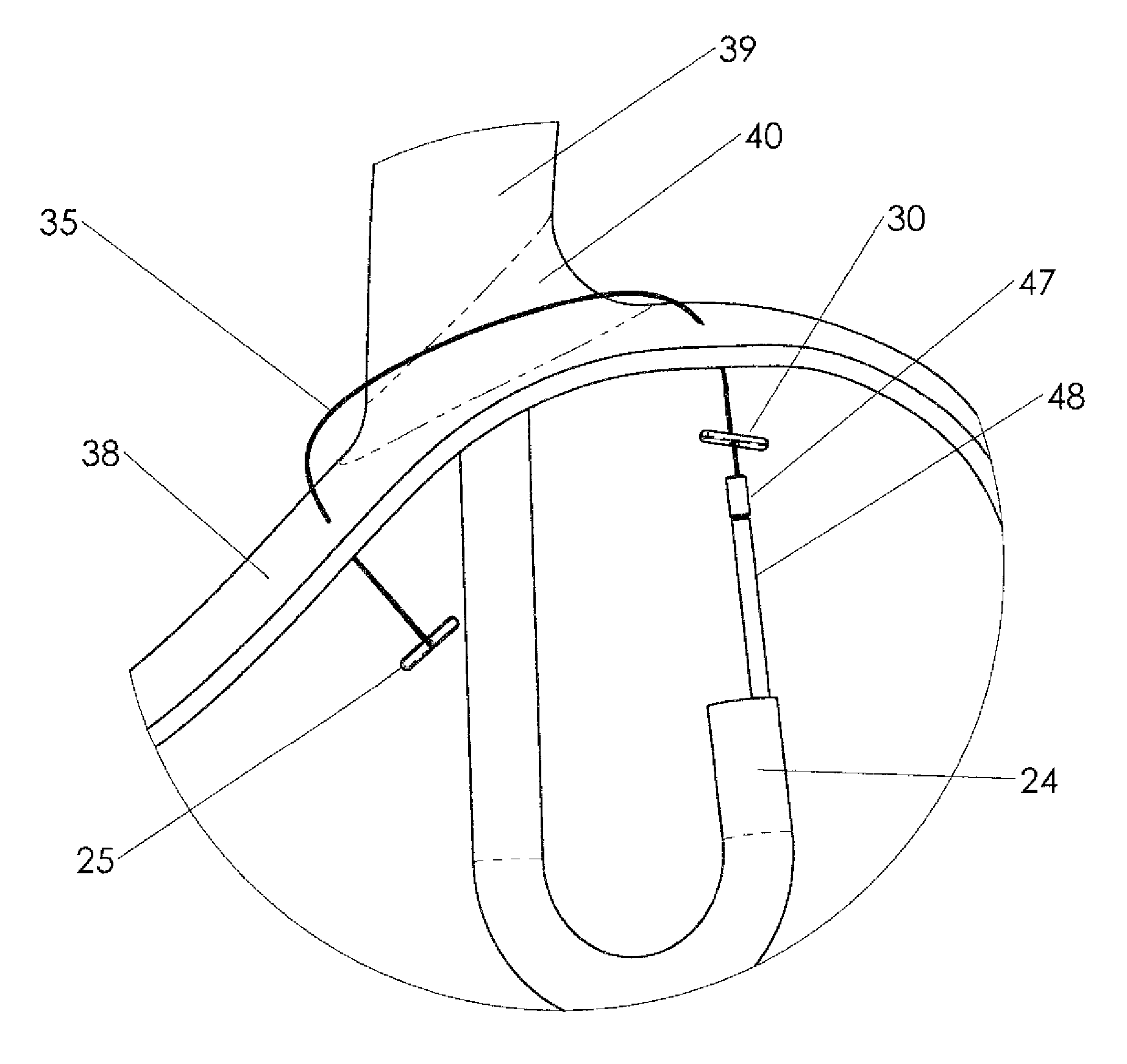
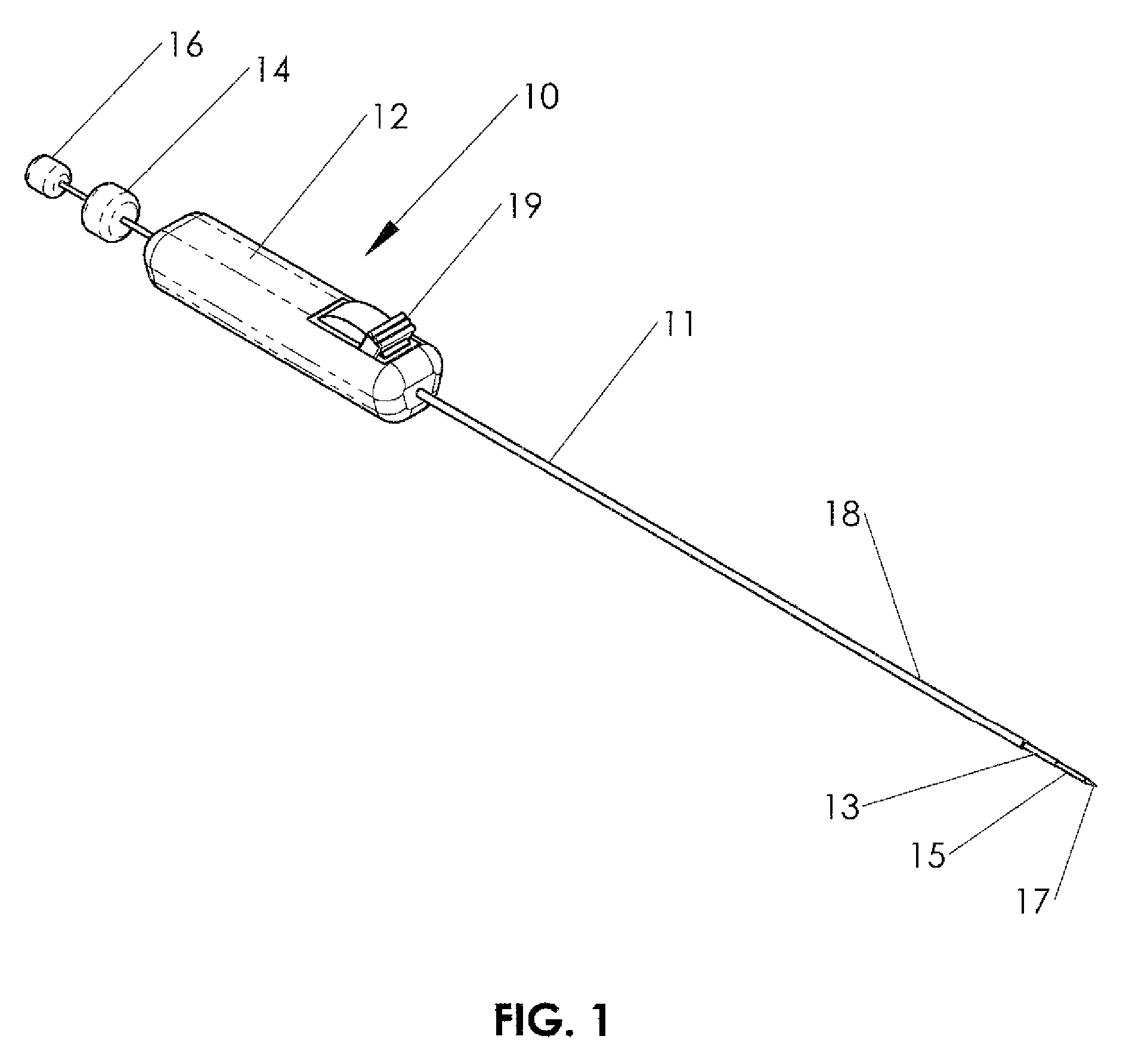
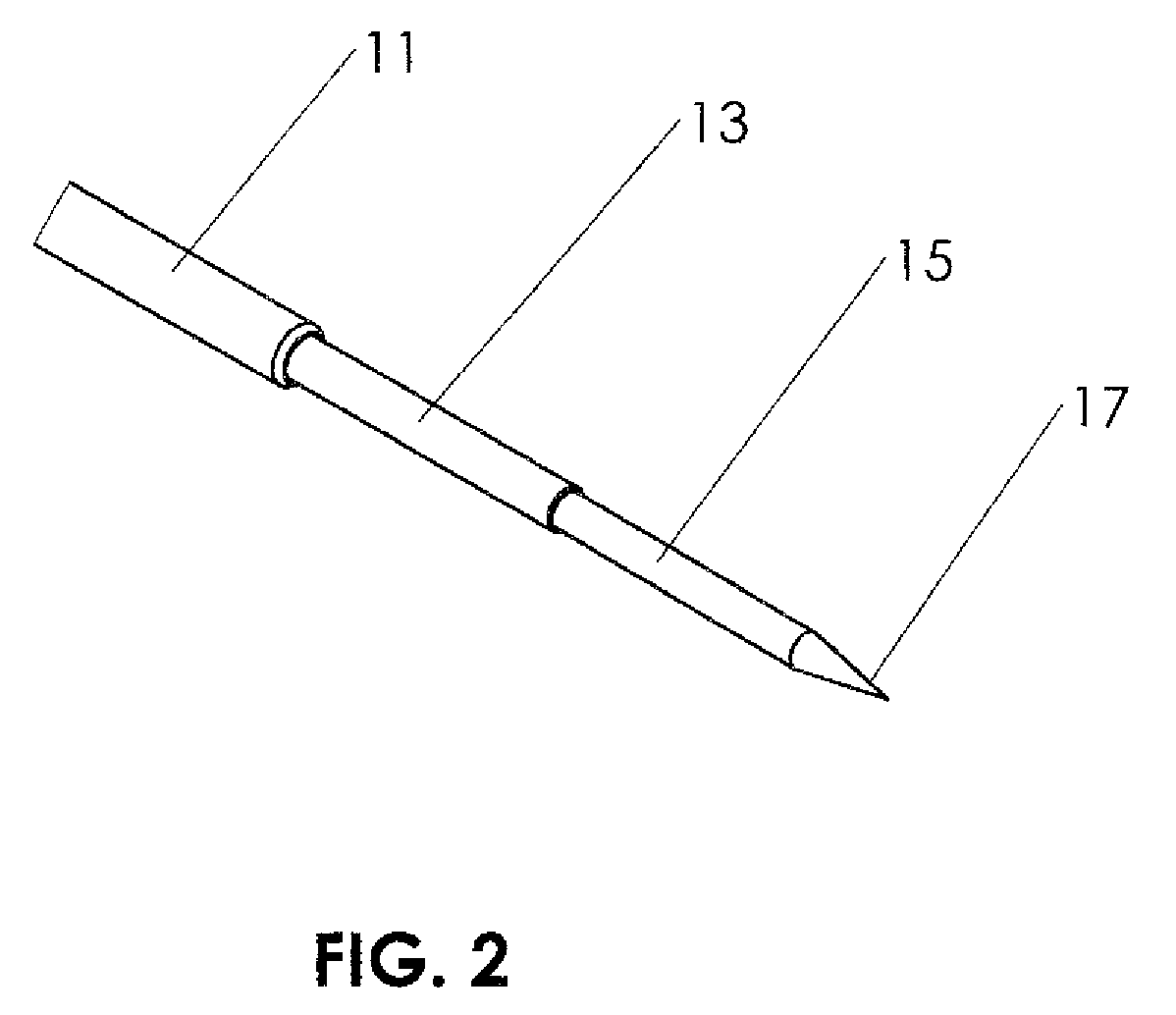
[0104]FIGS. 1-15 and 36-38 illustrate a preferred embodiment of the invention, and FIGS. 16-35 illustrate the novel method which is enabled by the invention. Alternate embodiments will be described in reference to each figure.

[0105]FIG. 1 shows an instrument 10 which is used to create and fixate a fold in the wall of a hollow organ. The instrument includes a shaft 11 which is tubular, with an internal lumen passing through the tube. Instrument shaft 11 is an elongated member with a proximal end and a distal end, and is sized to fit within the working channel of a flexible endoscope. In a preferred embodiment instrument...


PUM


Abstract

The present invention relates to devices and methods for the endoscopic treatment of hollow organs. The invention provides improved devices and methods for apposing and fixating multiple regions of the outer surface of a hollow organ. The invention is particularly useful for procedures in which multiple regions of the outer surface of the stomach are brought into apposition and fixated, for example procedures to treat gastroesophageal reflux disease (GERD) or procedures to treat obesity.

Description

CROSS-REFERENCE TO RELATED APPLICATION[0001]This application claims priority to provisional patent application No. 60 / 891,057, filed 22 Feb. 2007.FIELD OF THE INVENTION[0002]The present invention relates to devices and methods for the endoscopic treatment of hollow organs.BACKGROUND OF THE INVENTION[0003]The present invention enables the endoscopic treatment of organs. It can be used to manipulate the wall of a hollow organ, and to fixate two or more regions of the wall of the hollow organ to each other. An example of a hollow organ for which this invention applies is the stomach. This invention is applicable to any procedure in which regions of the stomach are manipulated and affixed to one another. This invention is particularly useful for procedures in which multiple regions of the outer surface of the stomach are brought into apposition and fixated. Numerous procedures are enabled by this invention, for example procedures to treat gastroesophageal reflux disease (GERD) or proced...

Claims


Application Information

Patent Timeline
Patent Type & Authority Patents(United States)
IPC IPC(8): A61B17/10
CPCA61B1/313A61B5/06A61B5/4211A61B17/06109A61B17/0487A61B17/1114A61B2017/00278A61B2017/003A61B2017/0409A61B2017/0417A61B2017/0462A61B2017/06095A61B5/065
Inventor CERIER, JEFFREY C.
Owner CERIER JEFFREY C
Learn More