
Stem cells of the islets of langerhans and their use in treating diabetes mellitus

a technology of stem cells and islets, applied in the field of stem cells and their differentiation, can solve the problems of unknown fractions of stem cells and what contaminating cell types may be presen

Inactive Publication Date: 2012-02-07
THE GENERAL HOSPITAL CORP
View PDF1 Cites 8 Cited by
  • Summary
  • Abstract
  • Description
  • Claims
  • Application Information

AI Technical Summary

Benefits of technology

This method allows for the effective treatment of diabetes by differentiating stem cells into insulin-producing cells, overcoming graft rejection issues and enabling successful transplantation across immunological barriers, thereby addressing the need for cellular replacement therapy.

Problems solved by technology

Even if such preparations contain genuine stem cells, it is unknown what fraction represent stem cells and what contaminating cell types may be present.

Method used


Image

  • Stem cells of the islets of langerhans and their use in treating diabetes mellitus
  • Stem cells of the islets of langerhans and their use in treating diabetes mellitus
  • Stem cells of the islets of langerhans and their use in treating diabetes mellitus

Examples


example 1

Isolation of Nestin-Positive Stem Cells from Rat Pancreas.

[0212]Rat islets were isolated from the pancreata of 2-3 month old Sprague-Dawley rats using the collagenase digestion method described by Lacy and Kostianovsky. Human islets were provided by the Diabetes Research Institute, Miami, Fla. using collagenase digestion. The islets were cultured for 96 hrs at 37° C. in 12-well plates (Falcon 3043 plates, Becton Dickinson, Lincoln Park, N.J.) that had been coated with concanavalin A. The culture medium was RPMI 1640 supplemented with 10% fetal bovine serum, 1 mM sodium pyruvate, 10 mM HEPES buffer, 100 μg / ml streptomycin, 100 units / ml penicillin, 0.25 μg / ml amphotericin B (GIBCO BRL, Life Science Technology, Gaithersburg, Md.), and 71.5 mM β-mercaptoethanol (Sigma, St. Louis, Mo.).

[0213]After 96 hrs, fibroblasts and other non-islet cells had adhered to the surface of concanavalin A coated wells and the islets remained floating (did not adhere to the surface). At this time, the media...

example 2

Differentiation of Pancreatic Stem Cells to Form Islet

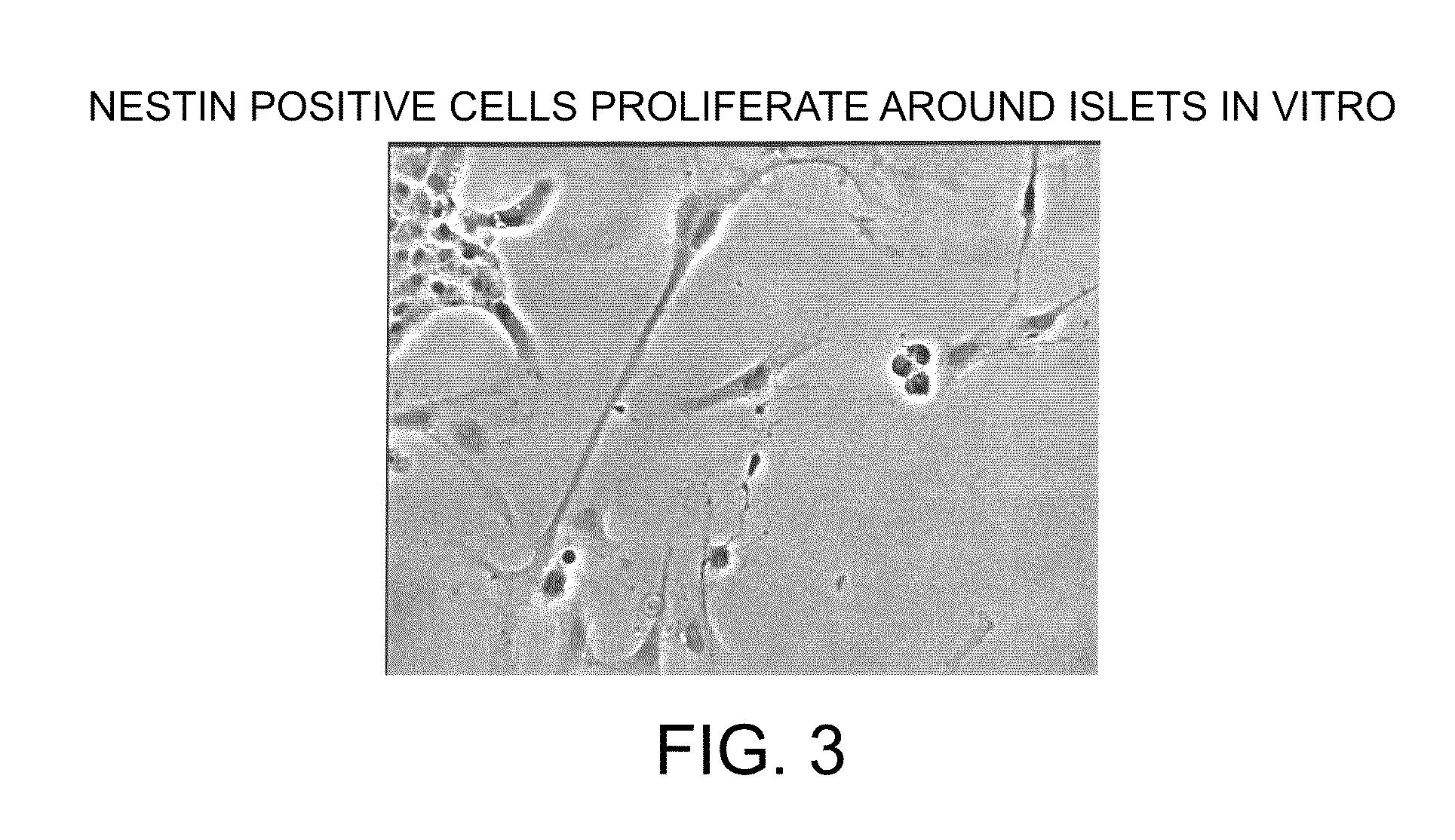
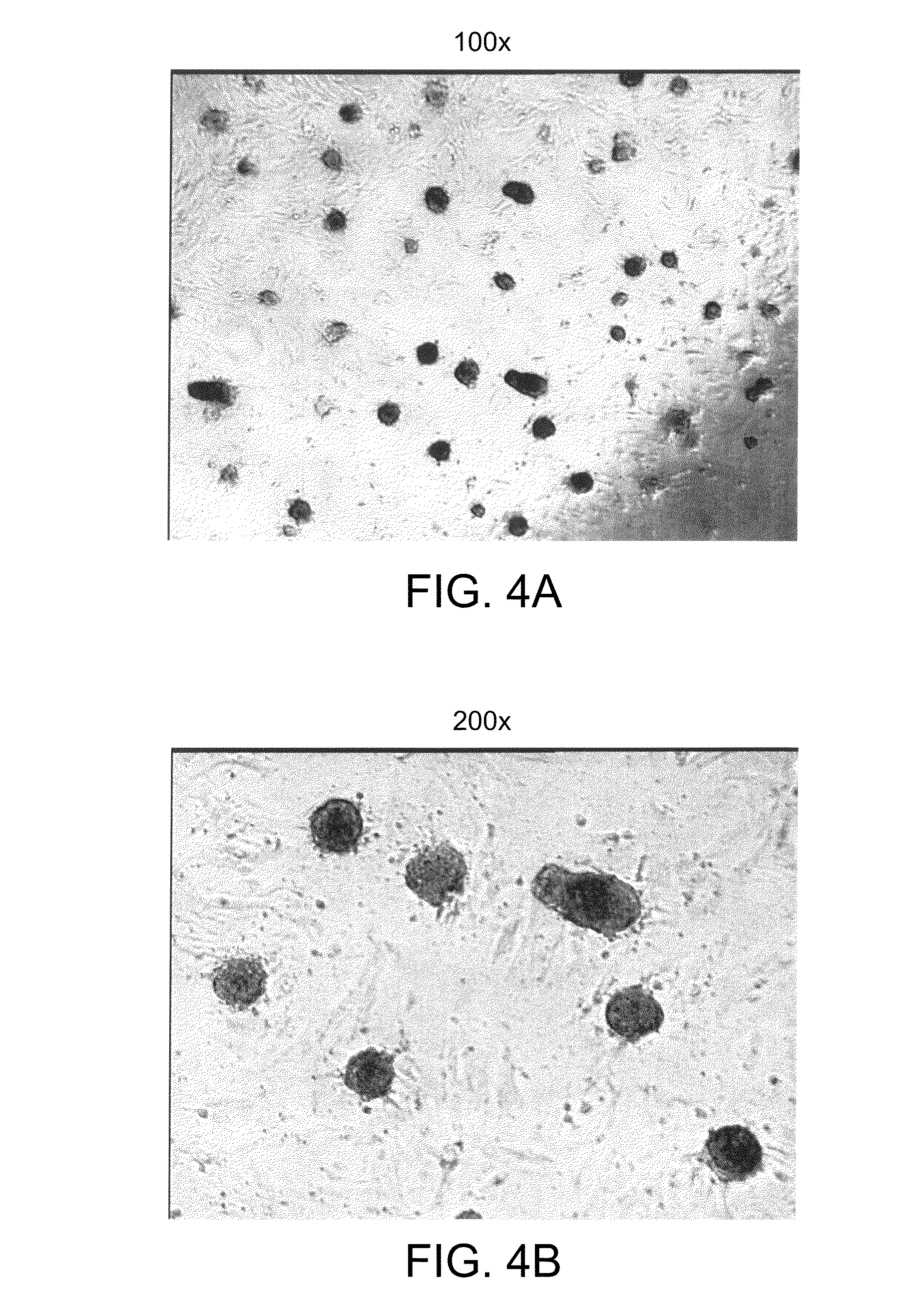
[0215]Pancreatic islets from rats were first cultured in RPMI medium containing 10% fetal bovine serum using concanavalin-A coated 12-well plates. The islets were maintained in culture for three days in the absence of added growth factors other than those supplied by fetal bovine serum. After this period, during which the islets did not attach, the islets were transferred to fresh plates without concanavalin A. The stem cells were then stimulated to proliferate out from the islets as a monolayer by exposing them to bFGF-2 (20 ng / ml) and EGF (20 ng / ml) for 24 days. After the 24 day period, the monolayer was confluent. Among them was a population of cells surrounding the islets. Cells from that population were picked and subcloned into new 12-well plates and again cultured in the medium containing bFGF and EGF. The subcloned cells proliferated rapidly into a monolayer in a clonal fashion, expanding from the center to the periphery....

example 3

Isolation and Culture of Human or Rat Pancreatic Islets

[0216]Human pancreatic islets were isolated and cultured. Human islet tissue was obtained from the islet distribution program of the Cell Transplant Center, Diabetes Research Institute, University of Miami School of Medicine and the Juvenile Diabetes Foundation Center for Islet Transplantation, Harvard Medical School, Boston, Mass. Thoroughly washed islets were handpicked, suspended in modified RPMI 1640 media (11.1 mM glucose) supplemented with 10% fetal bovine serum, 10 mM HEPES buffer, 1 mM sodium pyruvate, 100 U per mL penicillin G sodium, 100 μg per mL streptomycin sulfate, 0.25 ng per mL amphotericin B, and 71.5 μM β-mercaptoethanol, and added to Falcon 3043 12-well tissue culture plates that had been coated with Concanavalin A (ConA). The islet preparation was incubated for 96 hrs at 37° C. with 95% air and 5% CO2. In these conditions, many islets remained in suspension (floated), whereas fibroblasts and other non-islet c...


PUM

PropertyMeasurementUnit
diameteraaaaaaaaaa
diameteraaaaaaaaaa
cell doubling timeaaaaaaaaaa

Abstract

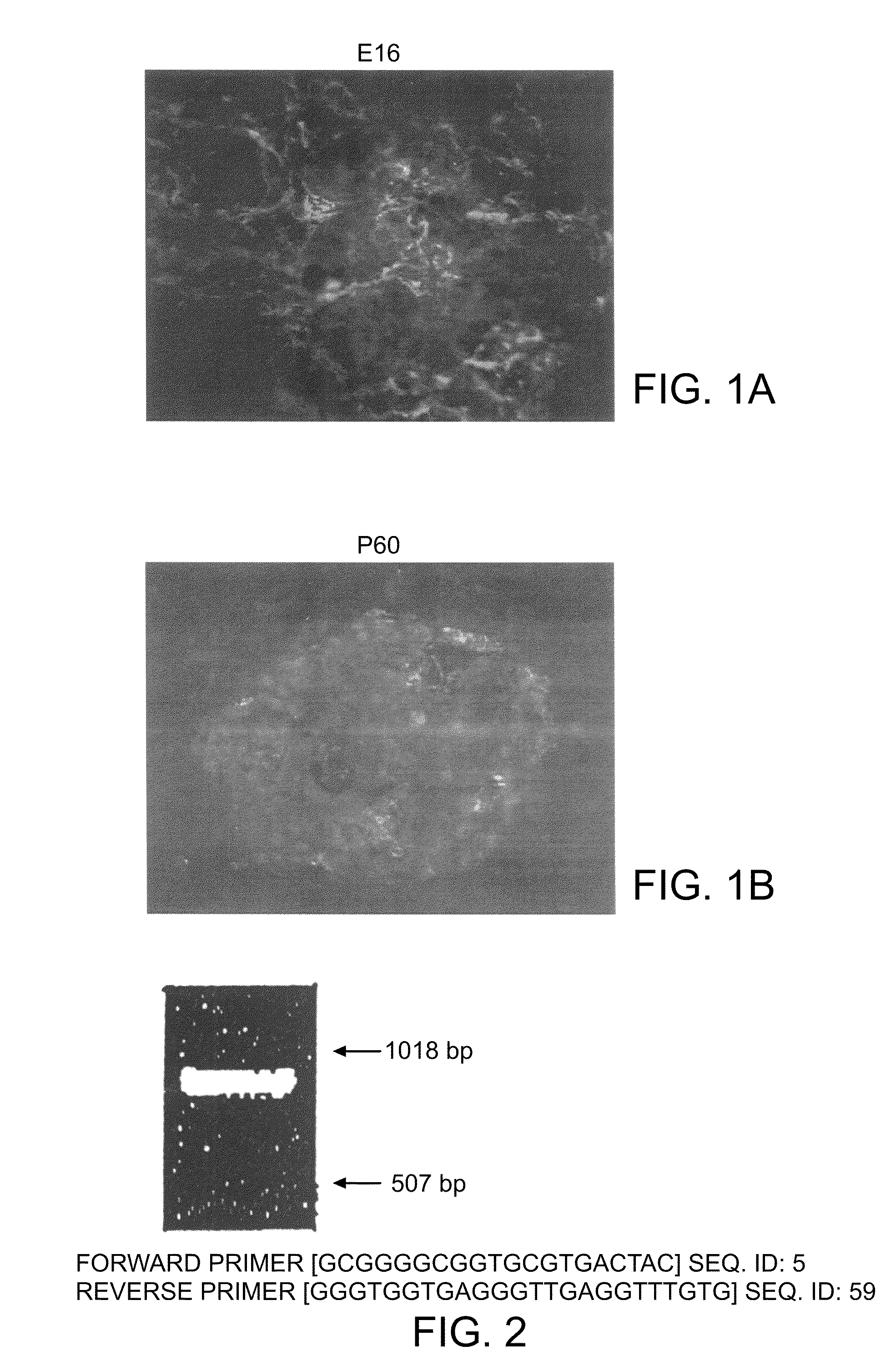
Methods and compositions are described for the treatment of type I insulin-dependent diabetes mellitus and other conditions using newly identified stem cells that are capable of differentiation into a variety of pancreatic islet cells, including insulin-producing beta cells, as well as hepatocytes. Nestin and ABCG2 have been identified as molecular markers for pancreatic stem cells, while cytokeratin-19 serves as a marker for a distinct class of islet ductal cells. Methods are described whereby nestin and / or ABCG2-positive stem cells can be isolated from pancreatic islets and cultured to obtain further stem cells or pseudo-islet like structures. Methods for ex vivo differentiation of the pancreatic stem cells are disclosed. Methods are described whereby pancreatic stem cells can be isolated, expanded, and transplanted into a patient in need thereof, either allogeneically, isogeneically or xenogenically, to provide replacement for lost or damaged insulin-secreting cells or other cells.

Description

PRIORITY[0001]The present application is a continuation of U.S. application Ser. No. 10 / 120,687, filed Apr. 11, 2002 which is a continuation-in-part application of U.S. application Ser. No. 09 / 731,261, filed Dec. 6, 2000, and is also a continuation-in-part application of U.S. application Ser. No. 09 / 963,875, filed Sep. 26, 2001. The present application also claims priority to U.S. Application Ser. No. 60 / 169,082, filed Dec. 6, 1999, U.S. Application Ser. No. 60 / 215,109, filed Jun. 28, 2000, and U.S. Application Ser. No. 60 / 238,880, filed Oct. 6, 2000.SEQUENCE LISTING[0002]The instant application contains a Sequence Listing which has been submitted via EFS-Web and is hereby incorporated by reference in its entirety. Said ASCII copy, created on Aug. 4, 2010, is named 1235C203.txt and is 52,157 bytes in size.[0003]The invention was made at least in part using U.S. government funds, grants DK30457, DK30834, DK55365, DK60125, awarded by the National Institutes of Health, and therefore th...

Claims


Application Information

Patent Timeline
Patent Type & Authority Patents(United States)
IPC IPC(8): C12N5/071A01N65/00A01N63/00A01K67/027C12N15/09A61KA61K31/436A61K31/7088A61K35/12A61K35/39A61K38/00A61K38/22A61K45/00A61K48/00A61P3/10C12NC12N5/02C12N5/074C12Q1/00G01N33/53
CPCA01K67/0271C12N5/0678A61K35/12A61K2035/122C12N2500/34C12N2501/11C12N2501/115C12N2501/335C12N2510/02A61P3/10
Inventor HABENER, JOEL F.ZULEWSKI, HENRYKTHOMAS, MELISSA K.ABRAHAM, ELIZABETH J.VALLEJO, MARIOLEECH, COLIN A.NOLAN, ANNA LOUISELECHNER, ANDREAS
Owner THE GENERAL HOSPITAL CORP
Learn More