Method and system for determining the contribution of hemoglobin and myoglobin to in vivo optical spectra

a technology of hemoglobin and myoglobin, applied in the field of optical spectra, can solve the problems of difficult direct accounting of scattering coefficients, unknown optical path lengths, and inability to quantify chromophores in biological tissues, and achieve the effect of removing baseline offsets or other artifacts

Inactive Publication Date: 2012-02-28
UNIV OF WASHINGTON
View PDF15 Cites 7 Cited by
  • Summary
  • Abstract
  • Description
  • Claims
  • Application Information

AI Technical Summary

Problems solved by technology

Variation in the optical scattering properties of tissue results in an unknown optical path length and makes quantifying chromophores in biological tissues difficult.
Methods for accounting for the scattering coefficient directly, such as time-resolved or phase modulated spectroscopy, or indirectly, such as the use of the spectral absorption of water as an internal standard, are problematic in that they assume a constant scattering coefficient and rely on changes in the total heme chromophore signal (Hb+Mb) to determine changes in muscle perfusion, but provide little information on the resting conditions.
The inability to distinguish the Hb and Mb signals means that the contributions of the intracellular (Mb) and vascular (Hb) compartments cannot be separated using traditional methods of analysis of optical spectra.
Interpretation of these optical spectroscopy data is limited by the unknown contributions of Hb and Mb to the optical spectra.
Given these differences, the ability to quantitatively interpret optical data from intact tissue is severely limited by the lack of knowledge of the contributions of each to the signal.
Therefore, earlier methods are limited to use in animal models where the tissue could be frozen following the in vivo experiments and Hb and Mb concentrations could be measured from muscle homogenates.

Method used


Image

  • Method and system for determining the contribution of hemoglobin and myoglobin to in vivo optical spectra
  • Method and system for determining the contribution of hemoglobin and myoglobin to in vivo optical spectra
  • Method and system for determining the contribution of hemoglobin and myoglobin to in vivo optical spectra

Examples


Embodiment Construction

[0013]The following detailed description includes references to the accompanying drawings, which form a part of the detailed description. The drawings show, by way of illustration, specific embodiments in which the present subject matter may be practiced. These embodiments, which are also referred to herein as “examples,” are described in enough detail to enable those skilled in the art to practice the present subject matter. The embodiments may be combined, other embodiments may be utilized, or structural, logical and electrical changes may be made without departing from the scope of the present subject matter. The following detailed description is, therefore, not to be taken in a limiting sense, and the scope of the present subject matter is defined by the appended claims and their equivalents.

[0014]In this document, the terms “a” or “an” are used, as is common in patent documents, to include one or more than one. In this document, the term “or” is used to refer to a nonexclusive ...


PUM


Abstract

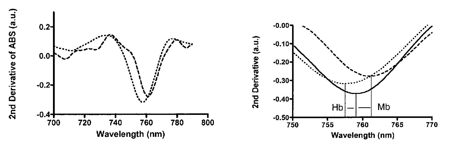
This document discusses, among other things, quantification of hemoglobin content, and therefore blood volume, of muscle. An analysis of the optical spectra can determine the ratio of hemoglobin (Hb) to myoglobin (Mb) content in intact muscle. The peak position of the in vivo optical spectra from intact tissue is used to determine the ratio of Hb to Mb contributing to the optical signal. The wavelength of the peak is a linear function of the percent contribution of Hb to the optical spectra. Such analysis in combination with known Mb concentrations yields a non-invasive measure of the Hb content for in vivo muscle.

Description

STATEMENT OF GOVERNMENT RIGHTS[0001]The invention was made with Government support under Contract or Grant No. AR41928 awarded by the National Institutes of Health. The Government has certain rights in this invention.TECHNICAL FIELD[0002]This document pertains generally to optical spectra, and more particularly, but not by way of limitation, to a method and system for analysis of the relative contributions of hemoglobin (Hb) and myoglobin (Mb) to in vivo optical spectra.BACKGROUND[0003]Optical spectroscopy is emerging as an important modality for assessment of the biologic state of living tissues. Determination of the concentration of hemoglobin in biologic tissue has importance in determining blood flow, and in determination of the oxygen availability to cells within tissues, among other things. Variation in the optical scattering properties of tissue results in an unknown optical path length and makes quantifying chromophores in biological tissues difficult. Methods for accounting...

Claims


Application Information

Patent Timeline
Patent Type & Authority Patents(United States)
IPC IPC(8): A61B5/1455
CPCA61B5/0059A61B5/1455G01N21/31A61B5/4519
Inventor MARCINEK, DAVID J.CONLEY, KEVINSCHENKMAN, KENNETH A.
Owner UNIV OF WASHINGTON
Why Eureka
Try Eureka