Method and apparatus for magnetic resonance imaging on the basis of a gradient echo sequence

a magnetic resonance imaging and gradient echo technology, applied in the field of magnetic resonance tomography, can solve the problems of significant time saving, image contrast and signal-noise ratio (snr) of the mrt image remain limiting factors, and the description of contrast limits is tolerated. , to achieve the effect of good contras

Active Publication Date: 2012-04-24
SIEMENS HEALTHCARE GMBH
View PDF12 Cites 15 Cited by
  • Summary
  • Abstract
  • Description
  • Claims
  • Application Information

AI Technical Summary

Benefits of technology

[0016]An object of the present invention is to provide a method and an apparatus for MRT imaging in which a good contrast is ensured both between grey and white brain matters and between grey brain matter and CSF.

Problems solved by technology

In spite of the technical progress in the design of MRT apparatuses, image contrast and signal-noise ratio (SNR) of the MRT image remain limiting factors for many applications of MRT in medical diagnostics.
Thus it is not necessary to wait as long for the relaxation of the magnetization, which leads to significant time savings.
Moreover, the consequence is that an error in the description of the contrast limits is tolerated.
This is a problem particularly at high field strengths, since the B1 homogeneity is generally poorer and makes it difficult to achieve completely automatic segmentation, in addition to causing dielectric resonance effects.

Method used


Image

  • Method and apparatus for magnetic resonance imaging on the basis of a gradient echo sequence
  • Method and apparatus for magnetic resonance imaging on the basis of a gradient echo sequence
  • Method and apparatus for magnetic resonance imaging on the basis of a gradient echo sequence

Examples


Embodiment Construction

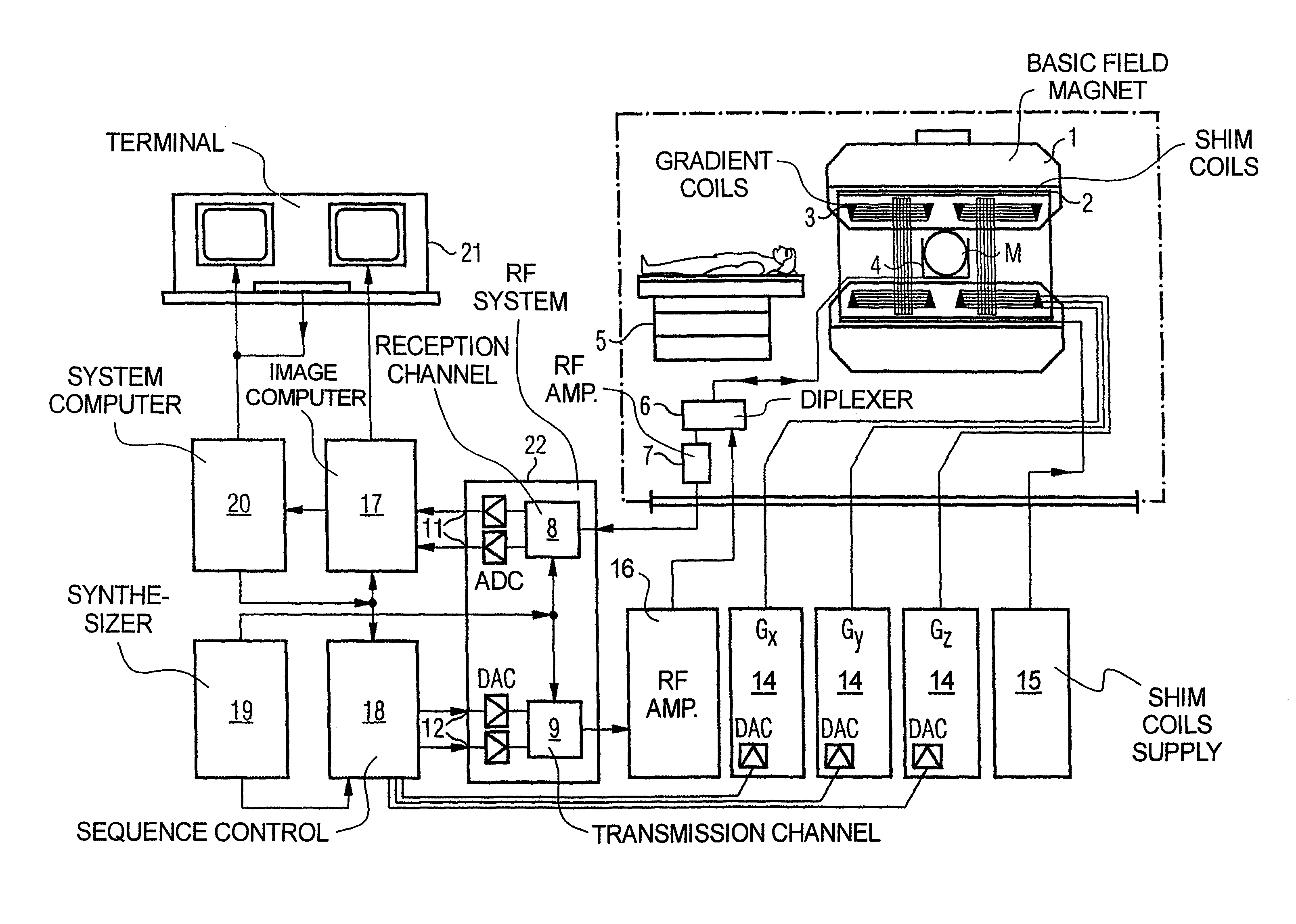
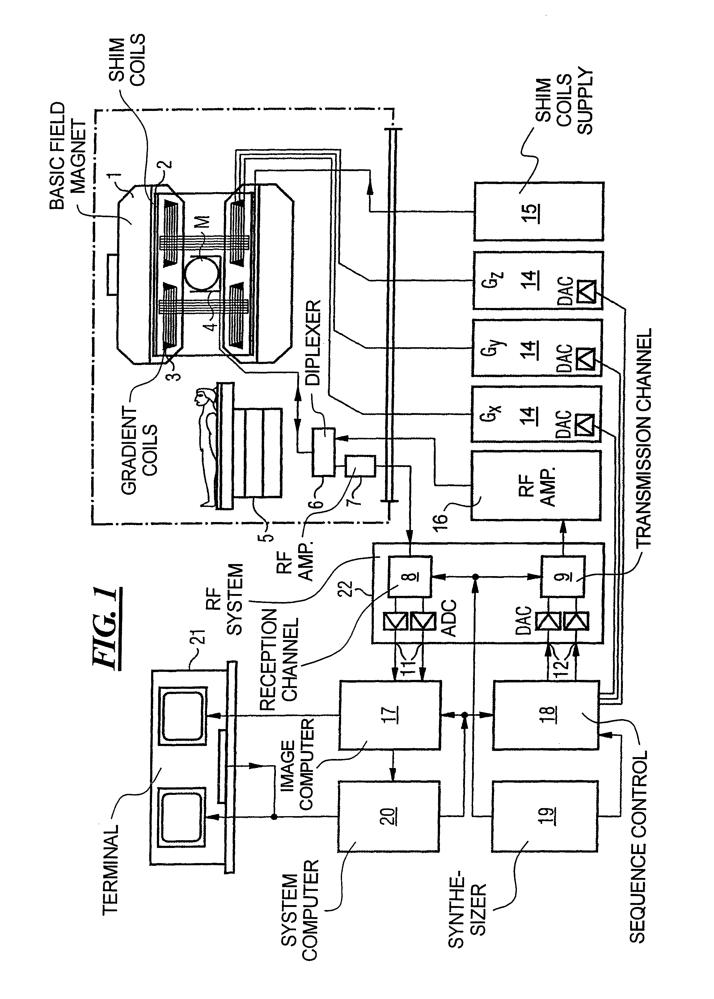
[0043]FIG. 1 is a schematic representation of a magnetic resonance imaging (magnetic resonance tomography) apparatus for generation of a magnetic resonance image of a subject according to the present invention. The design of the magnetic resonance tomography apparatus corresponds to the design of a conventional tomography apparatus, with the differences noted below. A basic field magnet 1 generates a temporally constant strong magnetic field for polarization or alignment of the nuclear spins in the examination region of the subject (such as, for example, a part of the human body to be examined). The high homogeneity of the basic magnetic field that is required for the magnetic resonance measurement is defined in a spherical measurement volume M into which the parts of the human body to be examined are introduced. Shim plates made from ferromagnetic material are mounted at suitable points to support the homogeneity requirements and in particular to eliminate temporally-invariable inf...


PUM


Abstract

In a method and apparatus for magnetic resonance imaging on the basis of a gradient echo sequence by excitation of nuclear spins and measurement of radio-frequency signals arising from the excited nuclear spins:(a) the magnetization of the spins is prepared by an inversion pulse;(b) a number of steps for spin excitation are implemented as well as acquisition of an RF response signal for a first image contrast, with the measurement data being acquired along a first two-dimensional slice, and this first two-dimensional slice being parallel to a plane spanned by two coordinate axes x, y standing orthogonal to one another;(c) implementation of a number of steps for spin excitation as well as acquisition of an RF response signal for a second image contrast, with the measurement data being acquired along the first two-dimensional slice that exist in (b); and(d) repetition of steps (a) through (c) for further two-dimensional slices that are offset parallel to the first two-dimensional slice along a third coordinate axis z that is orthogonal to the first two coordinate axes x and y.

Description

BACKGROUND OF THE INVENTION[0001]1. Field of the Invention[0002]The present invention generally concerns magnetic resonance tomography (MRT) as used in medicine for examination of patients. The present invention more particularly concerns a magnetic resonance tomography apparatus as well as a method for operation of such a magnetic resonance tomography apparatus, with which a high contrast can be achieved for imaging on the basis of a gradient echo sequence.[0003]2. Description of the Prior Art[0004]MRT is based on the physical phenomenon of nuclear magnetic resonance and has been successfully used for over 15 years as an imaging modality in medicine and in biophysics. In this examination modality the subject is exposed to a strong, constant magnetic field. The nuclear spins in the atoms in the subject, which were previously randomly oriented, are aligned.[0005]Radio-frequency energy can now excite these “ordered” nuclear spins to a specific oscillation. This oscillation generates t...

Claims


Application Information

Patent Timeline
Patent Type & Authority Patents(United States)
IPC IPC(8): A61B5/055
CPCG01R33/5613G01R33/5602
Inventor KRUEGER, GUNNAR
Owner SIEMENS HEALTHCARE GMBH
Why Eureka
Try Eureka