

Functionalized magnetic nanoparticles and methods of use thereof
a technology of magnetic nanoparticles and magnetic nanoparticles, applied in the field of functionalized magnetic nanoparticles, can solve the problems of patient discomfort, multiple simultaneous sources, and inability to meet the needs of patients,
- Summary
- Abstract
- Description
- Claims
- Application Information
AI Technical Summary
Benefits of technology
Problems solved by technology
Method used
Image

Examples
example 1
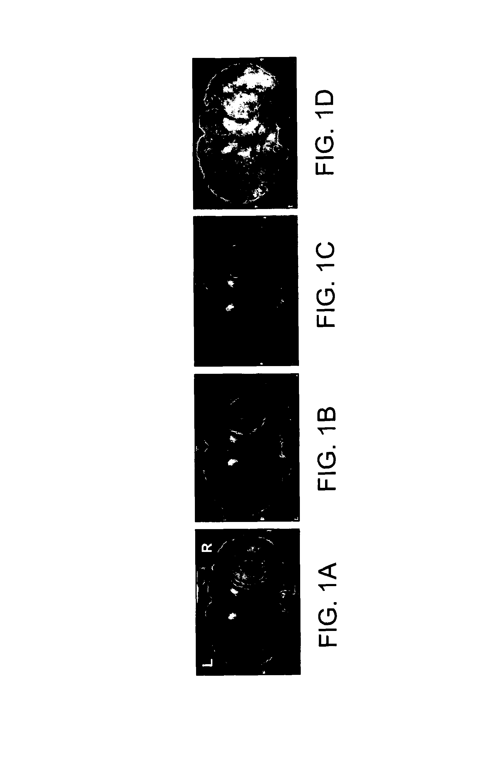
Imaging 2DG MNPs in an Animal Model of Intracranial Tumors
[0202]2DG-functionalized MNPs were applied to an animal model of intracranial tumors. MRI scans were acquired. Uptake of 2DG-functionalized MNPs by intracranial tumors was shown; the 2DG-functionalized MNPs were able to clearly delineate tumor tissues from the surrounding normal (non-cancerous) brain tissues.
Methods
[0203]Tumor studies—Nude mice were injected intracranially with glioblastoma cell line U87Rluc (U87 glioblastoma cell line (ATCC HTB14) genetically modified to express luciferase). Dextran-coated magnetic nanoparticles were functionalized with 2DG. The 2DG moiety was attached to the dextran via the 6-carbon of 2DG. Baseline MR scans were obtained prior to injection with 2DG-MNP. Immediately after baseline scans, the mice were injected with 2DG-MNP (7 mg particles / kg body weight; 1.7 mg Fe / kg body weight) through the tail vein. Scans were obtained at 2 hours, 6 hours, and 24 hours post-MNP injection.
Results
[0204]The...
example 2
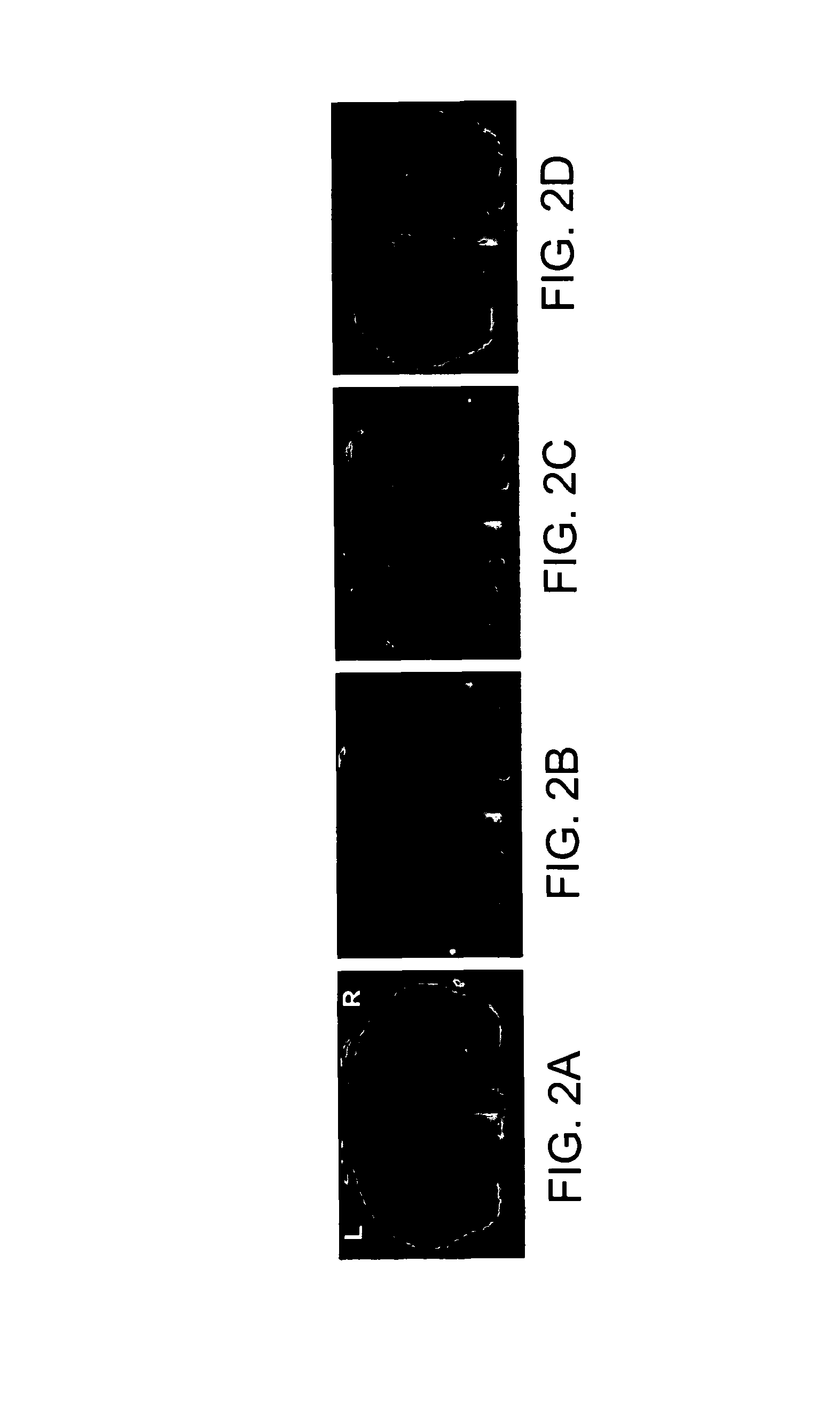
Imaging 2DG MNPs in an Animal Model of Epilepsy
[0207]Dextran-coated magnetic nanoparticles were functionalized with 2DG. The 2DG moiety was attached to the dextran via the 6-carbon of 2DG. 2DG-functionalized MNPs were applied to animal models of generalized epilepsy. MRI scans were acquired. It was shown that the brain distribution of 2DG-MNP in acute generalized epilepsy resembles that obtained by 2DG autoradiography.
Methods
[0208]Epilepsy studies—Baseline MRI scans were obtained from a healthy (naïve) Lewis rat. After the baseline scans, the rat was injected with pentanyl tetrazole (PTZ, 60 mg / kg, subcutaneous) to induce acute generalized seizures (Racine stage 2). The rat was then injected with 2DG-MNP (10 mg particles / kg body weight, intravenous, tail vein). Second scan was obtained 2 hours post MNP injection. After this scan, the rat was injected with an additional dose of PTZ (60 mg / kg, subcutaneous) to induce Racine Stage 4-5 seizures.
Results
[0209]The results are shown in FIGS...
example 3
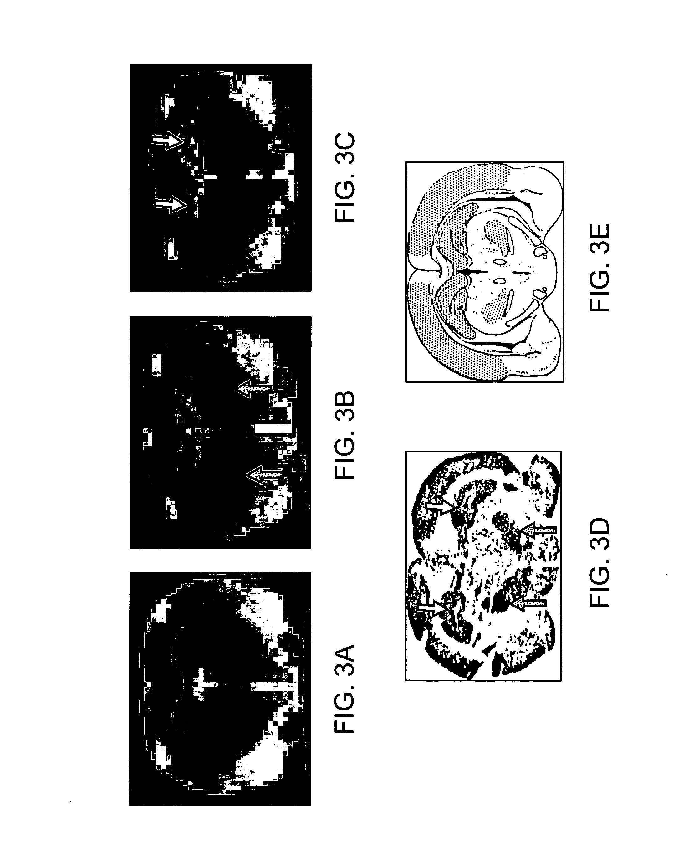
Studies with Pilocarpine-Induced Seizures
[0210]A naïve Lewis rat was injected with intraperitoneally (IP) pilocarpine (PILO; 30 mg / kg). The rat was injected with 2DG-MNP (15 mg / kg, i.v. tail) after stage 4 seizures developed, and anesthetized with pentobarbital 10 minutes thereafter. MRI images were acquired. The data are shown in FIGS. 4A-F.
[0211]FIGS. 4A-F. Panel a) shows the MR images of a naïve Lewis rat. Panels b), c), d), and e) show pattern of 2DG-MNP uptake, 1, 2, 2.5, and 3 hours, respectively after particle injection. The signal intensity in subiculum, dentate gyrus, and retrosplenial cortex, white ellipse, was measured and showed increased (negative) signal enhancement of about 22% in this period. Negative signal enhancement in the peri-thalamic ventricles also indicates the particles have crossed the BBB. Panel f) shows the approximate location of the slice on Paxinos atlas. Dashed lines outline some of the brain areas. Abbreviations are: nc—neocortex; rsc—retrosplenial ...
PUM
| Property | Measurement | Unit |
|---|---|---|
| diameter | aaaaa | aaaaa |
| diameter | aaaaa | aaaaa |
| diameter | aaaaa | aaaaa |
Abstract
Description
Claims
Application Information