Digital pathology system

a digital pathology and image technology, applied in the field of information system and image processing techniques, can solve the problems of manual handling of packages and their contents, inconvenient and time-consuming, physical handling and distribution of case packages, and the risk of mixing the contents of two or more case packages, so as to facilitate the automatic handling of digital pathology images, speed and efficiency of such handling, and speed and efficiency with microscopes

Active Publication Date: 2013-06-11
FUJIFILM HEALTHCARE AMERICAS CORPORATION
View PDF40 Cites 23 Cited by
  • Summary
  • Abstract
  • Description
  • Claims
  • Application Information

AI Technical Summary

Benefits of technology

[0011]According to an aspect of the present disclosure, the automated handling of digital pathology images, and also the speed and efficiency of such handling, are facilitated in part by addressing technical and also human issues associated with handling case package information. Pathologists are proficient with operating microscopes and have experience in dealing with case packages as discrete workload units. What is needed is a digital pathology system and / or process that provides an image viewing and case management experience that substantially resembles or even emulates the slide navigation tools and features that are familiar to pathologists who have experience using microscopes and traditional case package handling techniques. For example, pathologists' workflow organization comes from sorting and categorizing case packages, and their speed and efficiency with microscopes is due in part from an acquired ability to navigate slides without taking their eyes off the microscope image. An object of the present disclosure, therefore, is to provide digital pathology systems and / or processes that use some of the same skills, facilitating smooth transition from physical case package piles and queues to the benefits of digital pathology systems, digital images and networked data, and in all respects so serve the overall efficiency and effectiveness of pathologists at work.
[0019]The disclosed system and associated methods provide the capability to view multiple cases and / or slides simultaneously. Further, multiple images can be processed from a single slide.
[0020]One object of the digital pathology system and associated methods is to improve the overall efficiency, accuracy and effectiveness of pathologists. Another object is to overcome the physical constraints and comparative clumsiness of manual prior art pathology systems and methods that rely on handling paper files, glass slides and racks for holding the slides, while retaining benefits derived from user familiarity with these physical items.
[0021]Another object of the disclosed system and methods is to facilitate a smooth transition for pathologists and their organizations and workplaces, from physical microscope systems to digital pathology systems.
[0022]As additional benefits, the disclosed digital pathology system and methods facilitate and improve reporting processes and data handling. The system and methods provide mechanisms for easy data exchange and retrieval. The system is readily scalable as well as capable of integration into patient medical information databases, institutional or organizational data processing systems.

Problems solved by technology

Although case packages are discrete physical items that can be treated as units of workload, there are drawbacks associated with the need to handle the packages and their contents manually.
For example, the physical handling and distribution of case packages may be inefficient and error prone.
The physical nature of the slides and paperwork may result, for example, in breakage or misplacement of slides, “lost” case packages or package contents, a risk of mixing the contents of two or more case packages, inability to quickly find or assess the status of cases and so on.
The process is characterized by many of the problems associated with manually kept files, with the additional complication of having associated slides.
Manual tracking and reporting mechanisms on paper are inefficient to manage, store and monitor.
It is difficult to exchange information between interested parties, difficult to archive and quickly retrieve information, and documentation or slides can become missing or misplaced.
These problems reduce the pathologists' efficiency and may adversely affect patients waiting for diagnoses.
Despite the potential advantages, current digital slide viewer technology has not been widely accepted into day-to-day practice.

Method used


Image

  • Digital pathology system
  • Digital pathology system
  • Digital pathology system

Examples


Embodiment Construction

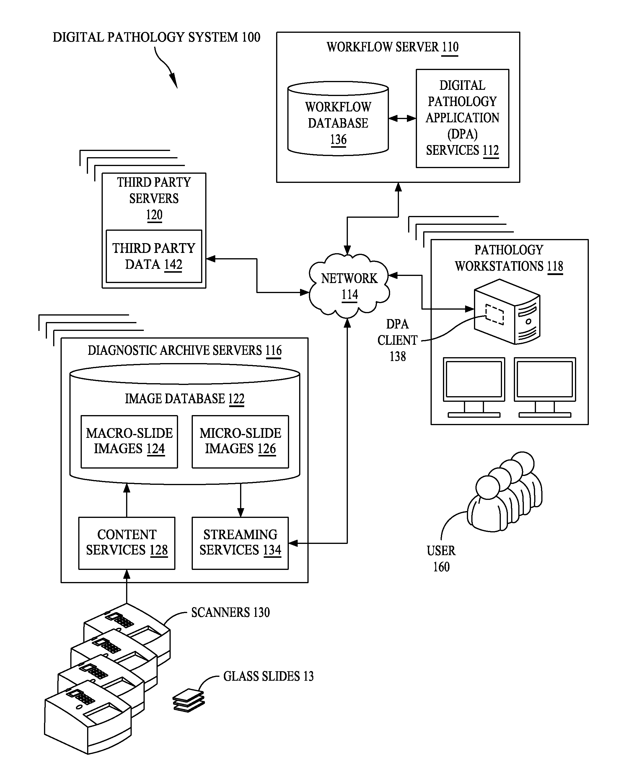
[0057]FIG. 1 is a functional block diagram showing an example of a digital pathology system 100 according to the present disclosure. Among other aspects, digital pathology system 100 is configured for manipulation by and interaction with pathologists, and supports graphic visualization elements that resemble the slide navigation tools and features that are familiar to pathologists who have used microscope systems. Such resemblance includes both appearance and function. At the same time, system 100 benefits from the speed, efficiency and computing power of a digital image and data processing system that reduces the need for pathologists to depend upon physical slides, optical microscopes and case package paperwork files.

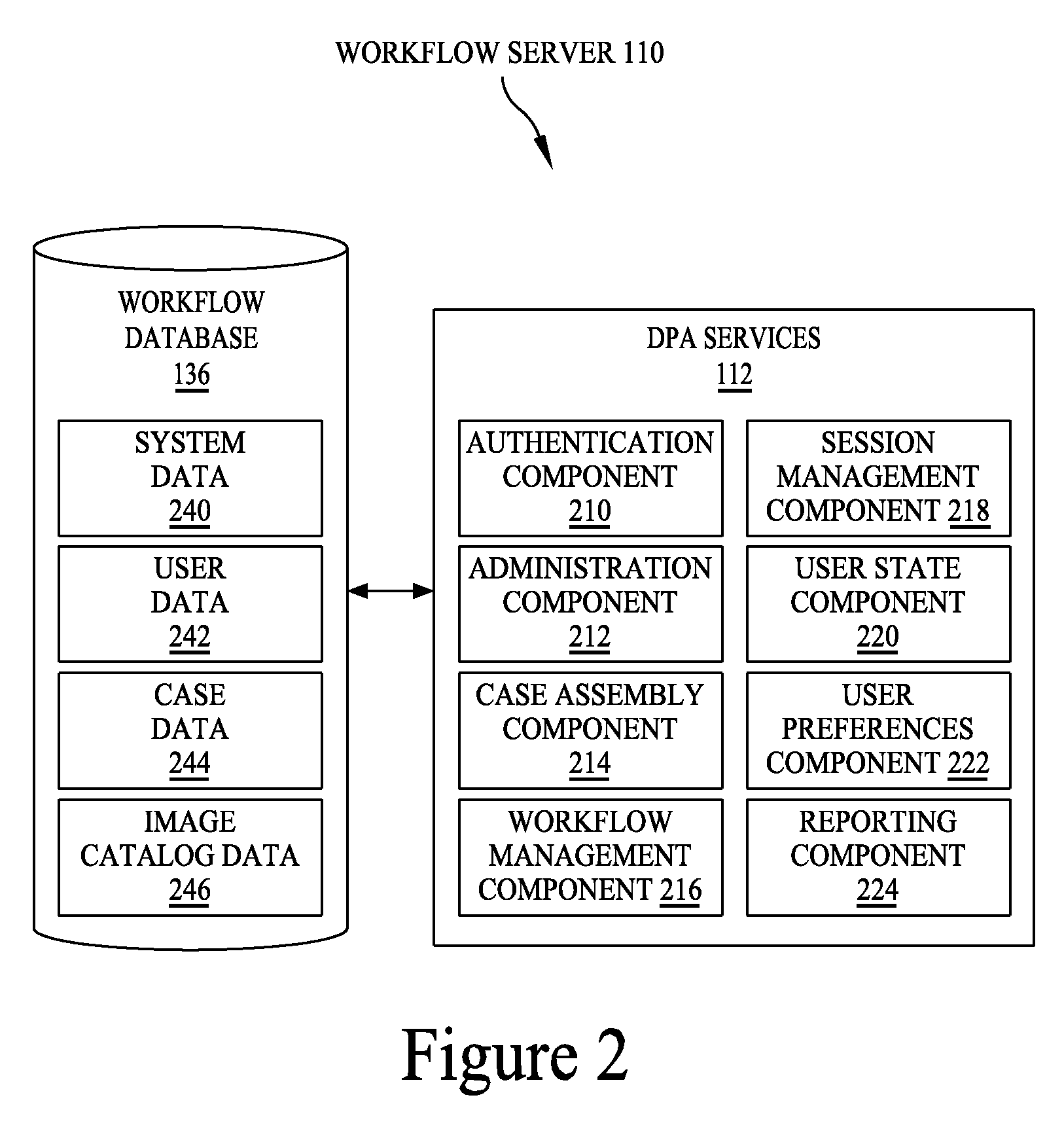
[0058]Digital pathology system 100 may include a workflow server 110, where “workflow” refers primarily to the pathologists' workflow. Workflow server 110 may be a centralized server for hosting digital pathology application (DPA) services 112. Via a network 114, work...


PUM


Abstract

A digital pathology system has a central workflow server hosting digital pathology application services and supporting one or more pathology workstations. The digital pathology system may include one or more image servers, providing digital images of sample specimen slides that are associated with medical cases. Residing at and executing on each pathology workstation is a digital pathology application client, which is the counterpart of digital pathology application services at the central workflow server. The combination of digital pathology application services at the central workflow server and digital pathology application client at each pathology workstation support a pathology workflow software module and a slide viewer software module. The present disclosure also describes a method of operation and / or use of the digital pathology system.

Description

BACKGROUND[0001]1. Field of the Disclosure[0002]This disclosure concerns an information system and techniques for processing images and associated data. In particular, an information system for handling microscopic digital images of pathology and histology samples and the like organizes and manages pathologists' workflow using a graphical user interface and visualization mechanisms that are efficiently computerized but resemble the look and feel of working with glass specimen slides, optical microscopes and patient paperwork files.[0003]2. Related Art[0004]Pathology is the study and diagnosis of disease through examination of organs, tissues, bodily fluids and even whole bodies (autopsies). Typically, tissue samples are taken, prepared and examined under magnification. Pathologists are physicians who diagnose and characterize disease by examining biopsies, bodily fluid, duct scrapings and similar samples. In a typical operation, tissue samples might be prepared by being sliced. The ...

Claims


Application Information

Patent Timeline
Patent Type & Authority Patents(United States)
IPC IPC(8): G06F17/30G16H10/40G16H30/20
CPCG06F3/0481G06F3/04845G06F19/321G06F19/3487G06Q10/10G16H15/00G16H10/40G16H30/20G16H30/00
Inventor EHLKE, JOSHUA TODDKHANDAVILLI, NARASIMHA RAOMCVAUGH, TIMOTHY MARKMELANSON, ANTHONY J.SELZER, STEVESTRATMAN, CURTISVIRNIK, MARINAWEIHNACHT, JOSHUA MICAHZENG, ZHEN
Owner FUJIFILM HEALTHCARE AMERICAS CORPORATION
Why Eureka
Try Eureka