
Systems and methods for generating fused medical images from multi-parametric, magnetic resonance image data

a multi-parametric, magnetic resonance imaging and imaging system technology, applied in image enhancement, instruments, reconstruction from projection, etc., can solve the problems of inability to integrate certain parameters of use in forming fusion images, laborious process for clinicians, lack of intuitiveness for clinicians, etc., to achieve flexibility in adapting to a clinician's specific protocol, the clinician's intuition is lacking

Active Publication Date: 2013-06-25
KONINKLJIJKE PHILIPS NV
View PDF4 Cites 29 Cited by
  • Summary
  • Abstract
  • Description
  • Claims
  • Application Information

AI Technical Summary

Benefits of technology

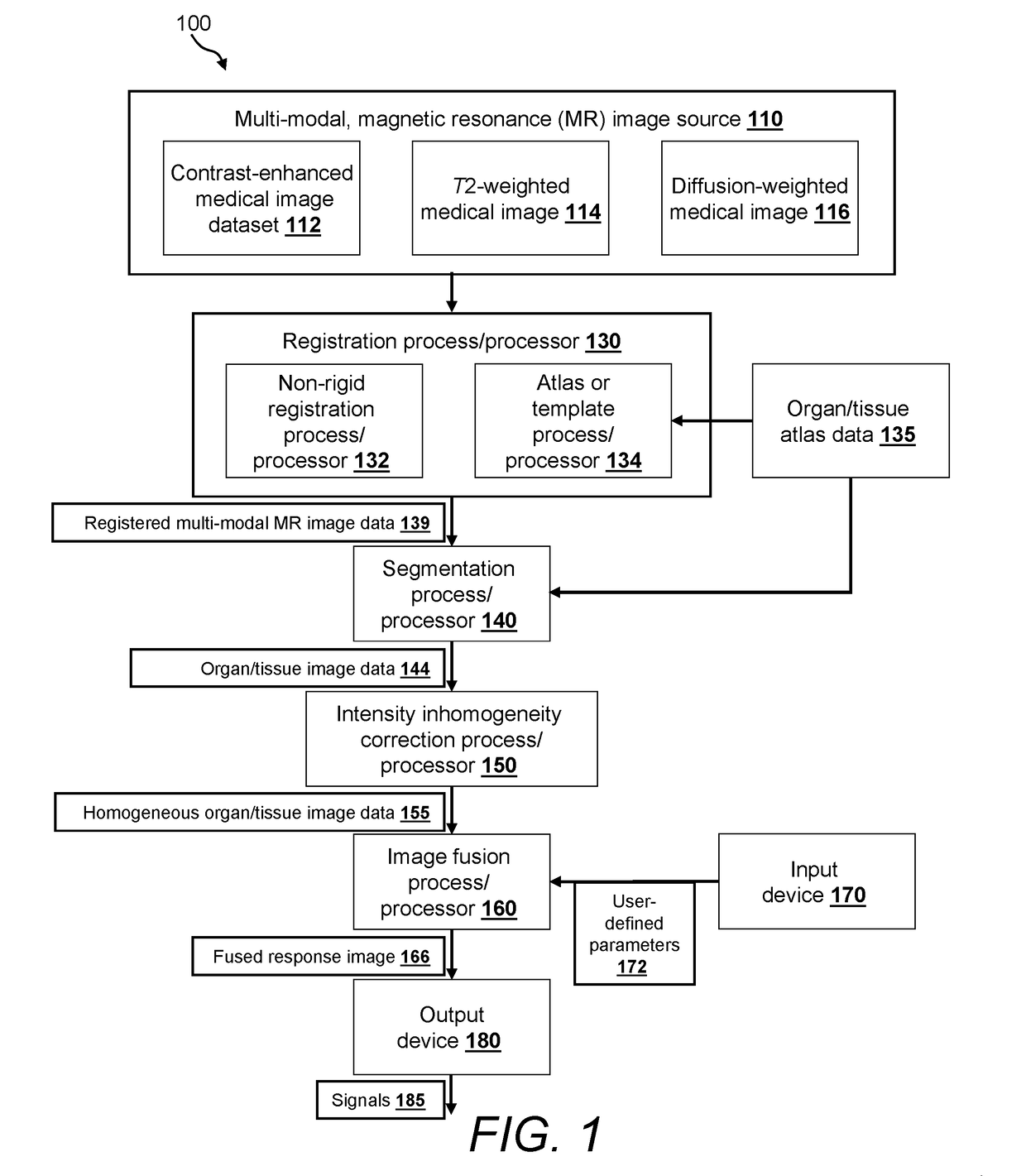
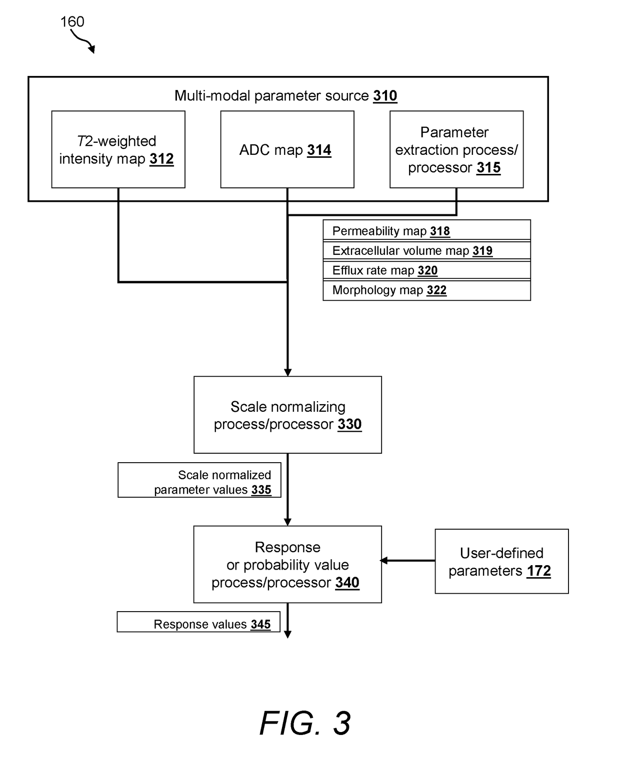
This patent describes a system and method for combining multiple medical images using different parameters to create a fused image that shows the visual enhancements of each image. This allows for the creation of a more useful image for the clinician. The system uses an image fusion processor that fuses the images based on user-defined parameters, such as apparent diffusion coefficient values or at least one other parameter. The fused image can also include color-enhanced or intensity-enhanced regions of interest. The system can also include a registration process to align the images, an intensity homogeneity correction process, and a scale normalizing process. The user can adjust the parameters during runtime to create a more precise image. Overall, this system provides a better way to combine medical images for diagnostic purposes.

Problems solved by technology

Synthesizing the information across images can be a laborious process for the clinician, especially due to the large number of thin slice images provided in each MR image dataset and the amount of spatial information in one dataset that might need to be correlated with information from one or more other datasets.
Disadvantageously, prior art fusion methods such as those described in Viswanath, provide preset solutions (e.g., fusion parameters) that are applied to all datasets regardless of the clinician's individual needs and desires and fail to integrate certain parameters of use in forming fusion images that aid clinician's in differentiating tissue types.
These approaches, thus, lack both intuitiveness for a clinician and flexibility in adapting to a clinician's specific protocol.

Method used


Image

  • Systems and methods for generating fused medical images from multi-parametric, magnetic resonance image data
  • Systems and methods for generating fused medical images from multi-parametric, magnetic resonance image data
  • Systems and methods for generating fused medical images from multi-parametric, magnetic resonance image data

Examples


Embodiment Construction

[0023]In the present disclosure, the terms “pixels” and “voxels” can be used interchangeably to refer to an element in an image. Image data is generally represented in units of picture elements (pixels). A pixel generally refers to the information stored for a single grid in an image or a basic unit of the composition of an image, usually in a two-dimensional space, for example, x-y coordinate system. Pixels can become volumetric pixels or “voxels” in three-dimensional space (x, y, z coordinates) by the addition of at least a third dimension, often specified as a z-coordinate. A voxel thus refers to a unit of volume corresponding to the basic element in an image that corresponds to the unit of volume of the tissue being scanned. It should be appreciated that this disclosure can utilize pixels, voxels and any other unit representations of an image to achieve the desired objectives presented herein. Both pixels and voxels each contain a discrete intensity and / or color, which is typica...


PUM


Abstract

This invention provides a system and method for fusing and synthesizing a plurality of medical images defined by a plurality of imaging parameters that allow the visual enhancements of each image data set to be selectively combined with those of other image datasets. In this manner, a user-defined parameter set can be generated in the final response image dataset. This final response image dataset displays visual data represents a form particularly useful to the clinician. In an illustrative embodiment, the system for fusing and synthesizing the plurality of medical images provides an image fusion process / processor that fuses a plurality of magnetic resonance imaging (MRI) datasets. A first image dataset of the MRI datasets is defined by apparent diffusion coefficient (ADC) values. A second image dataset of the MRI datasets is defined by at least one parameter other than the ADC values. The image fusion processor generates a fused response image that visually displays a combination of image features generated by the ADC values combined with image features generated by the at least one parameter other than the ADC values. The fused response image can illustratively include at least one of color-enhanced regions of interest and intensity-enhanced regions of interest.

Description

FIELD OF THE INVENTION[0001]This application relates generally to the processing of discrete groups of medical image data. More particularly, this application relates to automated techniques for fusing and / or synthesizing medical image data acquired by imaging tissue with multiple scanning parameters, sequences, or protocols.BACKGROUND OF THE INVENTION[0002]Early detection of disease and malignant tissue can lead to a better prognosis. The development of non-invasive methods for detection and characterization of tumors has an extreme importance in current medicine. Magnetic resonance imaging (MRI) is a noninvasive medical test that can help physicians diagnose and treat medical conditions. MR imaging uses a powerful magnetic field, radio frequency pulses and a computer to produce detailed pictures of organs, soft tissues, bone and virtually all other internal body structures. The images can be examined on a computer monitor, printed or copied to compact disc.[0003]By way of useful b...

Claims


Application Information

Patent Timeline
Patent Type & Authority Patents(United States)
IPC IPC(8): G06K9/00G06K9/32
CPCG06K9/6289G06T7/003G06K2209/051G06T2207/30096G06T2207/20076G06T2207/20128G06T2207/10096G06T7/337G06V2201/031G06V10/803G06F18/251G06T7/11A61B6/5235G06T5/50G06T11/003G06T2207/10088G06T2207/20221
Inventor PERIASWAMY, SENTHIL
Owner KONINKLJIJKE PHILIPS NV
Learn More