Method for reconstructing a fluorescent image of the interior of a turbid medium and device for imaging the interior of a turbid medium

a technology for interiors and turbid mediums, applied in the field of reconstructing fluorescence images of devices for imaging the interiors of turbid mediums, can solve the problems of not knowing how light interacts with turbid mediums, the size of the receiving portion may not match the size of the turbid medium perfectly, and the approximation cannot hold, so as to achieve the effect of more accurate determination

Inactive Publication Date: 2013-08-27
KONINK PHILIPS ELECTRONICS NV
View PDF11 Cites 19 Cited by
  • Summary
  • Abstract
  • Description
  • Claims
  • Application Information

AI Technical Summary

Benefits of technology

[0008]It is an object of the present invention to provide a method for reconstructing a fluorescence image of the interior of a turbid medium and a device for imaging the interior of turbid media with which the concentrations of a fluorescent contrast agent at different positions in the turbid medium can be determined more accurately from the reconstructed image. Further, this shall be achieved fast and efficiently without requiring additional hardware.
[0009]This object is attained by a method for reconstructing a fluorescence image of the interior of a turbid medium according to an embodiment. The method comprises the step: accommodating a turbid medium to which a fluorescent contrast agent has been administered in a measurement volume. The fluorescent contrast agent is capable of emitting light in a first range of wavelengths upon irradiation with light. The method further comprises: performing attenuation measurements at a plurality of different wavelengths by subsequently irradiating the turbid medium with light from a plurality of different source positions and detecting light emanating from the turbid medium in a plurality of detection positions for each source position. The method further comprises: reconstructing absorption properties as a function of the position in the interior of the turbid medium for the plurality of different wavelengths from the attenuation measurements; calculating absorption properties as a function of the position in the interior of the turbid medium for wavelengths of the first range of wavelengths; performing a fluorescence measurement by subsequently irradiating the turbid medium with light causing the fluorescent contrast agent to emit light in the first range of wavelengths from the plurality of source positions and detecting the light emanating from the fluorescent contrast agent in the plurality of detection positions for each source position; and reconstructing a fluorescence image of the spatial distribution of the fluorescent contrast agent in the interior of the turbid medium from the fluorescence measurement using the calculated absorption properties. As a result, the interaction of light having a wavelength in the first range of wavelengths with the turbid medium is estimated more correctly and the concentration and spatial distribution of the contrast agent in the turbid medium can be reconstructed with a higher degree of accuracy. Further, the attenuation measurements performed for discrete wavelengths can be used to achieve this higher degree of accuracy such that no additional hardware is required. “Absorption properties as a function of the position” means that absorption properties are determined in dependency on the position in the turbid medium. A functional dependency (in the mathematical sense) does not necessarily have to be established. The calculated absorption properties are used as input information in the step of reconstructing the fluorescence image.
[0011]According to an aspect, the absorption coefficients are calculated by considering the turbid medium as a linear combination of a plurality of substances and determining the local concentrations of the plurality of substances from the attenuation measurements at the plurality of different wavelengths. In this case, the absorption properties with respect to wavelengths in the first range of wavelengths can be calculated with satisfactory accuracy such that the concentration of the fluorescent contrast agent can be visualized more correctly in the reconstructed fluorescence image.
[0012]If the absorption properties for wavelengths of the first range of wavelengths are calculated using a known spectral behavior of the plurality of substances, the absorption properties with respect to wavelengths of the first range can be calculated with satisfactory accuracy. As a result, the reconstructed fluorescence image provides reliable information about the concentrations of the fluorescent contrast agent at the different positions. Thus, a fluorescence image can be provided which allows distinction of malignant lesions from other abnormalities.
[0014]Preferably, the reconstructed fluorescence image is fed back as an input to the step of reconstructing absorption properties as a function of the position in the interior of the turbid medium, and the steps of extrapolating the absorption properties and reconstructing a fluorescence image are iteratively repeated. In this case, the concentration of the fluorescent contrast agent can be determined with improved accuracy even if locations with high concentrations occur in the turbid medium.
[0015]The object is further attained by a device for imaging the interior of a turbid medium according to an embodiment. The device comprises: a measurement volume adapted for accommodating a turbid medium to which a fluorescent contrast agent has been administered which is capable of emitting light in a first range of wavelengths upon irradiation; a light source unit adapted to subsequently irradiate the measurement volume with light from a plurality of different source positions; the light source unit being capable of selectively emitting light of a plurality of different wavelengths; and a detection unit adapted to detect light emanating from the measurement volume in a plurality of different detection positions. The device further comprises a control and reconstruction unit adapted to control the device to: perform attenuation measurements at a plurality of different wavelengths by subsequently irradiating the measurement volume with light from a plurality of different source positions and detect light emanating from the measurement volume in a plurality of detection positions for each source position; perform a fluorescence measurement by subsequently irradiating the measurement volume with light causing the fluorescent contrast agent to emit light in the first range of wavelengths from the plurality of source positions and detecting the light emanating from the fluorescent contrast agent in the plurality of detection positions for each source position; reconstruct absorption properties as a function of the position in the measurement volume for the plurality of different wavelengths from the attenuation measurements; calculate absorption properties as a function of the position in the measurement volume for wavelengths of the first range of wavelengths; and reconstruct a fluorescence image of the spatial distribution of the fluorescent contrast agent in the measurement volume from the fluorescence measurement using the calculated absorption properties. As a result, the interaction of light having a wavelength in the first range of wavelengths with the turbid medium is estimated more correctly and the concentration and spatial distribution of the contrast agent in the turbid medium can be reconstructed with a higher degree of accuracy. Further, the attenuation measurements performed for several discrete wavelengths can be used to achieve this higher degree of accuracy such that no additional hardware is required.If a filter is provided in the device introducible in the light path between the measurement volume and the detection unit for the fluorescence measurement, the reconstruction can be achieved with a limited amount of hardware.

Problems solved by technology

Due to different sizes of the turbid media to be examined, the size of the receiving portion may not perfectly match the size of the turbid medium, i.e. a space remains between the boundary of the receiving volume and the turbid medium.
Thus, for the relevant wavelengths of the fluorescence measurement, there arises the problem that it is not known how the light interacts with the turbid medium.
However, this approximation does not hold in general and the quality of this approximation differs depending on e.g. different patients and the constitution of their tissue.
As a result, current methods for reconstructing fluorescence images of the interior of turbid media, i.e. for reconstructing an image of the spatial distribution of the fluorescent contrast in the turbid medium, comprise the problem that concentrations of the fluorescent contrast agent at different positions in the turbid medium cannot be determined to a satisfactory degree from the reconstructed image.

Method used


Image

  • Method for reconstructing a fluorescent image of the interior of a turbid medium and device for imaging the interior of a turbid medium
  • Method for reconstructing a fluorescent image of the interior of a turbid medium and device for imaging the interior of a turbid medium
  • Method for reconstructing a fluorescent image of the interior of a turbid medium and device for imaging the interior of a turbid medium

Examples


first embodiment

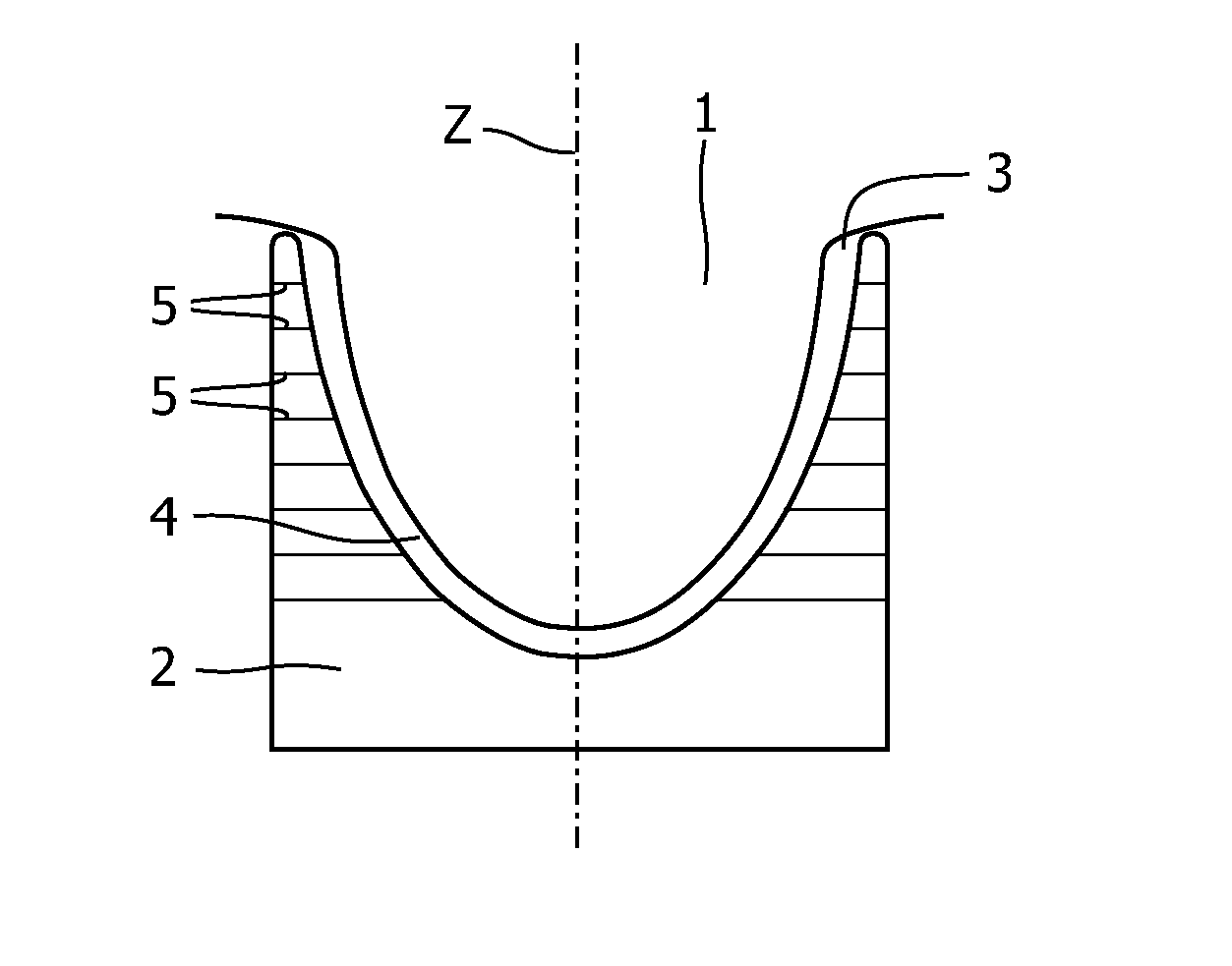
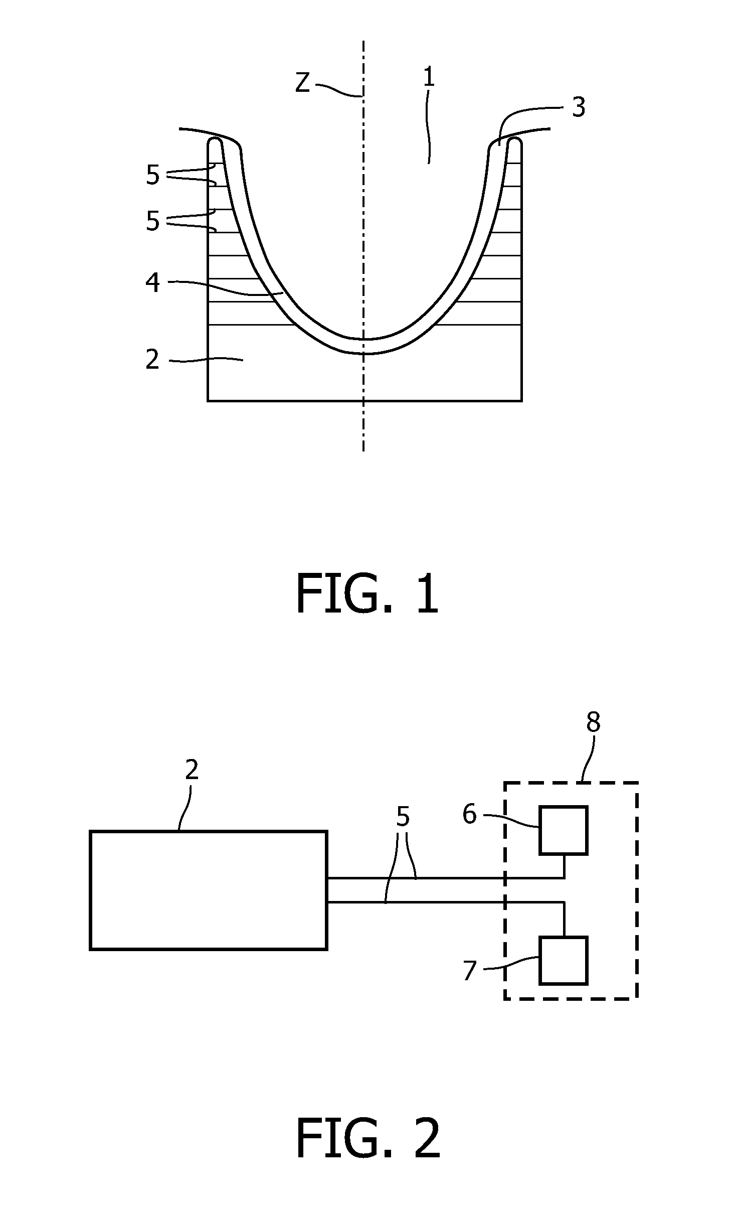
[0020]An embodiment of the present invention will now be described with reference to FIGS. 1 and 2. The device for imaging the interior of a turbid medium according to the embodiment is a device for diffuse optical tomography (DOT). In particular, the device is adapted for examination of female breasts. The overall construction of such a device is known in the art. The device comprises a bed (not shown) on which the person under examination is lying in a prone position. An opening is formed in the bed below which a measurement volume 4 extends. The measurement volume 4 is shown in FIG. 1.

[0021]In the device shown in FIG. 1, the turbid medium 1 to be examined is a female human breast. The measurement volume 4 is bounded by a receiving portion 2 adapted to receive the turbid medium 1, as schematically indicated in FIG. 1. The receiving portion 2 has a cup-like shape and is provided with an opening 3. As can be seen in FIG. 1, the turbid medium 1 to be examined is placed in the measure...

second embodiment

[0036]A second embodiment will be described in the following. The second embodiment substantially corresponds to the first embodiment described above and only differs in that the fluorescent contrast agent is considered as one of the constituents the concentration of which is determined from the attenuation measurements in the second range of wavelengths. Thus, according to the second embodiment, the self-absorption of the fluorescent contrast agent for the fluorescence light is taken into account for. This leads to improved fluorescence images, in particular in case of high concentrations of the fluorescent contrast agent.

[0037]According to the second embodiment, the concentration cf(r) of the fluorescent contrast agent and its absorption coefficient μaf are comprised in the equation

[0038]μa⁡(λ,r)=∑i⁢ci⁡(r)·μai⁡(λ,r),

i.e. the term cf(r) μaf(λ, r) is taken into account in the summation. Thus, the concentration of the fluorescent contrast agent is already (pre-) calculated from the a...


PUM


Abstract

A method for reconstructing a fluorescence image of the interior of a turbid medium is provided. The method comprises the step: accommodating a turbid medium (1) to which a fluorescent contrast agent has been administered in a measurement volume (4). The fluorescent contrast agent is capable of emitting light in a first range of wavelengths upon irradiation with light. The method further comprises: performing attenuation measurements at a plurality of different wavelengths (λi, . . . , λk) by subsequently irradiating the turbid medium (1) with light from a plurality of different source positions and detecting light emanating from the turbid medium (1) in a plurality of detection positions for each source position; reconstructing absorption properties (μa(r, λ) as a function of the position in the interior of the turbid medium (1) for the plurality of different wavelengths from the attenuation measurements; calculating absorption properties as a function of the position in the interior of the turbid medium (1) for wavelengths of the first range of wavelengths; performing a fluorescence measurement by subsequently irradiating the turbid medium (1) with light causing the fluorescent contrast agent to emit light in the first range of wavelengths from the plurality of source positions and detecting the light emanating from the fluorescent contrast agent in the plurality of detection positions for each source position; and reconstructing a fluorescence image of the spatial distribution of the fluorescent contrast agent in the interior of the turbid medium (1) from the fluorescence measurement using the calculated absorption properties.

Description

FIELD OF INVENTION[0001]The present invention relates to a method for reconstructing a fluorescence image of the interior of a turbid medium and to a device for imaging the interior of a turbid medium.BACKGROUND OF THE INVENTION[0002]In the context of the present application, the term turbid medium is to be understood to mean a substance consisting of a material having a high light scattering coefficient, such as for example intralipid solution or biological tissue. Further, light is to be understood to mean electromagnetic radiation, in particular electromagnetic radiation having a wavelength in the range from 180 nm to 1400 nm. The term “optical properties” covers the reduced scattering coefficient μ′s and the absorption coefficient μa. Furthermore, “matching optical properties” is to be understood as having a similar reduced scattering coefficient μ′s and a similar absorption coefficient μa.[0003]A method for imaging the interior of turbid media, e.g. for breast cancer screening,...

Claims


Application Information

Patent Timeline
Patent Type & Authority Patents(United States)
IPC IPC(8): G06K9/00
CPCA61B5/0091A61B5/4312G01N21/4795G01N21/6428
Inventor ZIEGLER, RONNYZIEGLER, ANDYNIELSEN, TIM
Owner KONINK PHILIPS ELECTRONICS NV
Why Eureka
Try Eureka