Method and apparatus for pulmonary ventilation imaging using local volume changes

a pulmonary ventilation and volume change technology, applied in the field of pulmonary ventilation imaging using local volume changes, can solve the problems of not considering differences in pulmonary ventilation and perfusion, and the intrinsic spatial resolution of the detector is the major limit of the spatial resolution of the pet system

Inactive Publication Date: 2013-09-17
UNIV OF SOUTH FLORIDA
View PDF3 Cites 6 Cited by
  • Summary
  • Abstract
  • Description
  • Claims
  • Application Information

AI Technical Summary

Problems solved by technology

However, in current practice of thoracic cancer radiotherapy, the differences in pulmonary ventilation and perfusion are not considered when generating treatment plans.
The intrinsic spatial resolution of the detectors is the major limit of the spatial resolution of a PET system.
Due to the complexity of MRI based ventilation imaging, the clinical application of this technique is not common.
The problem related to the HU change method is that the fluctuation of the HU in a CT image makes the ventilation image noisy and affects the HU-based ventilation images directly.
The other problem with this method is the edge artifact.
The mismatched regions on the low-high density interface, such as the interface between the lung tissues and blood vessels, and between the lung and chest wall, cause artifact of high ventilation spikes.
This would make the spatial resolution of ventilation images coarser, losing the advantage of CT images over nuclear medicine ventilation images.
The partial volume effect in CT images makes the CT voxel intensity higher or lower, which in turn creates artifact in the ventilation images.
Any voxel-to-voxel mismatch at the edge of high-low intensity interface, such as blood vessels and lung tissue interface, chest wall and lung interface, would cause spikes in ventilation images.
Even with averaging technique, these spikes would still introduce false high ventilation volumes.

Method used


Image

  • Method and apparatus for pulmonary ventilation imaging using local volume changes
  • Method and apparatus for pulmonary ventilation imaging using local volume changes
  • Method and apparatus for pulmonary ventilation imaging using local volume changes

Examples


Embodiment Construction

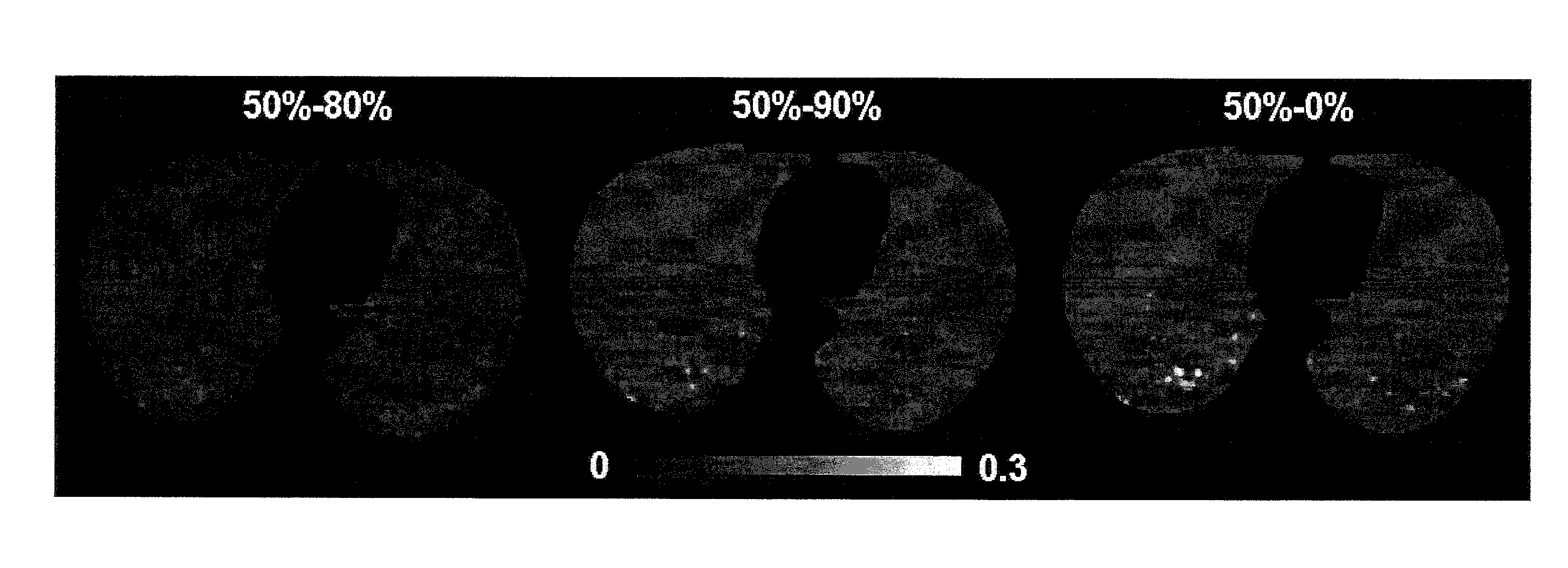
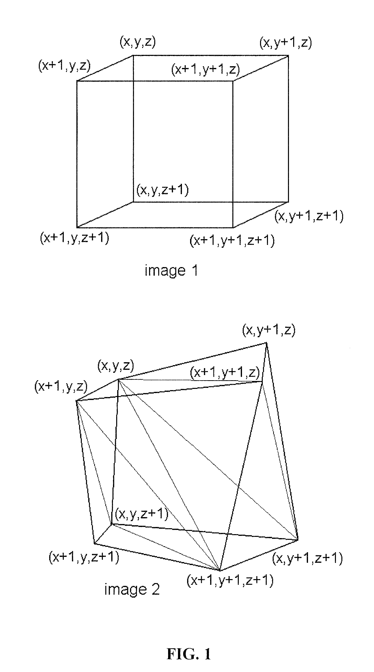
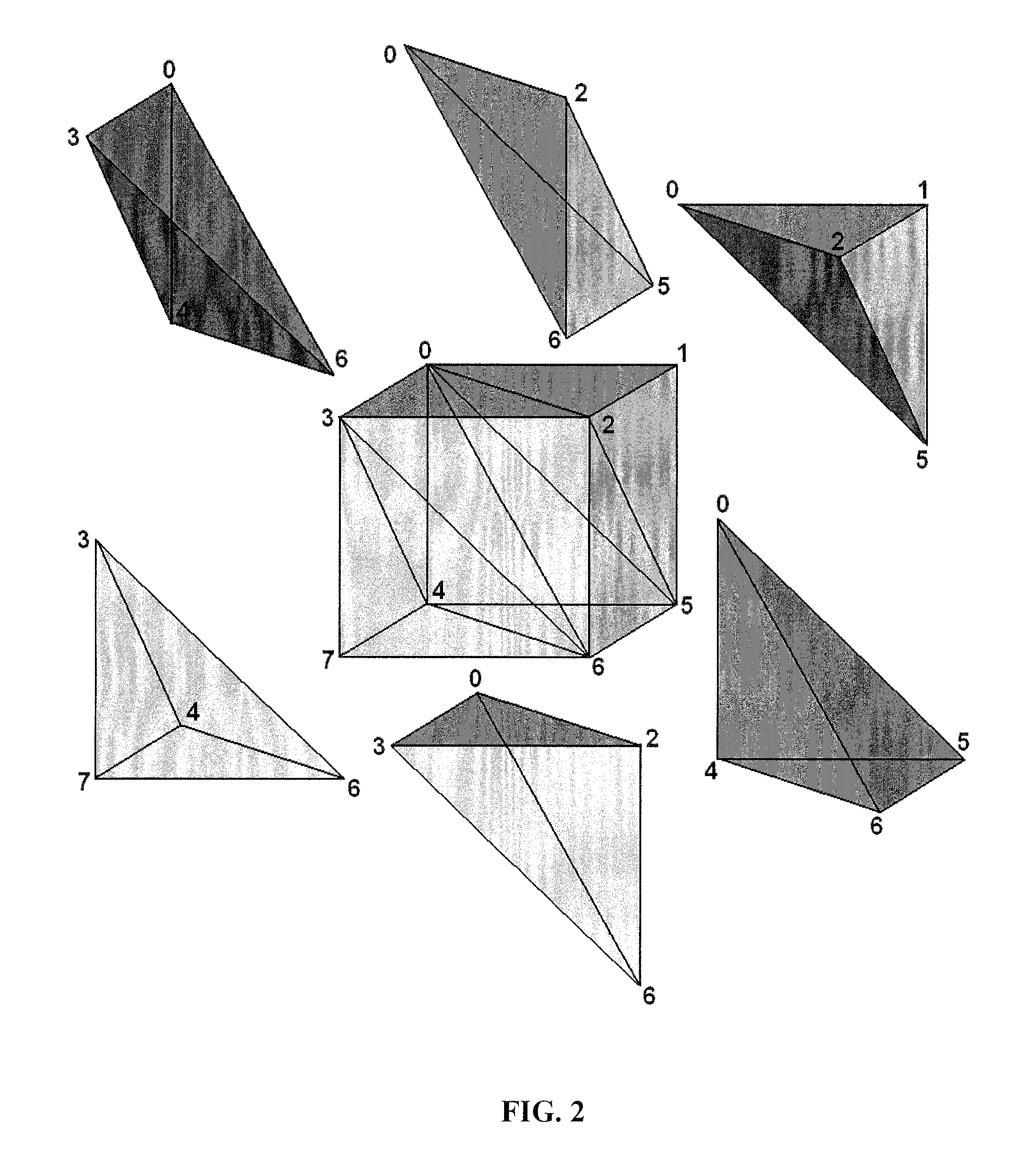
[0024]Embodiments of the subject invention relate to a method and apparatus for ventilation imaging. In a specific embodiment, a method and apparatus for use in ventilation computed tomography (CT) imaging is provided. Specific embodiments pertain to a method and apparatus for high resolution pulmonary ventilation imaging by using deformable image registration and local volume change to calculate ventilation. Since 4-D CT is less expensive than nuclear medicine, including SPECT and PET, and easier to perform, certain implementations of pulmonary ventilation imaging using nuclear medicine can be replaced by an embodiment of the subject invention using 4-D CT, unless concurrent perfusion imaging is required. Embodiments of the invention using 4-D CT can be used in radiological studies for pulmonary fibrosis, emphysema, or other indications of reduced lung function.

[0025]In lung cancer radiotherapy, 4-D CT images are often used to assess lung tumor motion. Using embodiments of the subj...


PUM


Abstract

The invention provides a novel method of high-resolution quantifiable pulmonary regional ventilation imaging using a package of computer programs, including deformable image registration and local volume change calculation on 4-D or breath-hold CT images. High resolution 3D ventilation images have been generated using this technique with superior quality. The deformation matrices between different respiration phases are calculated using deformable image registration and applied to calculate local volume change ΔV between respiration phases. The invention provides less image-noise and mismatch sensitivity than other techniques based on CT images, and is less expensive than nuclear medicine imaging. Implemented clinically, the method can derive a patient's pulmonary ventilation information from the same set of 4-D CT images, providing a cheaper but more accurate method to generate clinical lung ventilation image. Such information can be utilized in treatment planning sparing functional lung volumes without additional procedure and cost for lung functional imaging.

Description

CROSS-REFERENCE TO RELATED APPLICATIONS[0001]The present application is the U.S. National Stage Application of International Patent Application No. PCT / US2009 / 041252, filed on Apr. 21, 2009, which claims the benefit of U.S. Provisional Application Ser. No. 61 / 046,605, filed Apr. 21, 2008, and claims the benefit of U.S. Provisional Application Ser. No. 61 / 058,023, filed Jun. 2, 2008, all of which are hereby incorporated by reference herein in their entirety, including any figures, tables, or drawings.BACKGROUND OF THE INVENTION[0002]Pulmonary ventilation and perfusion typically vary when pulmonary diseases are involved. (Nakagawa T, et al., J Magnetic Resonance Imaging 2001; 14:419-424). However, in current practice of thoracic cancer radiotherapy, the differences in pulmonary ventilation and perfusion are not considered when generating treatment plans. Lung volumes are deemed equal when placing radiation beams in planning. Clinicians and researchers have proposed to include normal l...

Claims


Application Information

Patent Timeline
Patent Type & Authority Patents(United States)
IPC IPC(8): G06K9/00
CPCA61B5/08A61B5/1075A61B5/113A61B6/50A61B6/5217G06T7/0012G06T2207/10076G06T2207/10081G06T2207/30061G06T7/62G16H50/30A61B5/091
Inventor ZHANG, GEOFFREY G.FORSTER, KENNETH M.
Owner UNIV OF SOUTH FLORIDA
Why Eureka
Try Eureka