Method and system to reconstruct treatment dose to a patient from integrated exit-transit images of radiation fields taken during treatment

a radiation field and integrated technology, applied in the field of radiation therapy, can solve the problems of not describing the method of reverse calculation, several problems not addressed, and the energy dependence of imaging devices

a radiation field and integrated technology, applied in the field of radiation therapy, can solve the problems of not describing the method of reverse calculation, several problems not addressed, and the energy dependence of imaging devices

US8605857B1Active Publication Date: 2013-12-10LIFELINE SOFTWARE INC

Image

  • Method and system to reconstruct treatment dose to a patient from integrated exit-transit images of radiation fields taken during treatment
  • Method and system to reconstruct treatment dose to a patient from integrated exit-transit images of radiation fields taken during treatment
  • Method and system to reconstruct treatment dose to a patient from integrated exit-transit images of radiation fields taken during treatment

Examples


example

Application to Clinical Exit Images

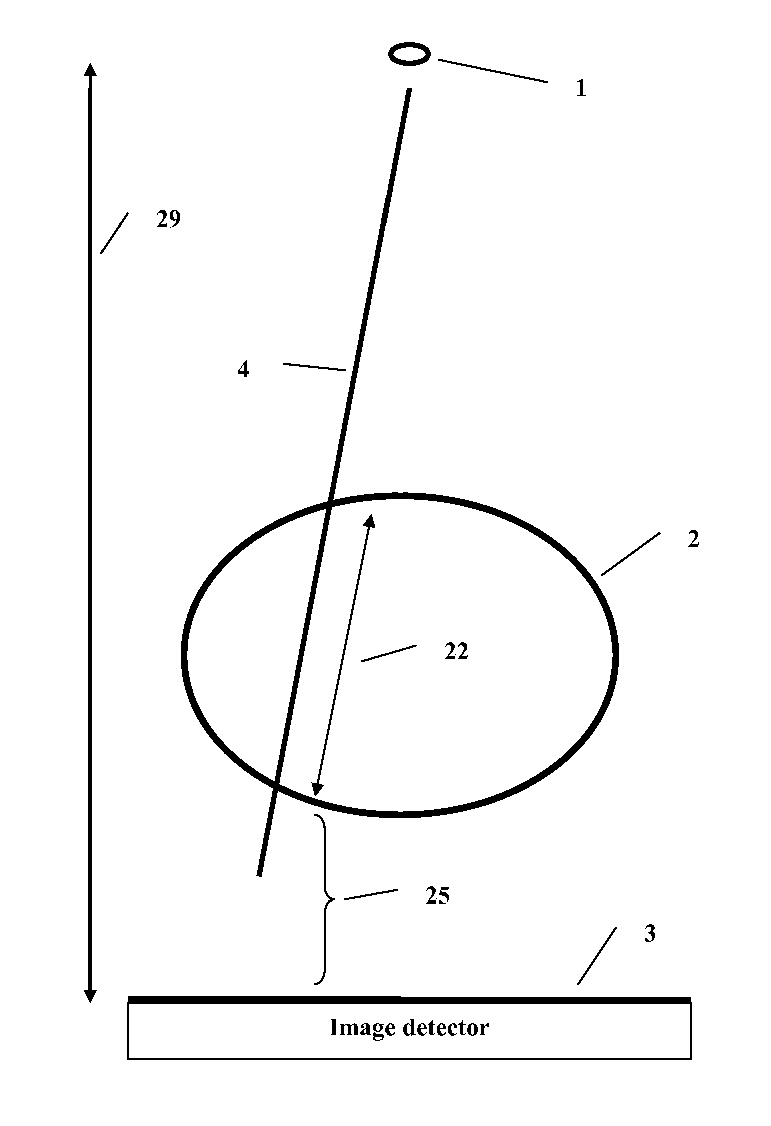
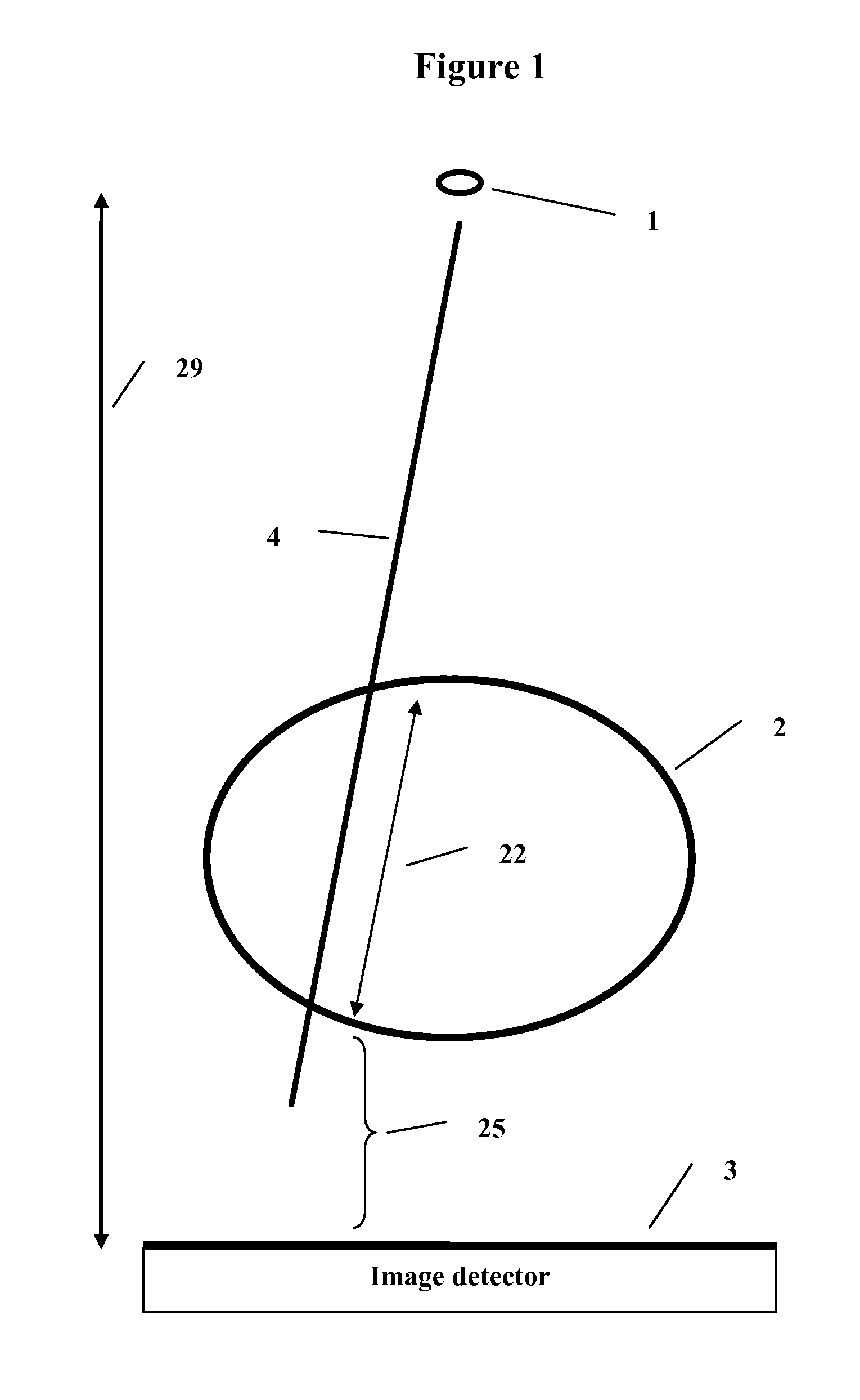
[0107]The data in this example includes images taken for a range of thickness, field size, and air gap. On a pixel by pixel basis, given particular values of the air gap distance (25) and thickness (22) at a pixel, and a field size to be defined below, a correction factor is interpolated from the corresponding three dimensional air gap correction table and applied to that pixel value.

[0108]Clinical images, however, are not typically of square open field sizes. They tend to comprise modulated fields. The field size used to look up the correction factor must be generated by a suitable metric that accounts for the amount of scatter generated in the patient from the radiation field in such manner that different size square fields will generate different amounts of scatter. For each field image, the maximum intensity is determined, and all other pixels are divided by that maximum value. The pixels are then summed and multiplied by the area per pixel. A ...


PUM


Abstract

A method and system to compute the dose to a patient (2) given a captured integrated exit-transit image (5) of the radiation rays (4) traveling from the source of x-rays (1) through the patient (2) to the imaging device (3) to product the exit-transit image (5). Each radiation field image (5) is transformed (6,8,10,12) to multiple images (7,9,11,13) for each phantom thickness (26) that was measured with the imaging device (3) for a range of field sizes (21). Given the water equivalent path (22) through the patient for a ray (4) reaching a pixel (15, 16), the final pixel value (19) is interpolated from the images (9, 11) that bracket the water equivalent path through the patient (22).

Description

RELATED APPLICATION DATA[0001]This application is a Continuation-In-Part of co-pending patent application Ser. No. 12 / 977,907, filed 23 Dec. 2010.BACKGROUND OF THE INVENTION[0002]1. Field of the Invention[0003]This invention relates to the radiation therapy applied to patients who are typically treated for cancer or other ailments with high energy x-rays from medical linear accelerators.[0004]The invention is directed to a method and system to reconstruct the dose the patient has received using as input the radiation fields measured during treatment on the exit side of the patient with an imaging system capable of integration or a radiation detector array. This information may be used as a quality control measure of the treatment to avoid errors by comparing the reconstructed dose to the intended planned dose.[0005]2. Prior Art[0006]U.S. Pat. No. 5,754,622 describes a system for verifying the amount of radiation delivered to an object. However, this system does not describe the meth...

Claims


Application Information

Patent Timeline
10 Dec 2013
Publication
US8605857B1
IPC
A61N5/10
CPC
A61N5/10; A61N5/1071; A61N2005/1054
Inventors
RENNER, WENDEL DEAN