Synthetic osteochondral composite and method of fabrication thereof

a composite and osteochondral technology, applied in the field of surgical instruments and procedures, can solve the problems of limited tissue supply, immunorejection, and lack of current techniques, and achieve the effects of improving the quality of li

Inactive Publication Date: 2014-02-04
UNIVERSITY OF MISSOURI
View PDF9 Cites 11 Cited by
  • Summary
  • Abstract
  • Description
  • Claims
  • Application Information

AI Technical Summary

Problems solved by technology

All current techniques suffer from deficiencies, such as donor site morbidity, limited tissue supply, immunorejection, potential transmission of pathogens, and implant loosening.
The efficacy of existing tissue-engineered osteochondral composites has been hampered by the difficulty of bonding the softer material used for artificial cartilaginous extracellular matrix to the harder material used for the subchondral support material.
To date, existing tissue-engineered osteochondral composites have had limited success due to problems associated with the separation of the artificial cartilaginous extracellular matrix from the underlying subchondral support.

Method used


Image

  • Synthetic osteochondral composite and method of fabrication thereof
  • Synthetic osteochondral composite and method of fabrication thereof
  • Synthetic osteochondral composite and method of fabrication thereof

Examples


example 1

Mesenchymal Stem Cells were Obtained from Bone Marrow of NZW Rabbits

[0043]To obtain the chondrogenic cells to be incorporated into the various embodiments of the osteochondral composites discussed below, the following experiment was conducted. Bone marrow-derived mesenchymal stem cells (MSCs) were prepared from euthanized New Zealand White (NZW) rabbits, using established methods.

[0044]Marrow plugs were removed from the femur or humerus of NZW rabbits with intramedullary-pin drilling and a bone curette and then transferred to 50-ml centrifuge tubes containing 25-ml of a culture medium containing DMEM low glucose, 10% fetal bovine serum (FBS), and penicillin / streptomycin / amphotericin (p / s / a). After vortexing and spinning for 5 minutes at 1,500 rpm, the fat layer and medium were removed and the pellet was then resuspended in 8-ml of culture medium and vortexed briefly. The 8-ml suspension was drawn into a 10-ml syringe with an 18-gauge needle and transferred to a new 50-ml centrifuge ...

example 2

Bone Marrow Derived Mesenchymal Stem Cells were Cultured with Hydrogel Polymers

[0047]The bone marrow derived MSCs obtained using the methods described in Example 1 were trypsinized with 0.25% trypsin in 1 mM EDTA, counted with a hemocytometer, and then resuspended by mixing the MSCs with poly(ethylene glycol) diacrylate (PEGDA) / photoinitiator solution at a concentration of 5-10×106 cells / cm3. The cell culture medium was further supplemented with 10 ng / ml TGF-β. Cell cultures were incubated for one week in 95% air / 5% CO2 at 37° C. with a fresh medium change every 3-4 days. After one week of culture at these conditions, chondrogenic differentiation of MSCs was achieved.

example 3

Osteochondral Composites were Fabricated using Bioactive Glass and PEG Hydrogel

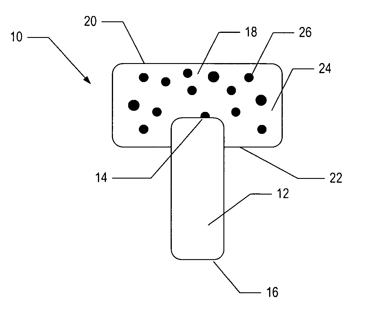
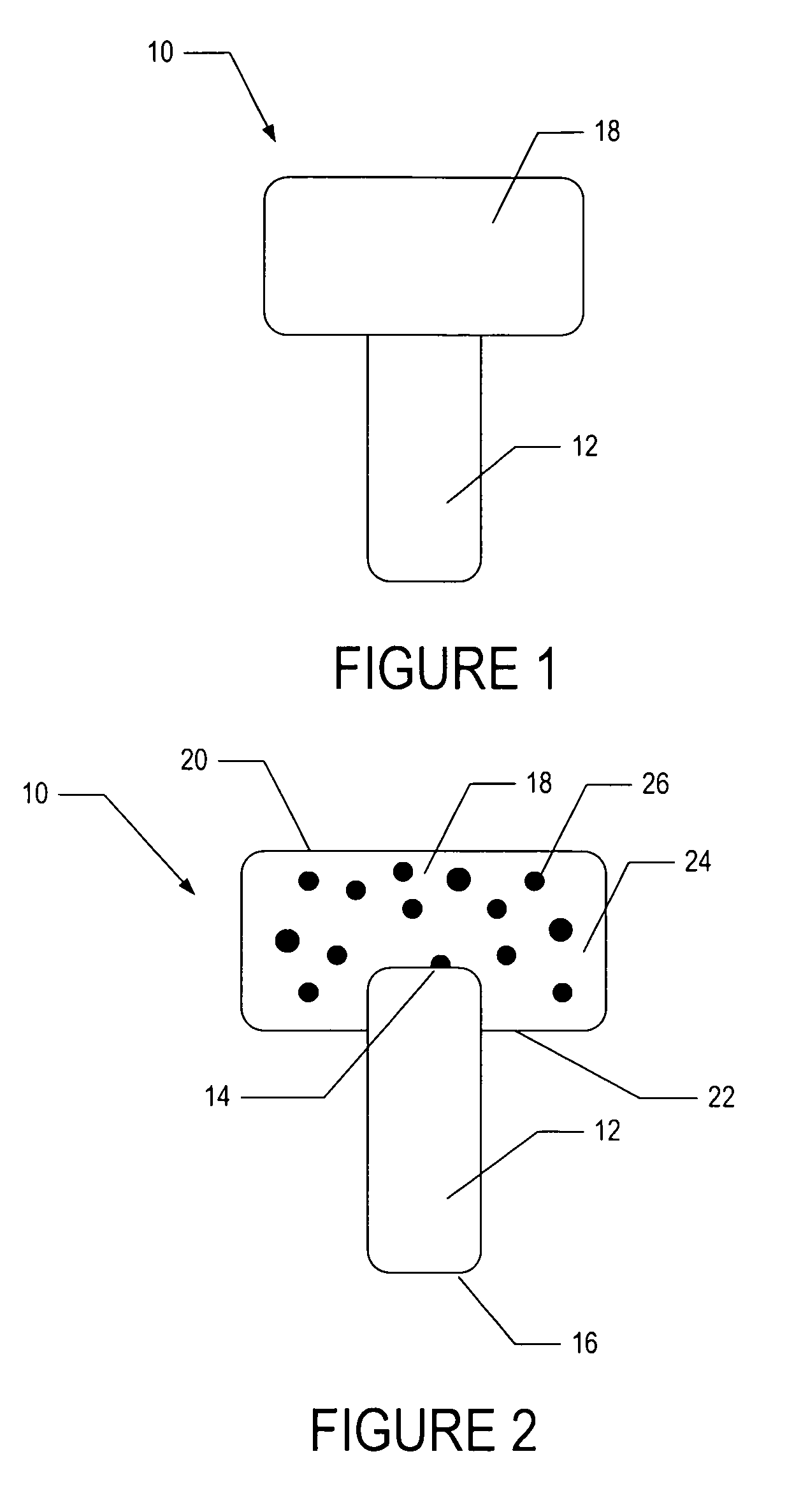
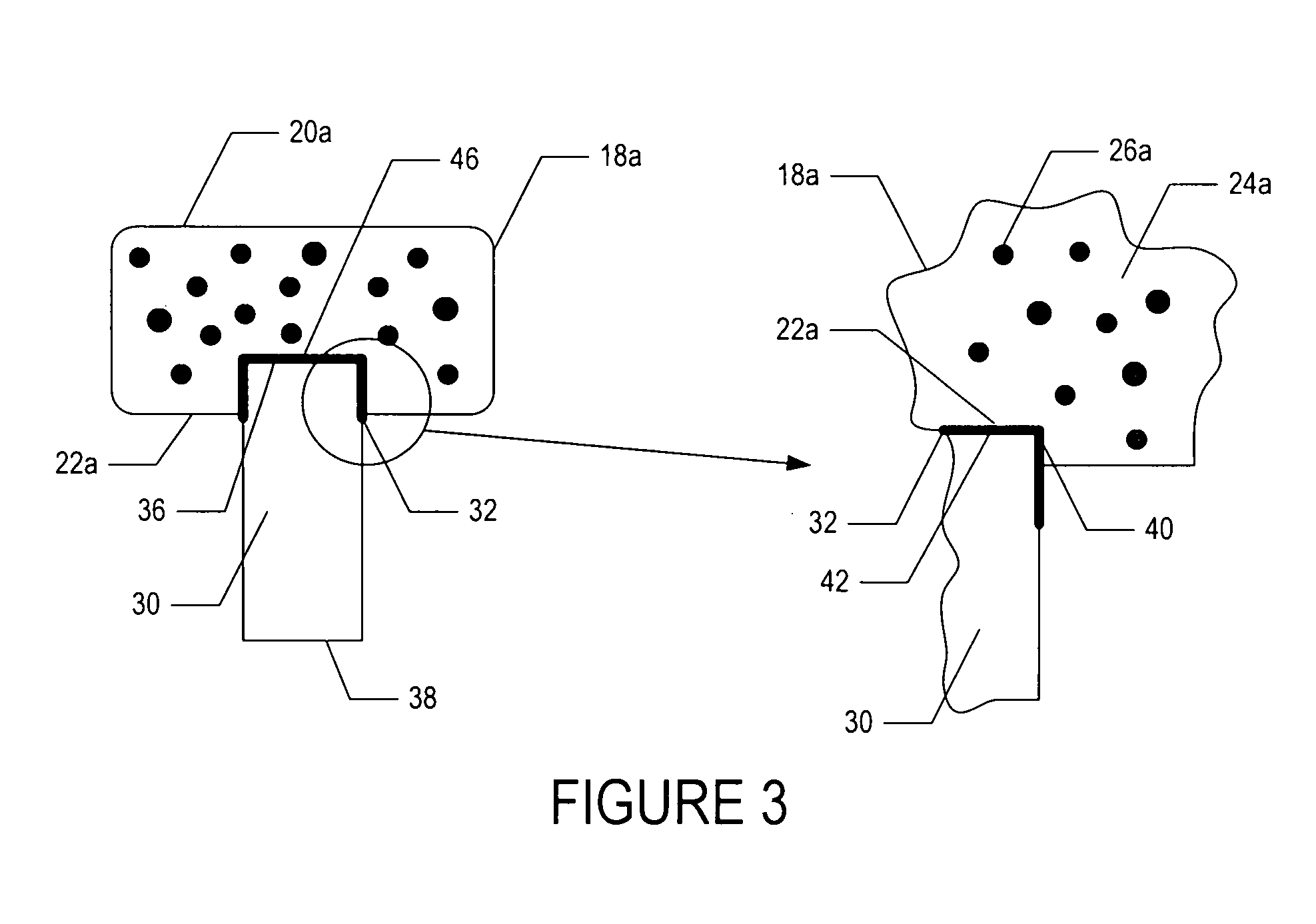
[0048]To demonstrate the feasibility of fabricating an osteochondral composite having a PEG hydrogel mechanically interlinked with a bioactive glass base, the following experiment was conducted.

[0049]In one embodiment, a bioactive glass, designated 13-93, with the composition 53 wt % SiO2, 20 wt % CaO, 6 wt % Na2O, 12 wt % K2O, 5 wt % MgO, and 4 wt % P2O5 was formed into cylindrical bases. Porous 13-93 bioactive glass cylinders (3 mm diameter×3.5 mm long) were prepared by pouring glass particles ranging in size from 212-325 μm into a graphite mold, and sintered for 15 min at 700° C. to form a network of particles.

[0050]PEG hydrogel was prepared by dissolving poly(ethylene glycol)diacrylate (PEGDA) (Shearwater, Huntsville, Ala.) in sterile phosphate buffer saline (PBS) supplemented with 1% penicillin and streptomycin (Gibco, Carlsbad, Calif.) to a final solution of 10% w / v. A biocompatible ultraviolet phot...


PUM

PropertyMeasurementUnit
diameteraaaaaaaaaa
thicknessaaaaaaaaaa
thicknessaaaaaaaaaa

Abstract

An osteochondral composite including a hydrogel with implanted biological cells joined to a porous supportive base at a mechanically-interlocked interface is disclosed. A method of fabricating the osteochondral composite and a method of treating damaged or diseased articular cartilage by implanting an osteochondral composite are also disclosed.

Description

CROSS REFERENCE TO RELATED APPLICATION[0001]This application claims priority from U.S. Provisional Patent Application Ser. No. 60 / 993,784 filed on Sep. 14, 2007 and is herein incorporated by reference in its entirety.FIELD OF INVENTION[0002]The present document relates to surgical apparatus and procedures, and more particularly to osteochondral composite and method of fabrication thereof.BACKGROUND OF INVENTION[0003]Osteochondral defects in adult knees are a common clinical finding. The limited reparative potential of such osteochondral defects has motivated numerous studies aimed at finding improved methods of treatment. Currently used procedures such as abrasion arthroplasty and microfracture, autologous chondrocyte implantation, osteochondral autografts and allografts, and partial or total joint arthroplasties have yielded varying levels of success. All current techniques suffer from deficiencies, such as donor site morbidity, limited tissue supply, immunorejection, potential tra...

Claims


Application Information

Patent Timeline
Patent Type & Authority Patents(United States)
IPC IPC(8): A61F2/02
CPCA61F2/2846A61L27/3834A61L2430/06A61L27/44A61F2002/30761A61F2/30756A61F2002/2839A61L27/3852A61F2002/30762A61L27/3817A61P19/00
Inventor RAHAMAN, MOHAMED NAUSHADBAL, BHAJANJIT SINGH
Owner UNIVERSITY OF MISSOURI
Why Eureka
Try Eureka