svegfr-2 and its role in lymphangiogenesis modulation
a growth factor receptor and vascular endothelial technology, applied in the direction of growth factor/regulator receptors, peptides, biological materials analysis, etc., can solve the problems of compromising corneal immunological privilege, affecting the clinical outcome of patients, so as to inhibit lymphangiogenesis and inhibit the proliferation of lymphatic epithelial cells
- Summary
- Abstract
- Description
- Claims
- Application Information
AI Technical Summary
Benefits of technology
Problems solved by technology
Method used
Image
Examples
example 1
sVEGFR-2 is the Product of Alternative Splicing of the VEGFR-2 Pre-mRNA
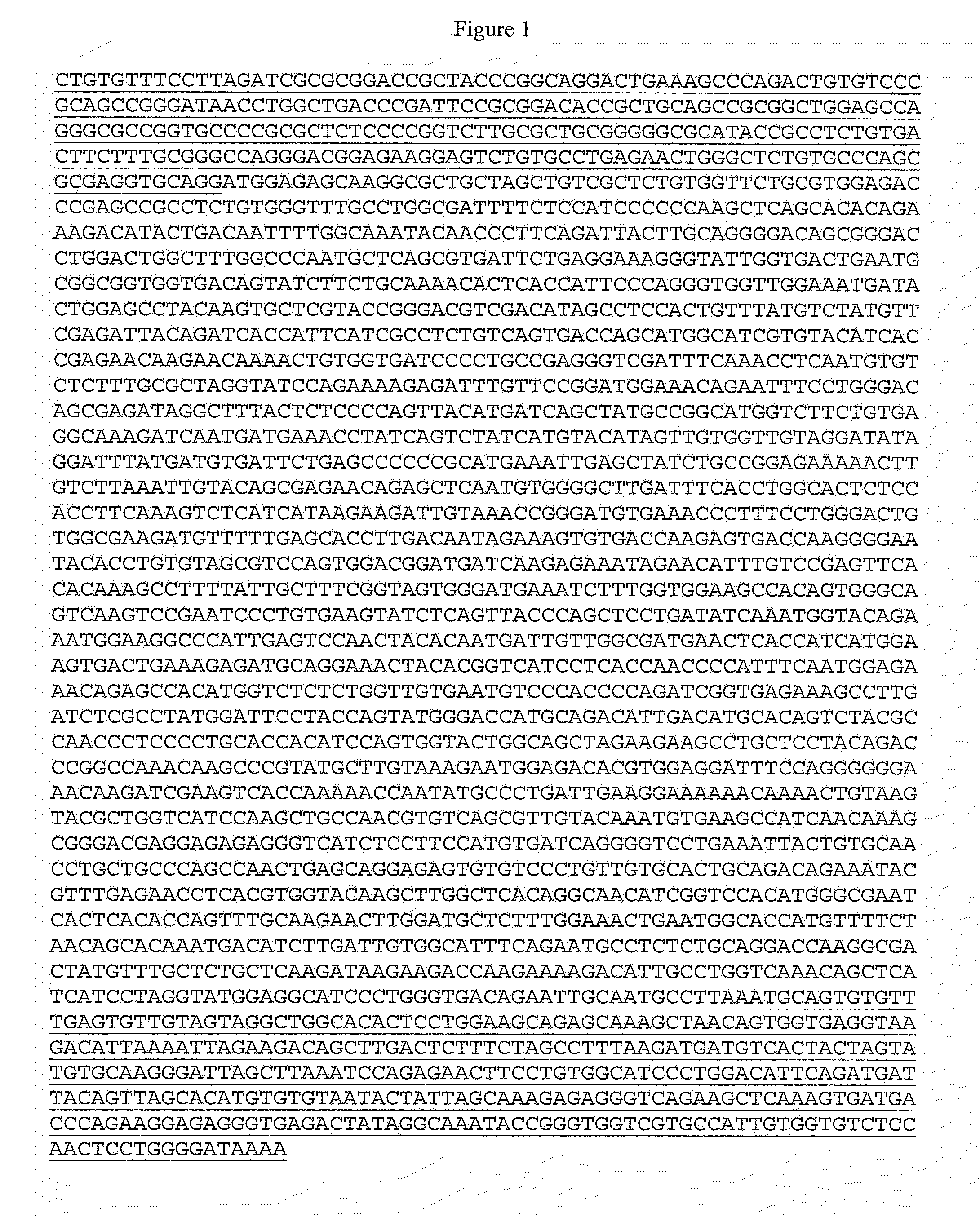
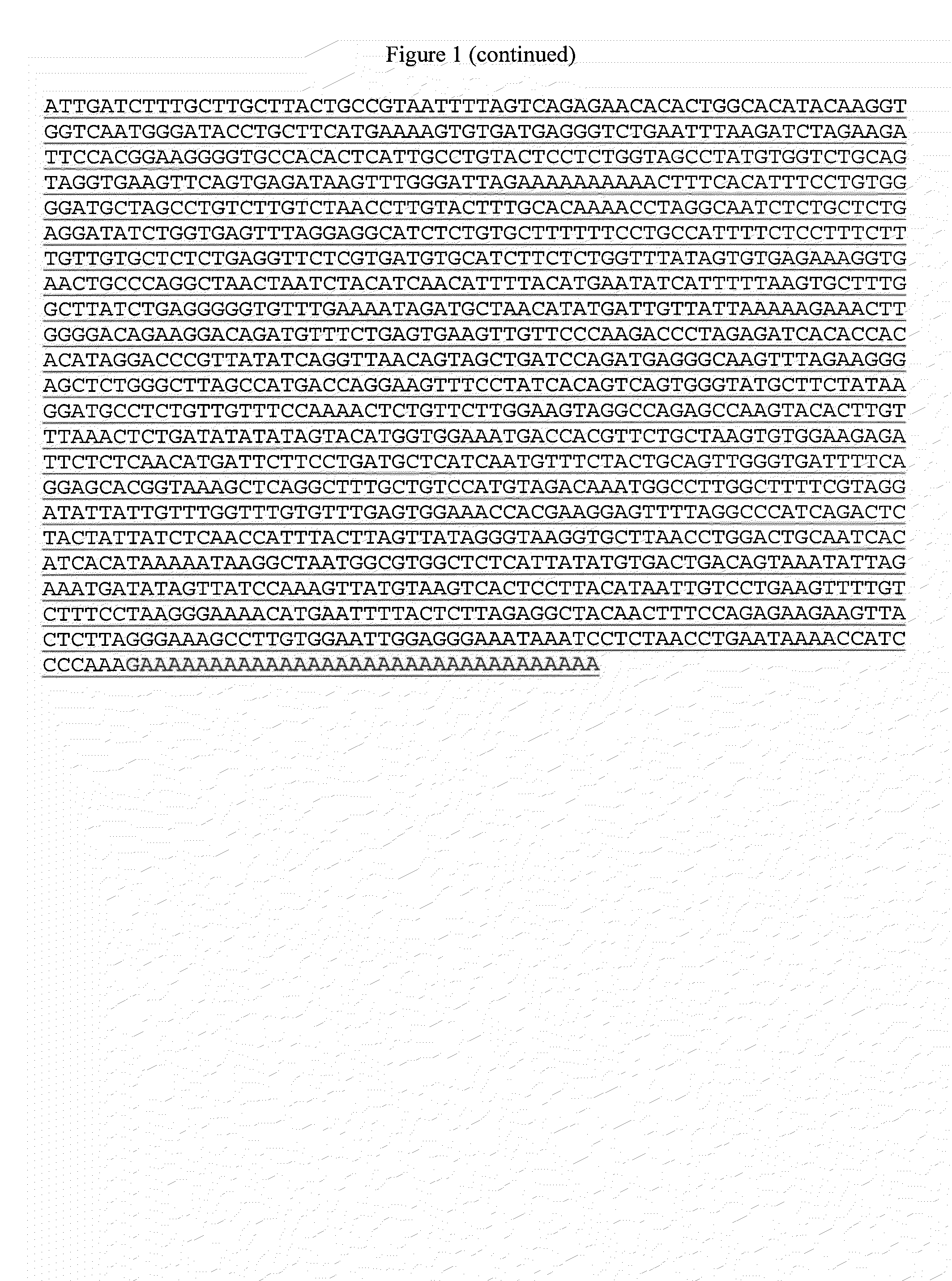
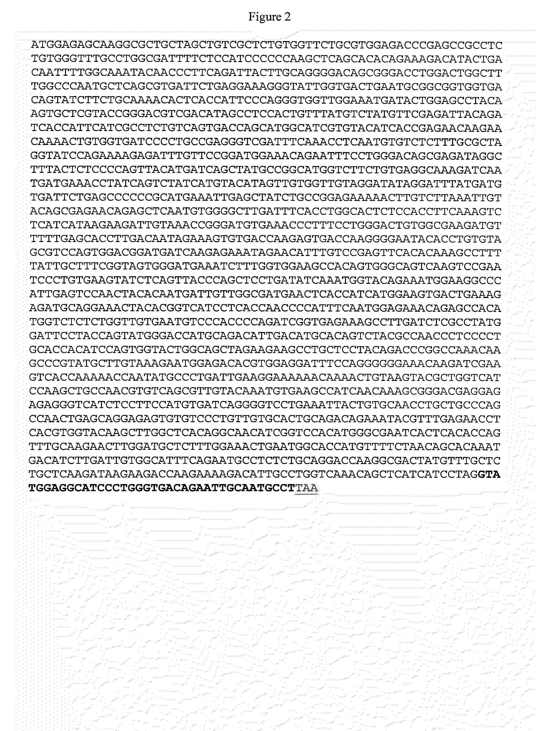
[0129]By modeling the alternative splicing event of sVEGFR-2 analogous to that of sVEGFR-1 (Kendall et al., Proc. Natl. Acad. Sci. U.S.A., 90(22): 10705-9, 1993), we determined that the retention of intron 13-14 yields a truncated transcript variant whose protein product lacks the transmembrane and intracellular tyrosine kinase domains present in the full length cell surface protein. This is because an early termination codon is present 39 nucleotide downstream (13 amino-acids) from this alternate exon / intron junction.
[0130]To demonstrate the presence of this novel soluble splicing variant in the mouse cornea and lung, we devised primers targeting an area of intron 13-14 and exon 12 (FIG. 14A). Targeting exon 12 allowed us to discern between amplification of cDNA derived from mRNA or from possible genomic DNA contamination based on amplicon size. PCR resulted in a 393 bp product encompassing the site where the al...
example 2
Corneal sVEGFR-2 mRNA Levels are Increased after Corneal Injury
[0132]The detection of sVEGFR-2 transcript in the cornea compared to lung (an organ known to express high levels of VEGFR-2 (Voelkel et al., Am. J. Physiol. Lung Cell Mol. Physiol., 290(2): L209-21, 2006)) was nearly negligible by RT-PCR (lane 1 from FIG. 14B). We investigated the effects of corneal injury (i.e., suture placement) on the levels of sVEGFR-2 mRNA by RT-PCR. RNA was isolated from untreated corneas (d.0) and from corneas 1, 2 and 5 days after suture placement. We observed a relative increase in the mRNA level of sVEGFR-2 as early as 1 day after suture placement (FIG. 16).
example 3
Corneal sVEGFR-2 Protein Levels are Undetectable in Normal Cornea, but Become Apparent after Corneal Injury
[0133]We have previously shown that under normal / non-inflammatory conditions, the levels of VEGFR-2 protein in the mouse cornea are below the detection threshold of a Western blotting technique using a rabbit derived polyclonal antibody that targets the extracellular domain of VEGFR-2 (Ambati et al., Nature, 443(7114): 993-7, 2006). See also the 2nd lane of FIG. 17. However, experiments aimed at probing for the presence of VEGFR-2 in corneal lysate 5 days after suture placement have unveiled an immunoreactive band migrating at approximately 80 kDa (3rd lane of FIG. 17), which is like the band of comparable molecular weight detected in mouse lung (1st lane of FIG. 17). The 230 kDa band corresponding to mbVEGFR-2 (evident in lung lane of FIG. 17) does not appear in the mouse cornea even 5 days following injury. This indicates that the cornea preferentially expresses a truncated f...
PUM
| Property | Measurement | Unit |
|---|---|---|
| length | aaaaa | aaaaa |
| pH | aaaaa | aaaaa |
| temperatures | aaaaa | aaaaa |
Abstract
Description
Claims
Application Information
 - R&D
- Intellectual Property
- Life Sciences
- Materials
- Tech Scout
- Unparalleled Data Quality
- Higher Quality Content
- 60% Fewer Hallucinations
Browse by: Latest US Patents, China's latest patents, Technical Efficacy Thesaurus, Application Domain, Technology Topic, Popular Technical Reports.
© 2025 PatSnap. All rights reserved.Legal|Privacy policy|Modern Slavery Act Transparency Statement|Sitemap|About US| Contact US: help@patsnap.com


