

Method and apparatus for tissue grafting and copying
a tissue grafting and tissue technology, applied in the field of tissue grafting and copying, can solve the problems of extensive healing time at the donor site, limited tissue availability for autografting, and large loss of sheet grafts after placement, etc., and achieve the effect of rapid healing of the donor si
- Summary
- Abstract
- Description
- Claims
- Application Information
AI Technical Summary
Benefits of technology
Problems solved by technology
Method used
Image

Examples
Embodiment Construction
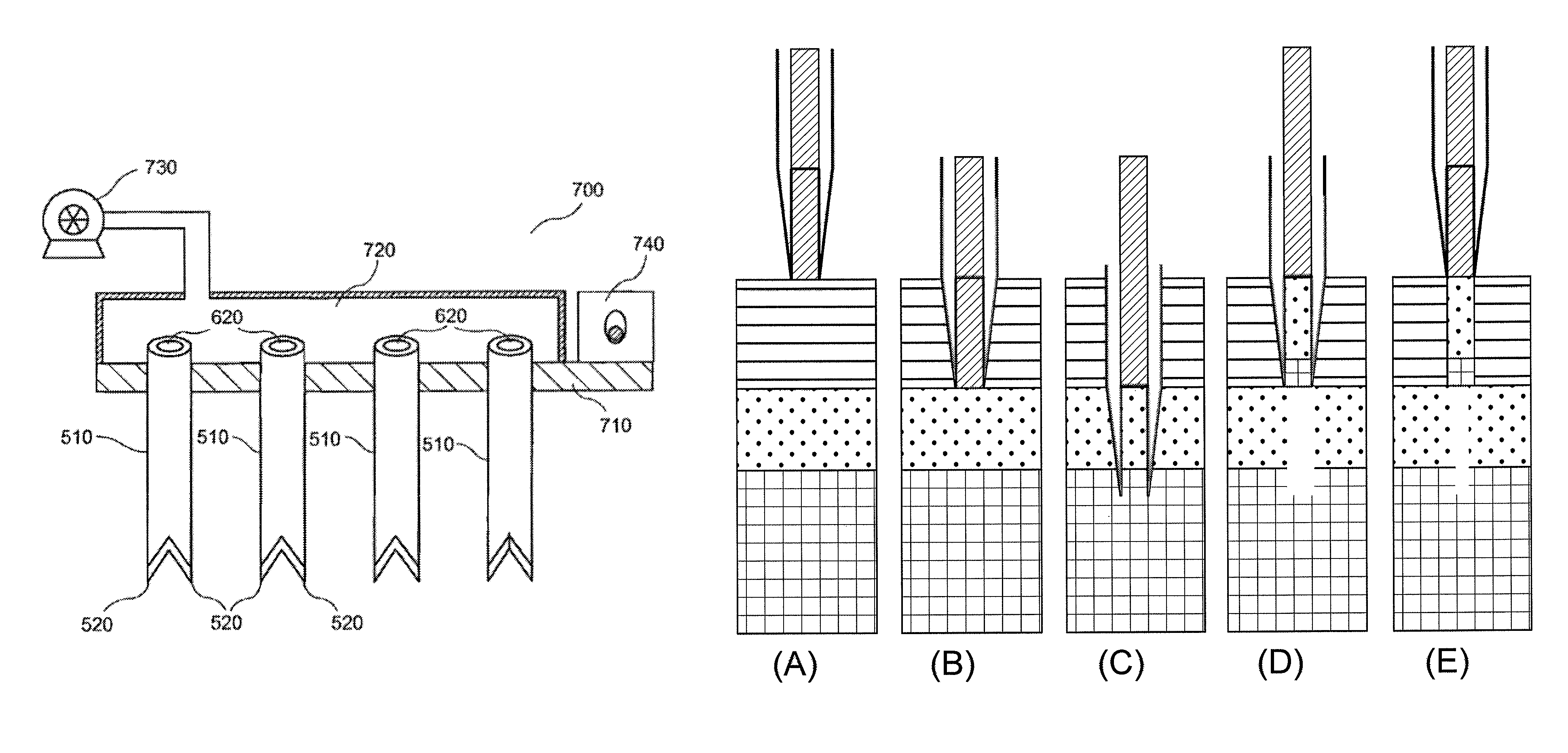
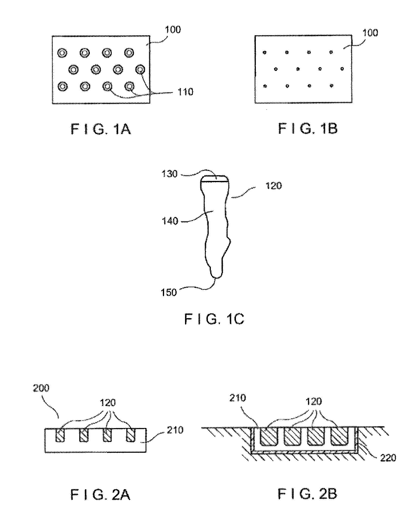
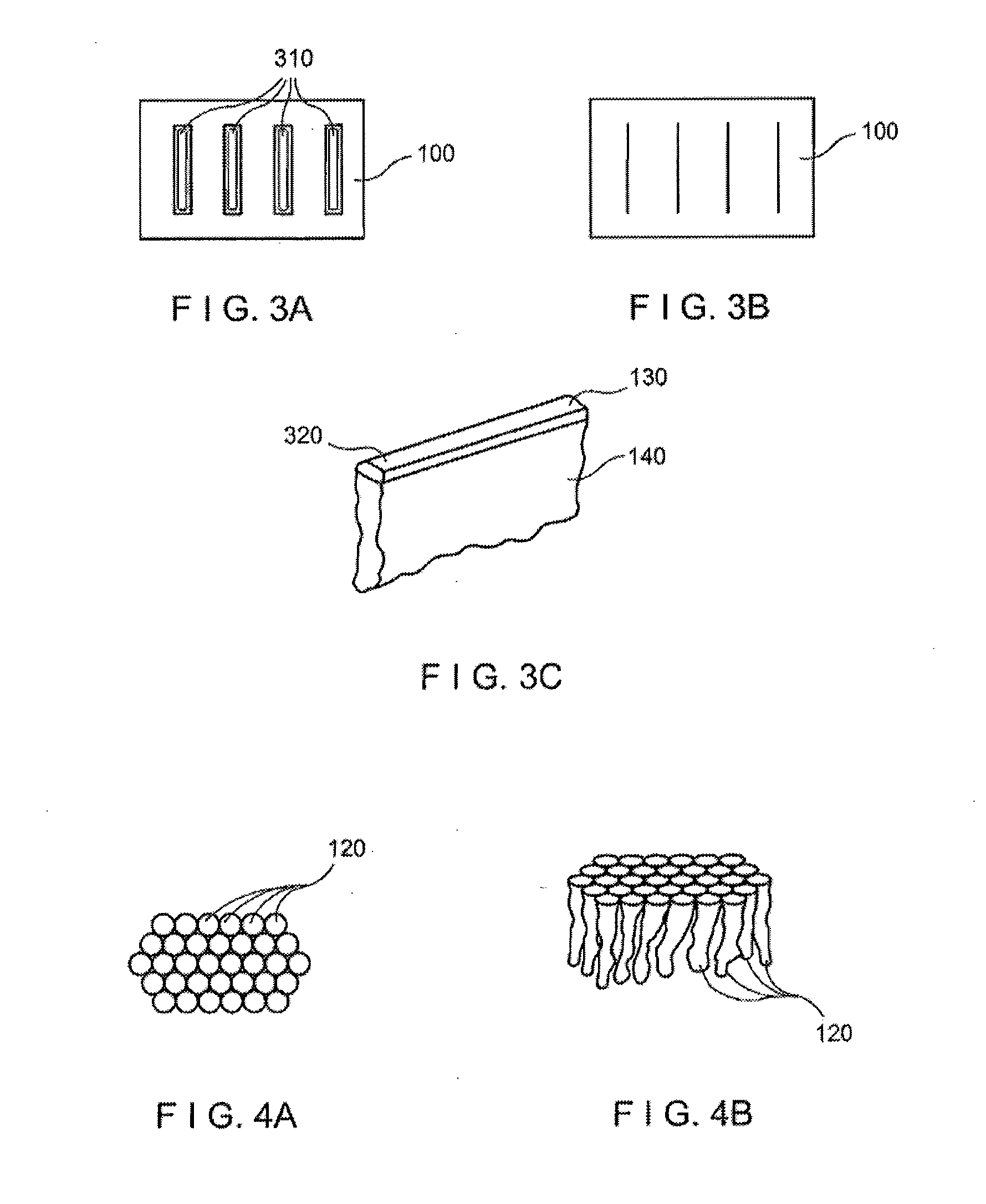
[0020]Exemplary embodiments of the present disclosure provide method and apparatus for obtaining small portions of graft tissue that can be accompanied by rapid healing of the donor site. For example, the exemplary embodiment of the method can be provided for obtaining skin graft tissue by harvesting small portions of the tissue, e.g., micrografts, from a donor site. Such micrografts can be used to form grafts or “copy” tissue to generate larger tissue structures from the small tissue samples.
[0021]Such micrografts can comprise skin tissue that can include, e.g., epidermal and dermal tissue, and / or tissue obtained from other body organs. The micrografts can have at least one dimension that is relatively small, e.g., less than about 1 mm, or less than about 0.5 mm, or optionally about 0.3 mm or less, or about 0.2 mm. Such exemplary small dimensions of the micrografts can facilitate both healing of the donor site following harvesting and viability of the micrografts by allowing greate...
PUM
 Abstract
Description
Claims
Application Information