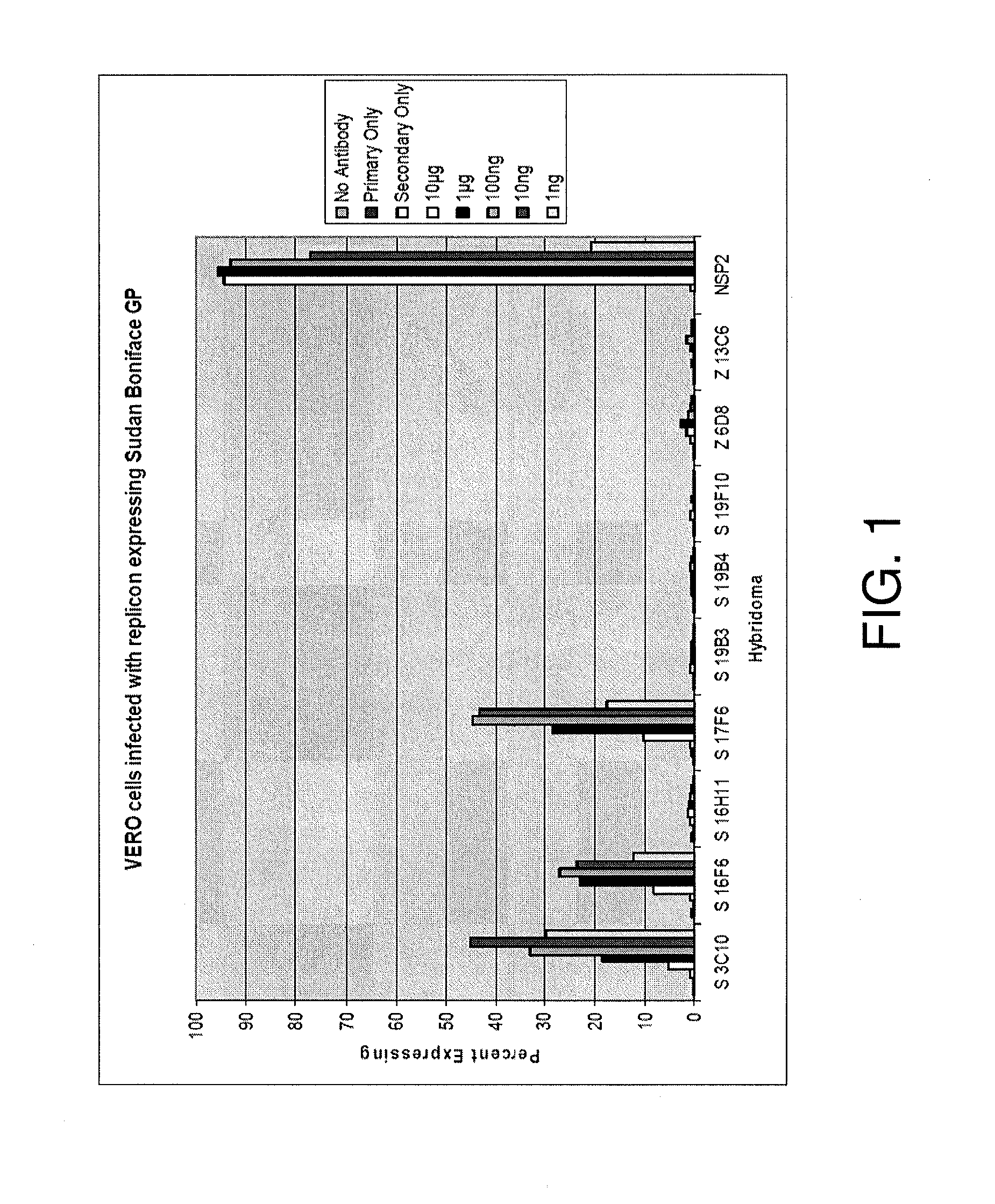
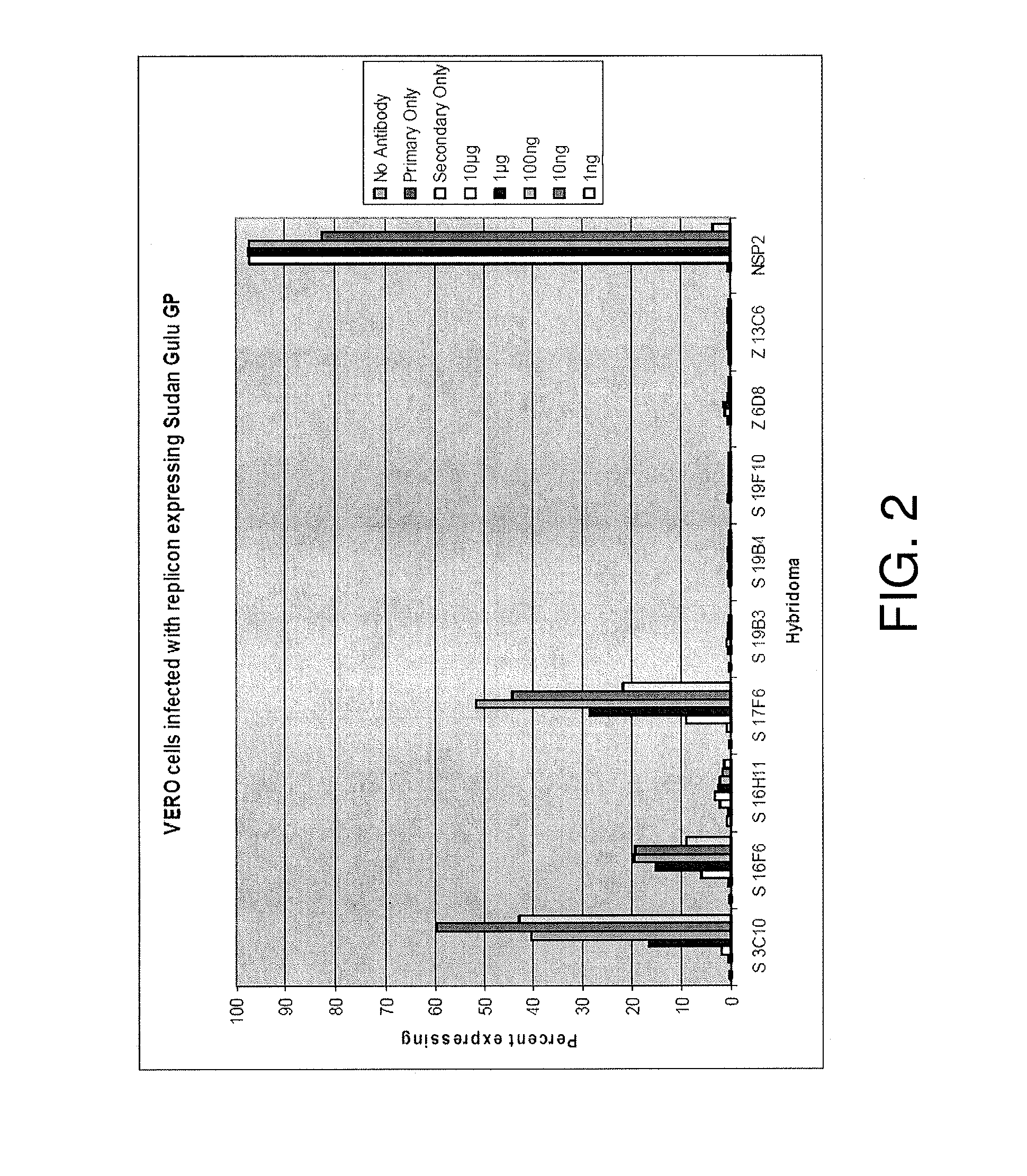
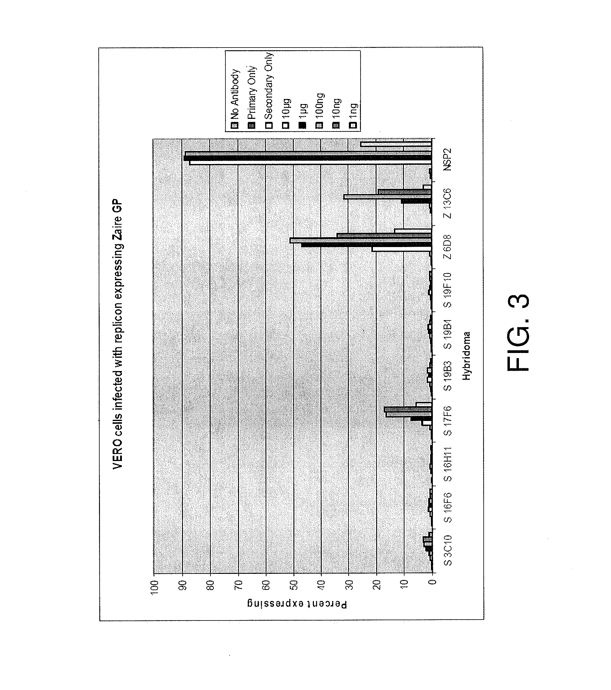
Monoclonal antibodies against glycoprotein of Ebola sudan boniface virus
a sudan boniface virus and monoclonal antibody technology, applied in the field of antibodies, can solve the problems of high mortality, severe hemorrhagic fever disease with high mortality rate, and no antibodies against the sudan boniface species of the ebola virus, and achieve the effect of facilitating the identification of the sudan boniface species
- Summary
- Abstract
- Description
- Claims
- Application Information
AI Technical Summary
Benefits of technology
Problems solved by technology
Method used
Image
Examples
example 1
Production and Characterization of Ebola GP MAbs
Materials and Methods
[0101]Animals.
[0102]Female BALB / c mice (5- to 8-weeks old) were obtained from the National Cancer Institute (Frederick, Md.) and housed under specific-pathogen free conditions. Research was conducted in compliance with the Animal Welfare Act and other federal statutes and regulations relating to animals and experiments involving animals and adhered to principles stated in the Guide for the Care and Use of Laboratory Animals (National Research Council, 1996). The facility where this research was conducted is fully accredited by the Association for the Assessment and Accreditation of Laboratory Animal Care International.
[0103]Vaccinations.
[0104]Balb / c mice were vaccinated subcutaneously in the dorsal neck region with Venezuelan equine encephalitis replicons (VRP) (2×10^6 focus forming units (ffu) / mouse) expressing the glycoprotein of Sudan Boniface. The mice were boosted three times over consecutive months with VRP (...
example 2
Competitive Binding of Ebola Mabs and Protective Efficacy of Ebola GP MAbs In Vivo
Materials and Methods
[0112]Animals.
[0113]Female BALB / c mice and severe combined immunodeficiency (SCID) mice (5- to 8-weeks old) were obtained from the National Cancer Institute, Frederick, Md. and housed under specific-pathogen-free conditions. Research was conducted in compliance with the Animal Welfare Act and other federal statutes and regulations relating to animals and experiments involving animals and adhered to principles stated in the Guide for the Care and Use of Laboratory Animals (National Research Council, 1996). The facility where this research was conducted is fully accredited by the Association for the Assessment and Accreditation of Laboratory Animal Care International.
[0114]Vaccinations for Antibody Generation.
[0115]Balb / c mice were vaccinated subcutaneously in the dorsal neck region with Venezuelan equine encephalitis replicons (VRP) (2×10^6 focus-forming units (ffu) / mouse) expressin...
example 3
Characterization of ESB MAbs Demonstrating In Vitro Neutralization of ESB Virus
Materials and Methods
[0128]Four Balblc mice were vaccinated by subcutaneous route with 1×10^7 ffu of Sudan Boniface glycoprotein (GP) expressed by Venezuelan equine encephalitis virus replicon developed and prepared at the United States Army Medical Research Institute of Infectious Diseases (USAMRIID). Mice were boosted with the same dose of the same construct three times at one month intervals. Mice were boosted one final time with irradiated Ebola Sudan Boniface virus by tail vein injection. Four days following final injection, mice were harvested and spleens were fused with an immortalized cell line to create hybridoma fusions. Hybridoma plasma cells secreting glycoprotein (GP) specific monoclonal antibodies were isolated and confirmed by Enzyme linked Immunosorbant Assay (ELISA) against irradiated Ebola Sudan Boniface virus. Eighteen Sudan OP-specific antibodies of the IgG isotype have been identified...
PUM
| Property | Measurement | Unit |
|---|---|---|
| volume | aaaaa | aaaaa |
| time | aaaaa | aaaaa |
| pH | aaaaa | aaaaa |
Abstract
Description
Claims
Application Information
 - R&D
- Intellectual Property
- Life Sciences
- Materials
- Tech Scout
- Unparalleled Data Quality
- Higher Quality Content
- 60% Fewer Hallucinations
Browse by: Latest US Patents, China's latest patents, Technical Efficacy Thesaurus, Application Domain, Technology Topic, Popular Technical Reports.
© 2025 PatSnap. All rights reserved.Legal|Privacy policy|Modern Slavery Act Transparency Statement|Sitemap|About US| Contact US: help@patsnap.com


