Methods for analysis of free and autoantibody-bound biomarkers and associated compositions, devices, and systems

a technology of autoantibody and biomarker, applied in the analysis of chemical indicators, laboratory glassware, instruments, etc., can solve the problems of affecting the assay used to detect the biomarker, the individual can be improperly treated for a condition, and the detection of a biomarker is problematic, so as to achieve high mass resolution and high mass accuracy

Active Publication Date: 2015-09-22
UNIV OF UTAH RES FOUND
View PDF7 Cites 15 Cited by
  • Summary
  • Abstract
  • Description
  • Claims
  • Application Information

AI Technical Summary

Problems solved by technology

In those cases where a biomarker is incorrectly identified in or its concentration is incorrectly determined in a biological sample, the individual can be improperly treated for a condition that is not present or can be left untreated for a condition that is present.
A variety of situations can make the detection of a biomarker problematic, both for the presence or absence of the biomarker, as well as the concentration level of the biomarker in the individual.
In such cases, the autoantibodies (AAb) can adversely affect the assay used to detect the biomarker.
One difficulty in using Tg as a biomarker is related to the presence of anti-Tg autoantibodies (Tg-AAb) in the blood of certain individuals.
Tg antibodies were first described by Stokinger and Heidelberger in 1937, and subsequent research to date has not generated an effective commercial technique to overcome interference of Tg-AAb in testing for Tg.
Approximately 25% of patients with TC and up to 10% of individuals without TC are positive for Tg-AAb, and thus the presence of autoantibodies in samples tested for Tg by IA could cause false-negative results when epitopes used by the capture or detection antibody are occupied by the Tg-AAb.
This can potentially cause misdiagnosis of the recurrence of TC.

Method used


Image

  • Methods for analysis of free and autoantibody-bound biomarkers and associated compositions, devices, and systems
  • Methods for analysis of free and autoantibody-bound biomarkers and associated compositions, devices, and systems
  • Methods for analysis of free and autoantibody-bound biomarkers and associated compositions, devices, and systems

Examples


example 1

Preparation of Reagents, Standards, and Quality Control Samples

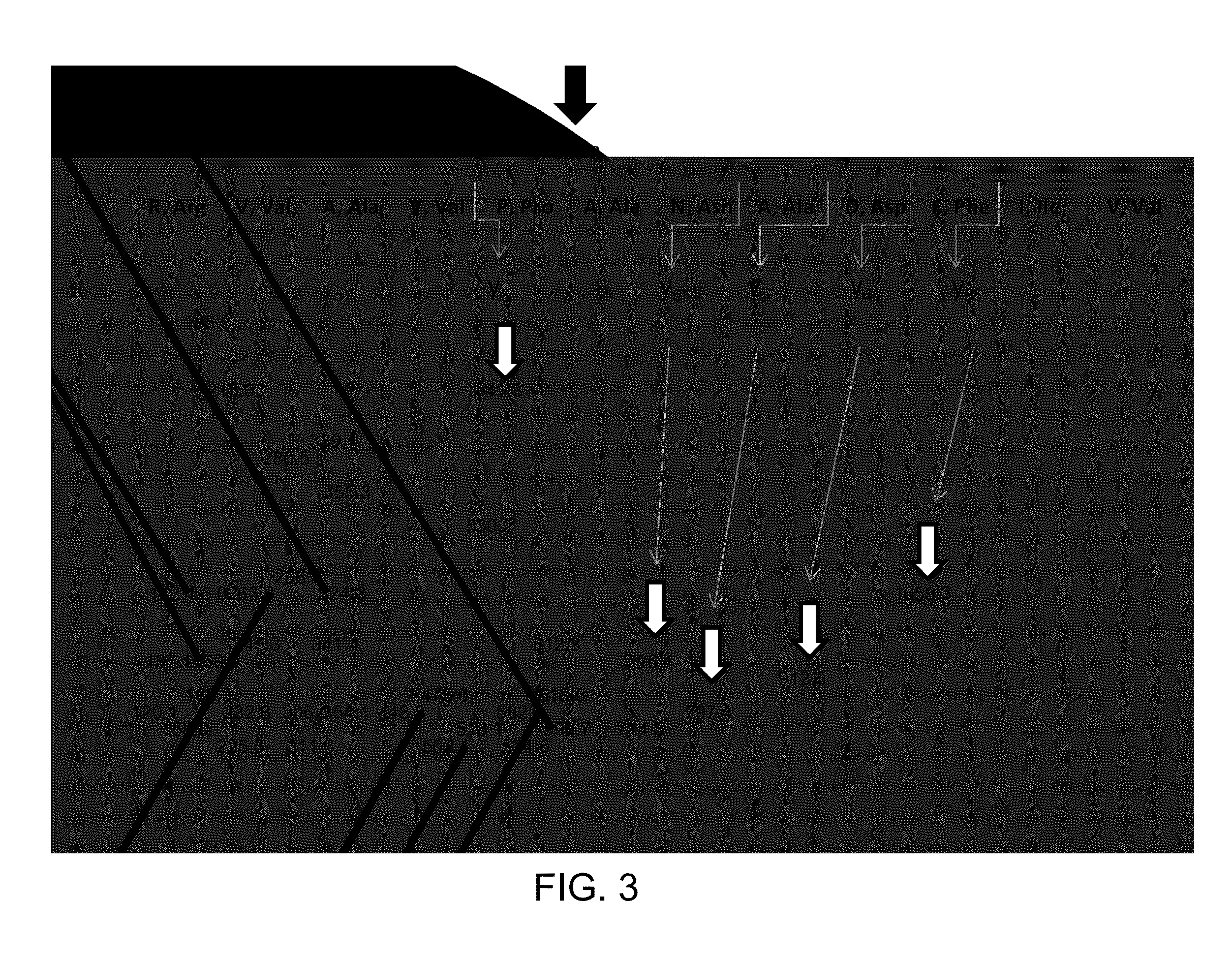
[0079]Standard of human Tg is purchased from AbD Serotec (Oxford, UK); stock solution of Tg is prepared at 1 mg / mL in 0.1% BSA, aliquoted in micro centrifuge tubes and stored at −70° C. Calibration standards of Tg are purchased from Beckman Coulter (Fullerton, Calif.); Tg concentrations in the standards are 0, 0.6, 6, 60, 150 ng / mL (0, 0.91, 9.1, 91 and 227 pmol / L). Rabbit polyclonal anti-Tg antibody is purchased from Covance (Princeton, N.J.) and diluted to 60 ng / μL with 0.1% BSA. Serum quality control samples are pooled human serum samples and contain 2, 6.5, and 170 ng / mL (3, 9.8, 258 pmol / L) of Tg. Working internal standard (IS) of “winged” peptide, sequence PVPESKVIFDANAPV*AVRSKVPDS (V* [13C5; 15N] (SEQ ID 005); mass shift 6 Da, RS synthesis Louisville, Ky.) is prepared at a concentration of 10 pg / μL (3.95 fmol / μL) in 20% acetonitrile in water.

[0080]Trypsin, formic acid (FA), dithiothreitol (DTT) and sodium deoxycho...

example 2

Conjugation of Antibody to Magnetic Beads

[0081]Custom polyclonal rabbit anti-peptide antibody (Covance, Princeton, N.J.) is conjugated to Tosyl activated magnetic beads (DynaBeads M280, Life Technologies, Carlsbad, Calif.) according to the manufacturer's recommendations. Briefly, the beads are washed with PBS (pH 7.4) and re-suspended in a 1M ammonium sulfate solution containing 20 μg of antibody per milligram of beads. Beads are incubated at 37° C. for 20 hours, washed and incubated for 1 hour with blocking buffer containing 0.5% BSA for one hour and reconstituted to a concentration of 20 μg / μL. Antibody content on the beads is approximately 0.2 μg / μL.

example 3

Trypsin Digestion Followed by Immunoaffinity Capture

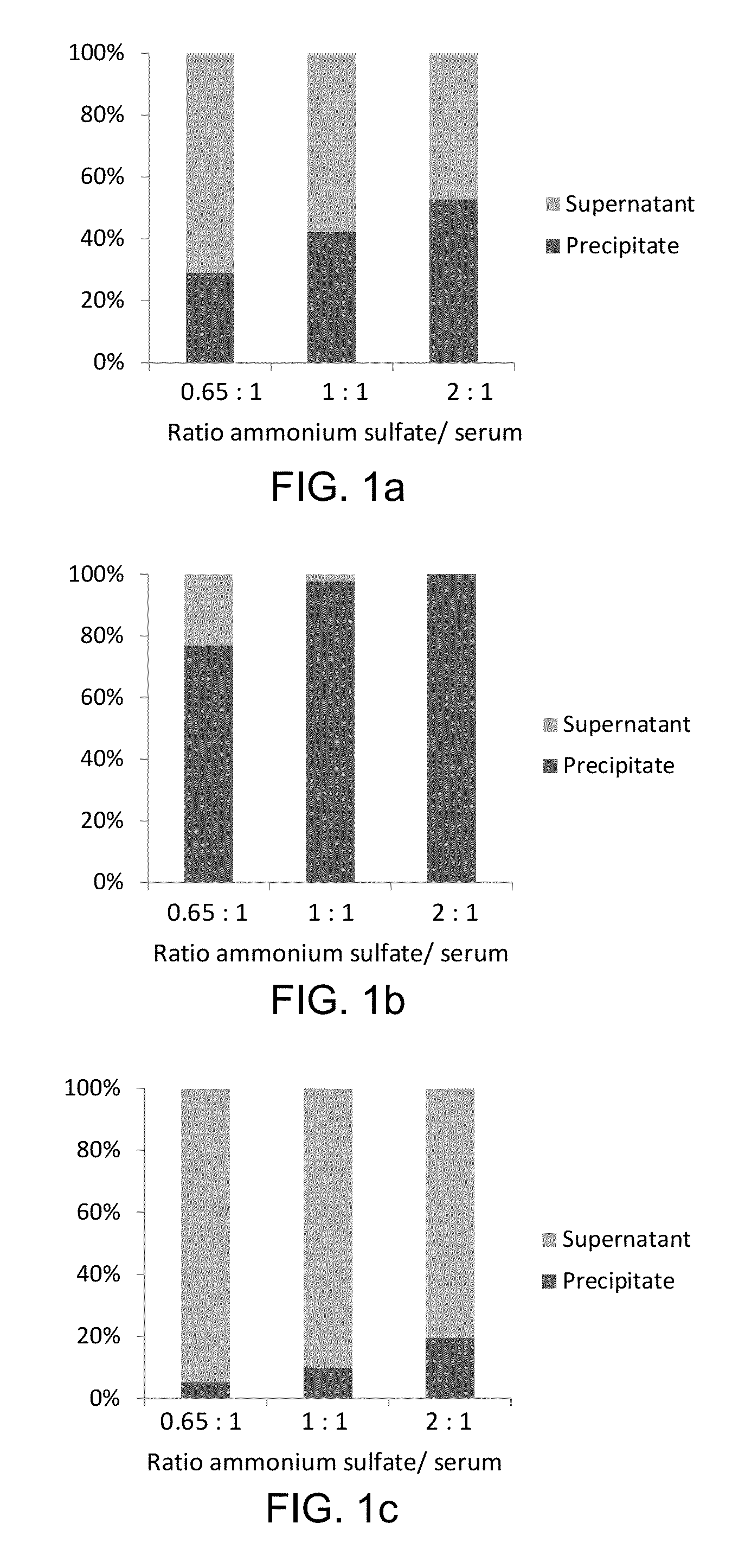
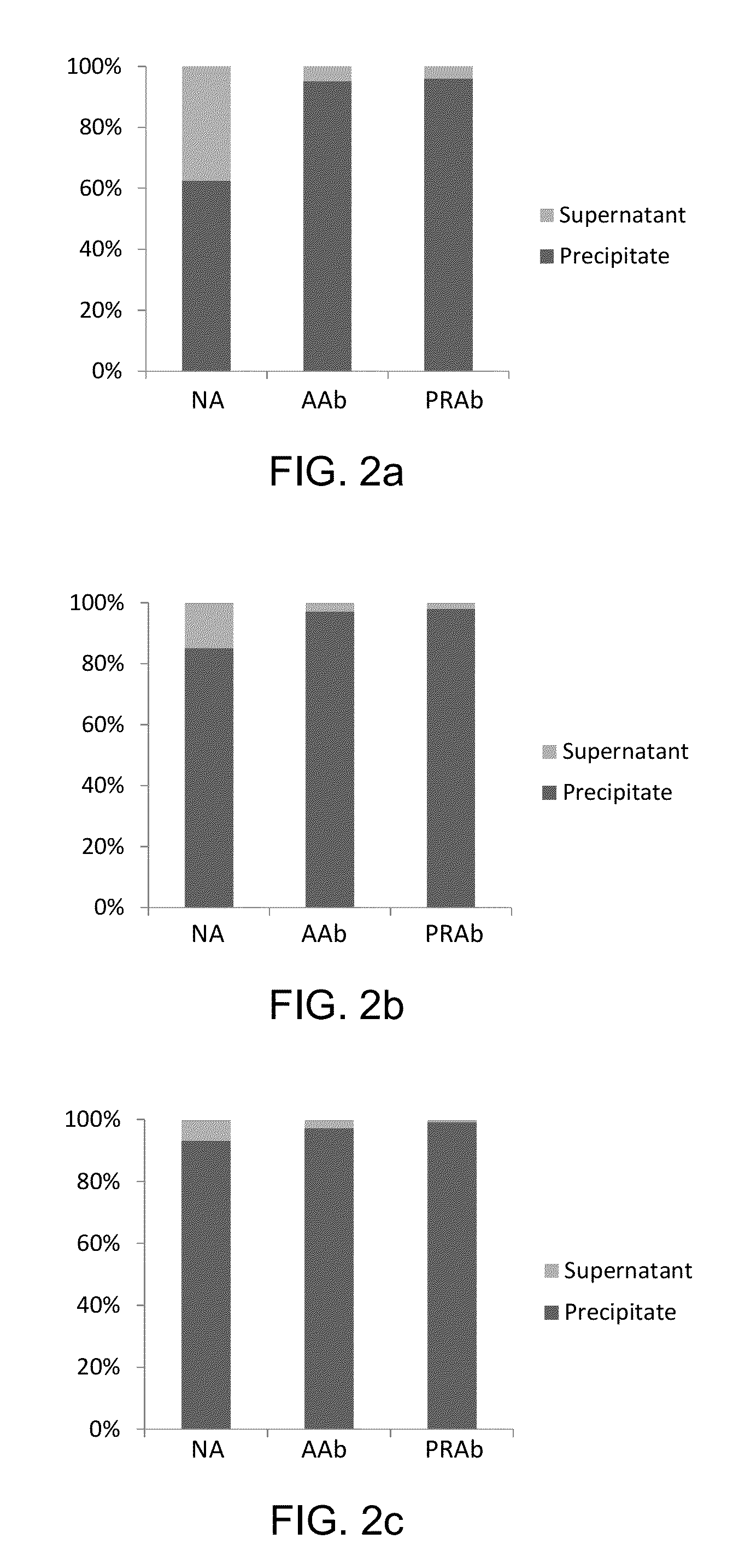
[0082]Sample preparation is performed on a liquid handler (epMotion, Eppendorf, Hamburg, Germany). 5 μL (60 ng / μL) of rabbit anti-Tg antibody is added to a 500 μL aliquot of serum or plasma sample and the samples are incubated for one hour with mixing at 20° C. After the incubation, 350 μL of saturated ammonium sulfate solution is added to the samples, and Tg is precipitated along with immunoglobulins (IG). The samples are vortex-mixed for 10 min, centrifuged for 5 min at 15000 g, and the supernatants are discarded. The precipitates are reconstituted with 300 μL of water, 10 μL of internal standard (IS), 10 μL of 20 mM dithiothreitol, and 30 μL of 5% sodium deoxycholate are added, after which the samples are incubated at 60° C. for 30 min. After the incubation, 400 μL of 25 mM ammonium bicarbonate and 10 μL of trypsin (4 μg / μL) are added to the samples, and the samples are incubated for 4 hours at 37° C.

[0083]Magnetic beads are pro...


PUM

PropertyMeasurementUnit
concentrationsaaaaaaaaaa

Abstract

The present invention provides methods, compositions, and kits associated with analyzing, enriching, and / or isolating a biomarker or analyte in a biological sample. In one aspect, for example, a method for determining a concentration of a biomarker in a biological sample can include binding any unbound biomarker with an antibody specific for the biomarker to form antibody-bound biomarker, enriching the antibody-bound biomarker and any endogenous autoantibody-bound biomarker to form an enriched fraction, identifying the biomarker in the enriched fraction, and determining the concentration of the biomarker in the biological sample. In one aspect, the concentration of the biomarker is derived from initially unbound biomarker and autoantibody-bound biomarker in the biological sample.

Description

PRIORITY DATA[0001]This application claims the benefit of U.S. Provisional Patent Application Ser. No. 61 / 632,125, filed on Jan. 18, 2012, which is incorporated herein by reference in its entirety.BACKGROUND OF THE INVENTION[0002]The presence or absence of specific biomarkers can be predictive in the diagnosis and / or determination of a variety of conditions in an individual. As such, the accuracy of detection and quantification of a particular biomarker can be important for the correct diagnosis and treatment of an associated condition. In those cases where a biomarker is incorrectly identified in or its concentration is incorrectly determined in a biological sample, the individual can be improperly treated for a condition that is not present or can be left untreated for a condition that is present.[0003]A variety of situations can make the detection of a biomarker problematic, both for the presence or absence of the biomarker, as well as the concentration level of the biomarker in ...

Claims


Application Information

Patent Timeline
Patent Type & Authority Patents(United States)
IPC IPC(8): G01N33/53G01N33/539G01N33/541
CPCG01N33/5306G01N33/539G01N33/541G01N33/564G01N2333/47G01N2560/00
Inventor KUSHNIR, MARK M.ROCKWOOD, ALAN L.MEIKLE, A. WAYNE
Owner UNIV OF UTAH RES FOUND
Why Eureka
Try Eureka