Self-resonant inductor wound portion of an implantable lead for enhanced MRI compatibility of active implantable medical devices

a self-resonant inductor and lead technology, applied in the field of medical leads, probes or catheters, can solve the problems of insufficient predictive sar level, latent problems of cardiac pacemakers, and the inability to fully predict the sar level, etc., and achieve the effect of rapid imaging

Active Publication Date: 2016-03-29
WILSON GREATBATCH LTD
View PDF450 Cites 1 Cited by
  • Summary
  • Abstract
  • Description
  • Claims
  • Application Information

AI Technical Summary

Problems solved by technology

Compatibility of cardiac pacemakers, implantable defibrillators and other types of active implantable medical devices with magnetic resonance imaging (MRI) and other types of hospital diagnostic equipment has become a major issue.
There have been reports of latent problems with cardiac pacemakers or other AIMDs after an MRI procedure sometimes occurring many days later.
Moreover, there are a number of recent papers that indicate that the SAR level is not entirely predictive of the heating that would be found in implanted lead wires or devices.
It is speculated that SAR level alone is not a good predictor of whether or not an implanted device or its associated lead wire system will overheat.
These antennas are not very efficient due to the damping effects of body tissue; however, this can often be offset by extremely high power fields (such as MRI pulsed fields) and / or body resonances.
There are a number of potential problems with MRI, including: (1) Closure of the pacemaker reed switch.
Worse yet, the reed switch may bounce or oscillate.
However, in patients with unstable conditions, such as myocardial ischemia, there is a substantial risk for ventricular fibrillation during asynchronous pacing.
However, it is generally felt that the reed switch will remain closed due to the powerful static magnetic field.
(2) Reed switch damage.
Direct damage to the reed switch is theoretically possible, but has not been reported in any of the known literature.
Some parts of pacemakers, such as the batteries and reed switch, contain ferrous magnetic materials and are thus subject to mechanical forces during MRI.
The effects of this heating are not readily detectable by monitoring during the MRI.
Such long term heating effects of MRI have not been well studied yet for all types of AIMD lead wire geometries.
There can also be localized heating problems associated with various types of electrodes in addition to TIP electrodes.
It has been observed that the RF field may induce undesirable fast pacing (QRS complex) rates.
The contribution of the time-varying gradient to the total strength of the MRI magnetic field is negligible, however, pacemaker systems could be affected because these fields are rapidly applied and removed.
Currents induced by time-varying magnetic gradient fields may lead to local heating.
There are additional problems possible with implantable cardioverter defibrillators (ICDs).
ICDs use different and larger batteries which could cause higher magnetic forces.
The programmable sensitivity in ICDs is normally much higher (more sensitive) than it is for pacemakers, therefore, ICDs may falsely detect a ventricular tachyarrhythmia and inappropriately deliver therapy.
There can also be heating problems of ICD leads which are expected to be comparable to those of pacemaker leads.
That is, during an MRI procedure, if they inadvertently sense the MRI fields as a dangerous ventricular arrhythmia, the ICD will attempt to charge up and deliver a high voltage shock.
This makes it highly unlikely that an ICD patient undergoing an MRI would receive an inappropriate high voltage shock therapy.
While ICDs cannot charge during MRI due to the saturation of their ferro-magnetic transformers, the battery will be effectively shorted and lose life.
This is a highly undesirable condition.
In summary, there are a number of studies that have shown that MRI patients with active implantable medical devices, such as cardiac pacemakers, can be at risk for potential hazardous effects.
These anecdotal reports are of interest, however, they are certainly not scientifically convincing that all MRI can be safe.
In a typical AM and FM radio in an automobile, these wavelength signals would not efficiently couple to the relatively short antenna of a cell phone.
There are certain implanted lead wire lengths that just do not couple efficiently with the MRI frequency and there are others that would couple very efficiently and thereby produce the worst case for heating.
Most experts still conclude that MRI for the pacemaker patient should not be considered safe.
As MRI technology progresses, including higher field gradient changes over time applied to thinner tissue slices at more rapid imagery, the situation will continue to evolve and become more complex.
However, some implantable pumps work on magneto-peristaltic systems, and must be deactivated prior to MRI.
There are newer (unreleased) systems that would be based on solenoid systems which will have similar problems.
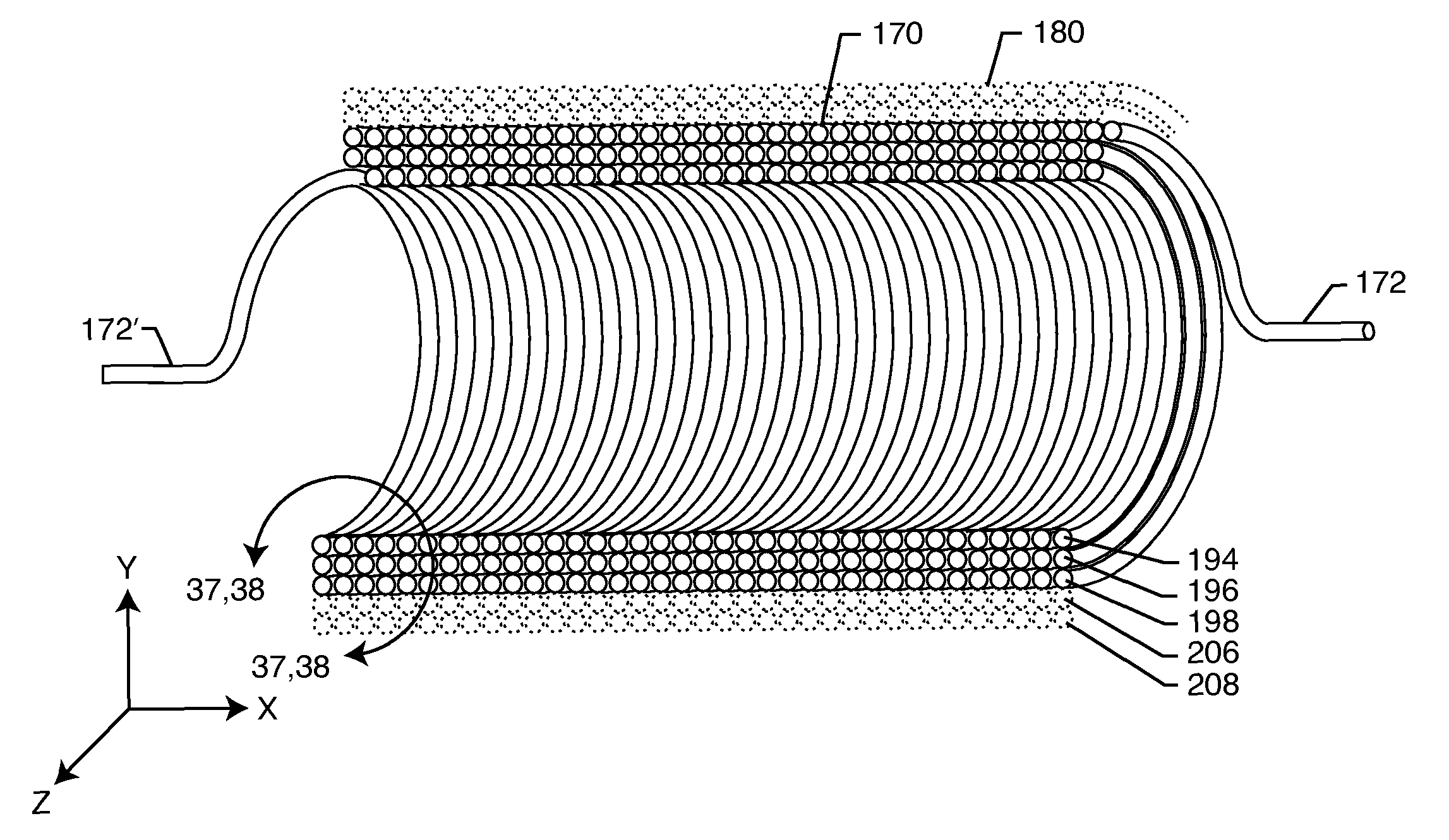
As one can see, many of the undesirable effects in an implanted lead wire system from MRI and other medical diagnostic procedures are related to undesirable induced currents in the lead wire system and / or its distal TIP (or RING).
This can lead to overheating either in the lead wire or at the body tissue at the distal TIP.

Method used


Image

  • Self-resonant inductor wound portion of an implantable lead for enhanced MRI compatibility of active implantable medical devices
  • Self-resonant inductor wound portion of an implantable lead for enhanced MRI compatibility of active implantable medical devices
  • Self-resonant inductor wound portion of an implantable lead for enhanced MRI compatibility of active implantable medical devices

Examples


Embodiment Construction

[0103]FIG. 1 illustrates various types of active implantable and external medical devices 100 that are currently in use. FIG. 1 is a wire formed diagram of a generic human body showing a number of implanted medical devices. 100A is a family of external and implantable hearing devices which can include the group of hearing aids, cochlear implants, piezoelectric sound bridge transducers and the like. 100B includes an entire variety of neurostimulators and brain stimulators. Neurostimulators are used to stimulate the Vagus nerve, for example, to treat epilepsy, obesity and depression. Brain stimulators are similar to a pacemaker-like device and include electrodes implanted deep into the brain for sensing the onset of the seizure and also providing electrical stimulation to brain tissue to prevent the seizure from actually happening. The lead wires that come from a deep brain stimulator are often placed using real time imaging. Most commonly such lead wires are placed during real time M...


PUM


Abstract

An implantable lead includes a lead conductor having a length extending from a proximal end to a distal end. A self-resonant inductor is connected in series along a portion of the length of the lead conductor. The self-resonant inductor includes a single length of conductive material including a dielectric coating substantially surrounding the single length of conductive material. The self-resonant inductor includes a first coiled or spiral conductor disposed along an inductor section spanning in a first direction from a first location to a second location. A second coiled or spiral conductor is disposed along the inductor section spanning in a second direction from the second location to the first location, where the second direction is opposite the first direction. A third coiled or spiral conductor is disposed along the inductor section spanning in the first direction from the first location to the second location.

Description

CROSS-REFERENCE TO RELATED APPLICATIONS[0001]This application claims priority to and is a continuation-in-part of U.S. application Ser. No. 12 / 891,292, filed on Sep. 27, 2010, now U.S. Pat. No. 8,437,865, the contents of which are incorporated herein by reference.DESCRIPTION[0002]1. Field of the Invention[0003]The present invention generally relates to medical leads, probes or catheters for surgery or implantation. More particularly, the present invention relates to a multilayer inductor connected in series along the lead, probe or catheter.[0004]2. Background of the Invention[0005]This invention relates generally to novel EMI tank filter assemblies, particularly of the type used in active medical devices (AMDs) such as cardiac pacemakers, cardioverter defibrillators, neurostimulators, externally worn Holter monitors and the like, which decouple lead wires and / or electronic components of the medical device from undesirable electromagnetic interference (EMI) signals at a selected fre...

Claims


Application Information

Patent Timeline
Patent Type & Authority Patents(United States)
IPC IPC(8): A61N1/05G01R33/36A61N1/08A61B18/00
CPCG01R33/3685A61N1/05A61B2018/00839A61B2090/374A61N2001/086A61N1/086
Inventor HALPERIN, HENRY, R.STEVENSON, ROBERT, A.KONDABATNI, KISHORE, KUMARFRYSZ, CHRISTINE, A.
Owner WILSON GREATBATCH LTD
Why Eureka
Try Eureka